-
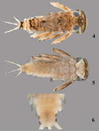
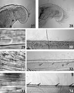
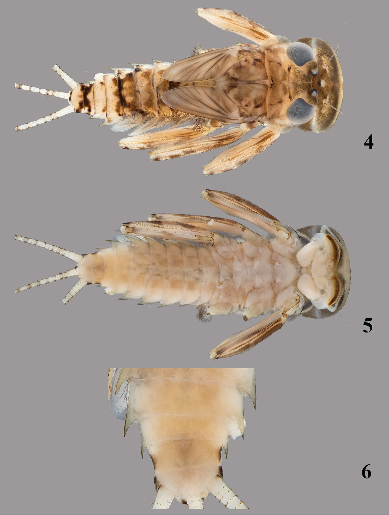
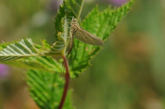
Figures 4–6.Thalerosphyrus sinuosus (Navás, 1933). 4 Habitus in dorsal view 5 Habitus in ventral view 6 Detail of abdominal segments VI–IX in ventral view.
-
Kongsvad Mølleå
-
Almind Sø, Jylland, Danmark
-
Djursland Midt
-
Boonsatien Boonsoong, Dietrich Braasch
Zookeys
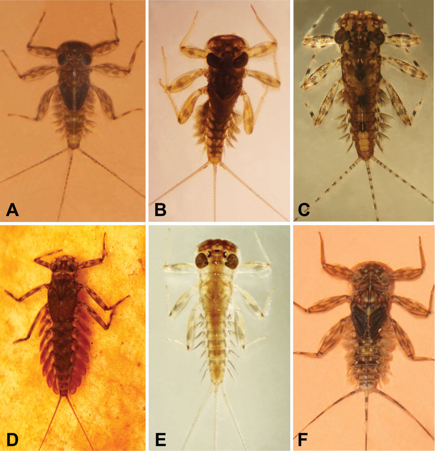
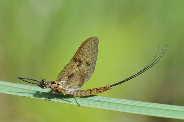
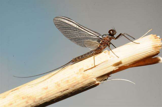
Figure 9.A Habitus of Asionurus namnaoensis Braasch & Boonsoong, 2010 B habitus of Asionurus primus Braasch & Soldán, 1986 Chabitus of Compsoneuria thienemanniUlmer, 1939 Dhabitus of Epeorus khayengensis Boonsoong & Braasch, 2010 E habitus of Rhithrogena tonkinensis Soldán & Braasch, 1986 F habitus of Thalerosphyrus sinuosus Navás, 1933.
-
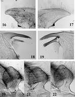
Figures 16–22.Mouthparts structure of Thalerosphyrus determinatus (20), Thalerosphyrus sinuosus (16, 21) and Thalerosphyrus lamuriensis (17, 18, 19, 22). 16–17 Hemi-labrum 18 Left mandible 19 Right mandible 20–22 Labial glossa.
-
Kongsvad Mølleå
-
Almind Sø, Jylland, Danmark
-
Djursland Midt
-
Boonsatien Boonsoong, Dietrich Braasch
Zookeys
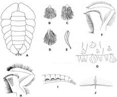
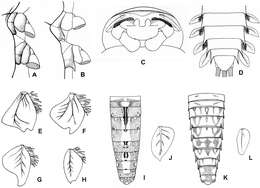
Figure 2.A Ventral view of abdomen of Rhithrogena siamensis Braasch & Boonsoong, 2009 B–E lamellae of gills 1 (B), 3 (C), 5 (D) and 7 (E) of Trichogenia maxillaris Braasch & Soldán, 1988 F ventral view of left maxilla of Trichogenia maxillaris Braasch & Soldán, 1988 G bristles on dorsal face of abdominal terga of Trichogenia maxillaris Braasch & Soldán, 1988 H ventral view of left maxilla of Compsoneuria langensis Braasch & Boonsoong, 2010 I-J abdominal terga (I) and tergum VII (J) of Notacanthurus baei Braasch & Boonsoong, 2009.
-
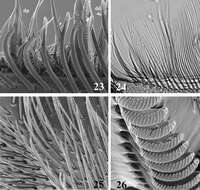
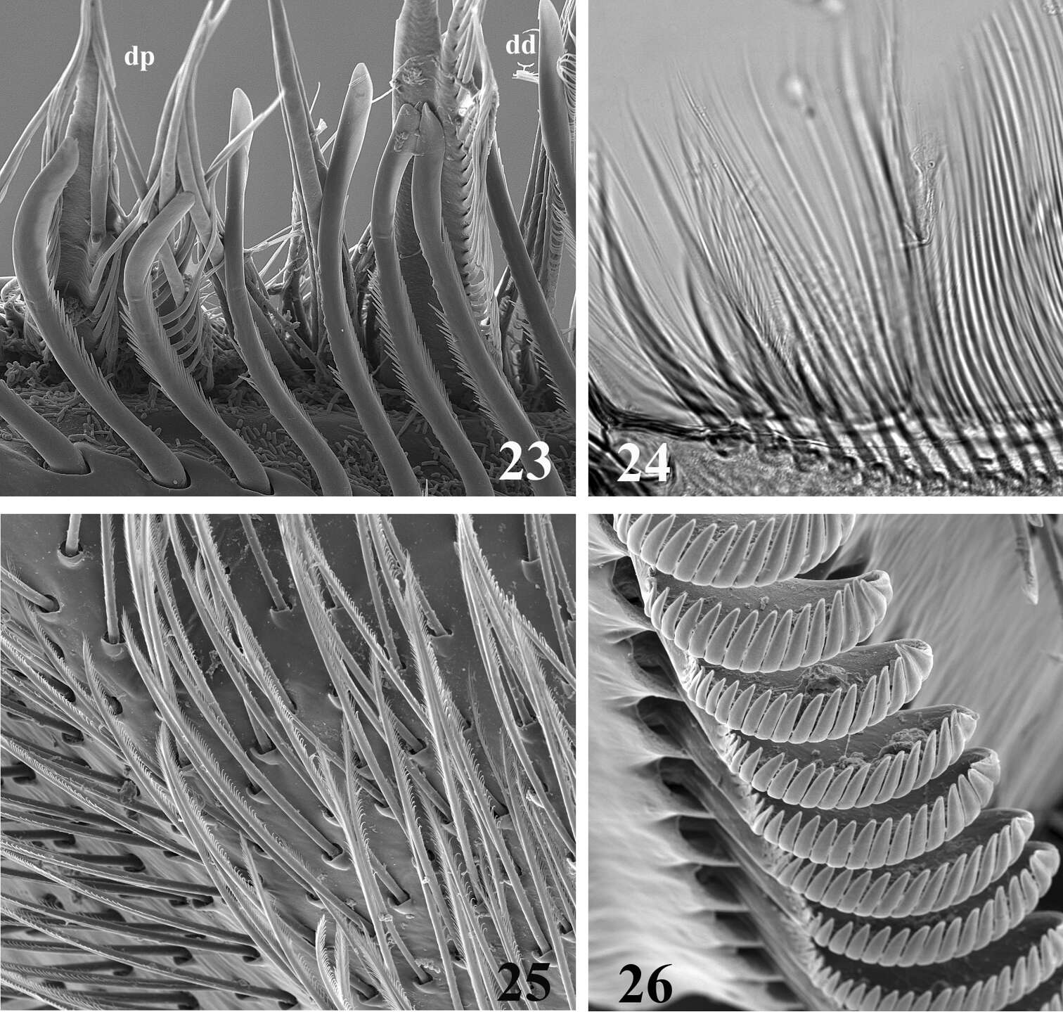
Figures 23–26.SEM (23, 25, 26) and optic (24) pictures of maxillar structure. 23–24 Dentisetae of Thalerosphyrus lamuriensis dp: proximal dentisetae, dd distal dentisetae 25 Scattered setae on the ventral face of the galea-lacinia of Thalerosphyrus sinuosus 26 Comb-shape setae on the crown of the galea-lacinia of Thalerosphyrus sinuosus.
-
Kongsvad Mølleå
-
Slåensø
-
Djursland Midt
-
Boonsatien Boonsoong, Dietrich Braasch
Zookeys
Figure 3.A Right side of thorax of Afronurus namnaoensis Braasch & Boonsoong, 2010 B–D right side of thorax (B), ventral view of head capsule (C) and ventral view of posterior abdomen (D) of Thalerosphyrus sinuosus Navás, 1933 E–H lamella of gills 3 (E), 4 (F), 5 (G), 6 (H) of Compsoneuria (Siamoneuria) kovaci Braasch, 2006 I–J dorsal view of abdomen (I), lamella of gills 7 (J) of Compsoneuria thienemanni Ulmer, 1939 K–L dorsal view of abdomen (K), lamella of gills 7 (L) of Compsoneuria langensis Braasch & Boonsoong, 2010.
-
Figures 27–34.Mouthpart (27–28) and thoracic (29–34) structures of Thalerosphyrus determinatus (27, 29, 30), Thalerosphyrus sinuosus (31, 32) and Thalerosphyrus lamuriensis (28, 33, 34). 27–28 Apex of superlingua of hypopharynx 29, 31, 33 Bristles on the dorsal face of hind femur 30, 32, 34 Outer margin of hind tibia.
-
Kongsvad Mølleå
-
Slåensø
-
Djursland Midt
-
Boonsatien Boonsoong, Dietrich Braasch
Zookeys
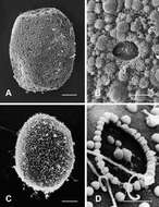
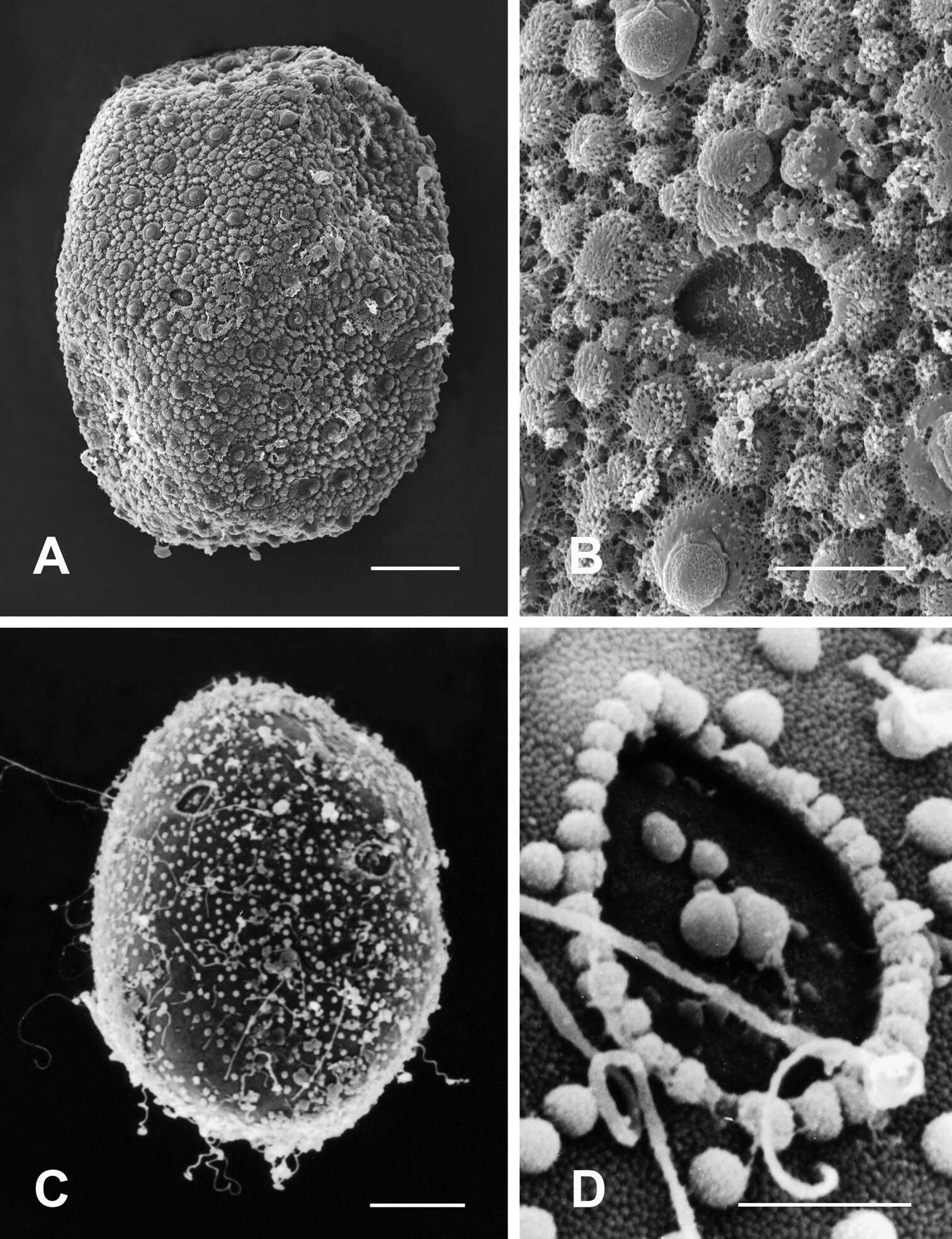
Figure 7.A–B General outline (A) and micropyle (B) of the egg of Compsoneuria thienemanniUlmer, 1939 C–D General outline (C) and micropyle (D) of the egg of Asionurus primus Braasch & Soldán, 1986. Scale bars 20 µm for A and C; 5 µm for B and D.
-
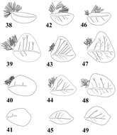
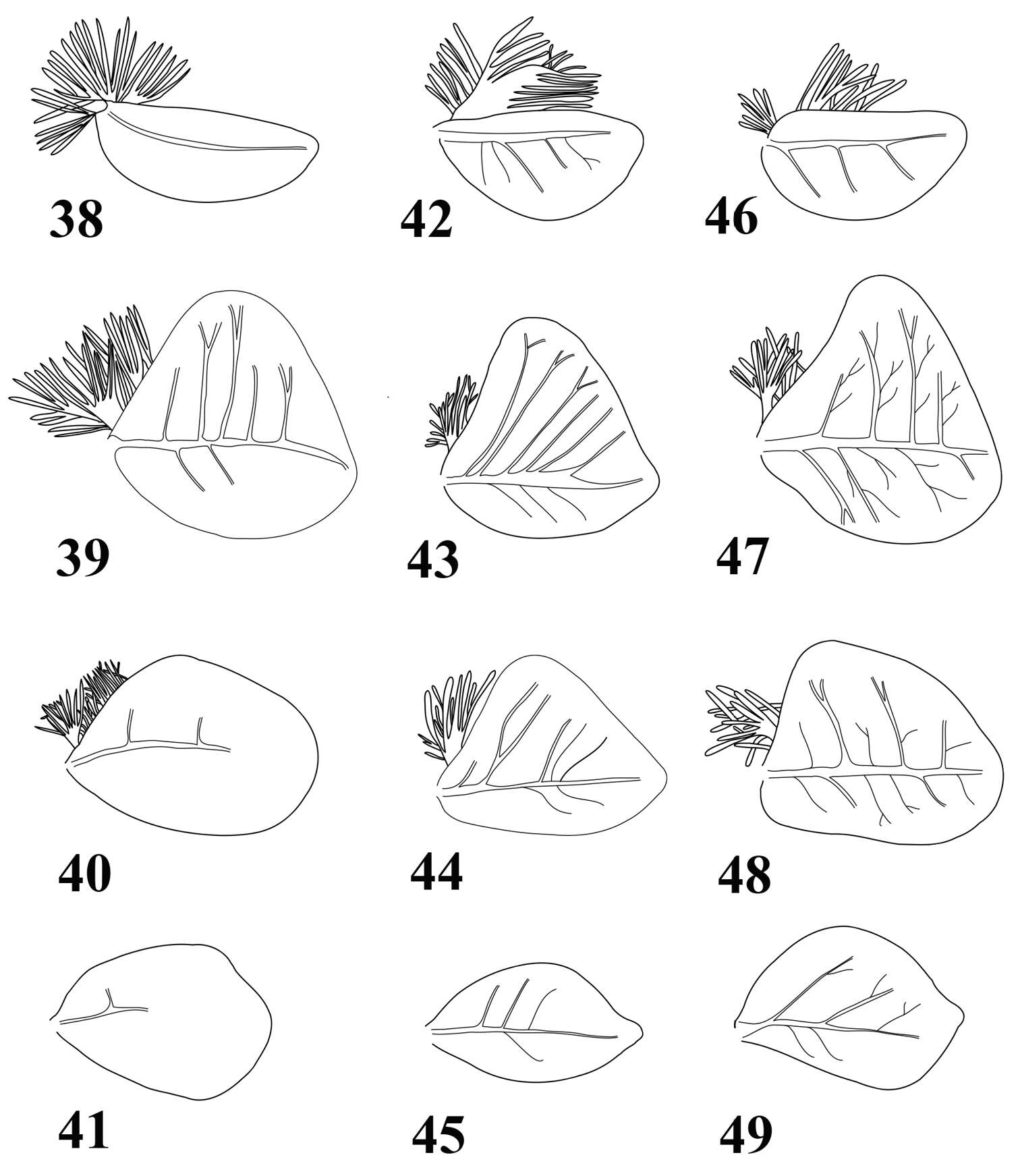
Figures 38–49.Gills of Thalerosphyrus determinatus (38–41), Thalerosphyrus sinuosus (42–45) and Thalerosphyrus lamuriensis (46–49). 38, 42, 46 Gill I 39, 43, 47 Gill IV 40, 44, 48 Gill VI 41, 45, 49 Gill VII.
-
Kongsvad Mølleå
-
Slåensø
-
Djursland Midt