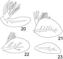
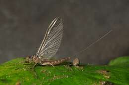
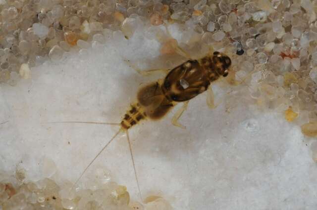
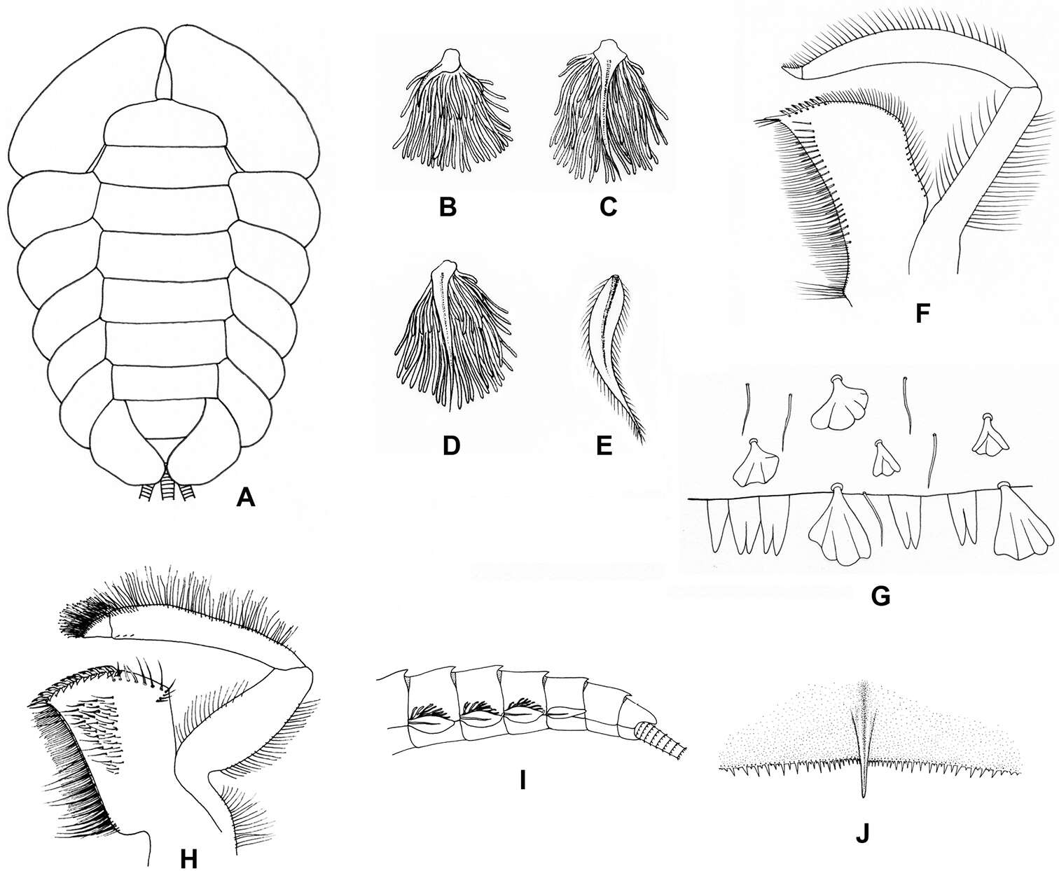
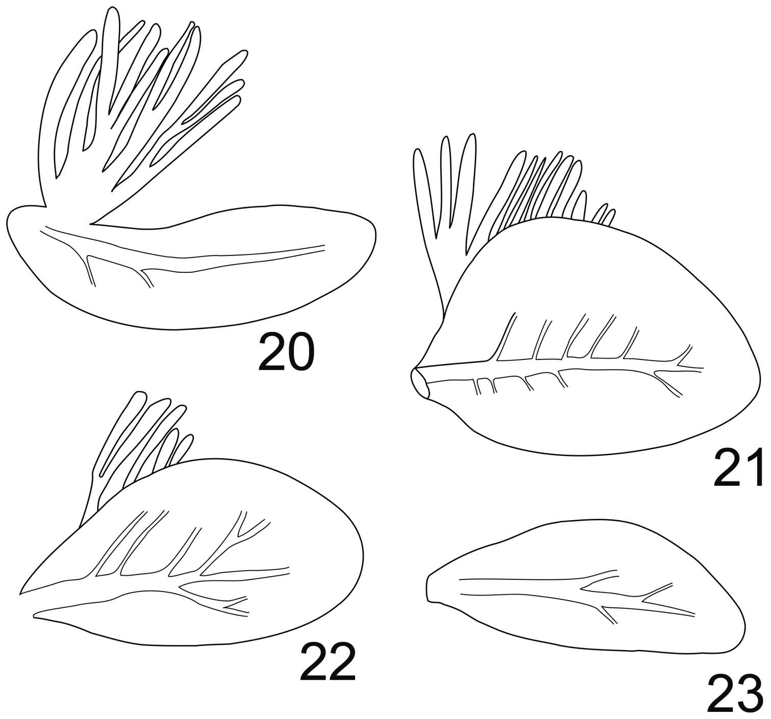
Figure 2.A Ventral view of abdomen of Rhithrogena siamensis Braasch & Boonsoong, 2009 B–E lamellae of gills 1 (B), 3 (C), 5 (D) and 7 (E) of Trichogenia maxillaris Braasch & Soldán, 1988 F ventral view of left maxilla of Trichogenia maxillaris Braasch & Soldán, 1988 G bristles on dorsal face of abdominal terga of Trichogenia maxillaris Braasch & Soldán, 1988 H ventral view of left maxilla of Compsoneuria langensis Braasch & Boonsoong, 2010 I-J abdominal terga (I) and tergum VII (J) of Notacanthurus baei Braasch & Boonsoong, 2009.
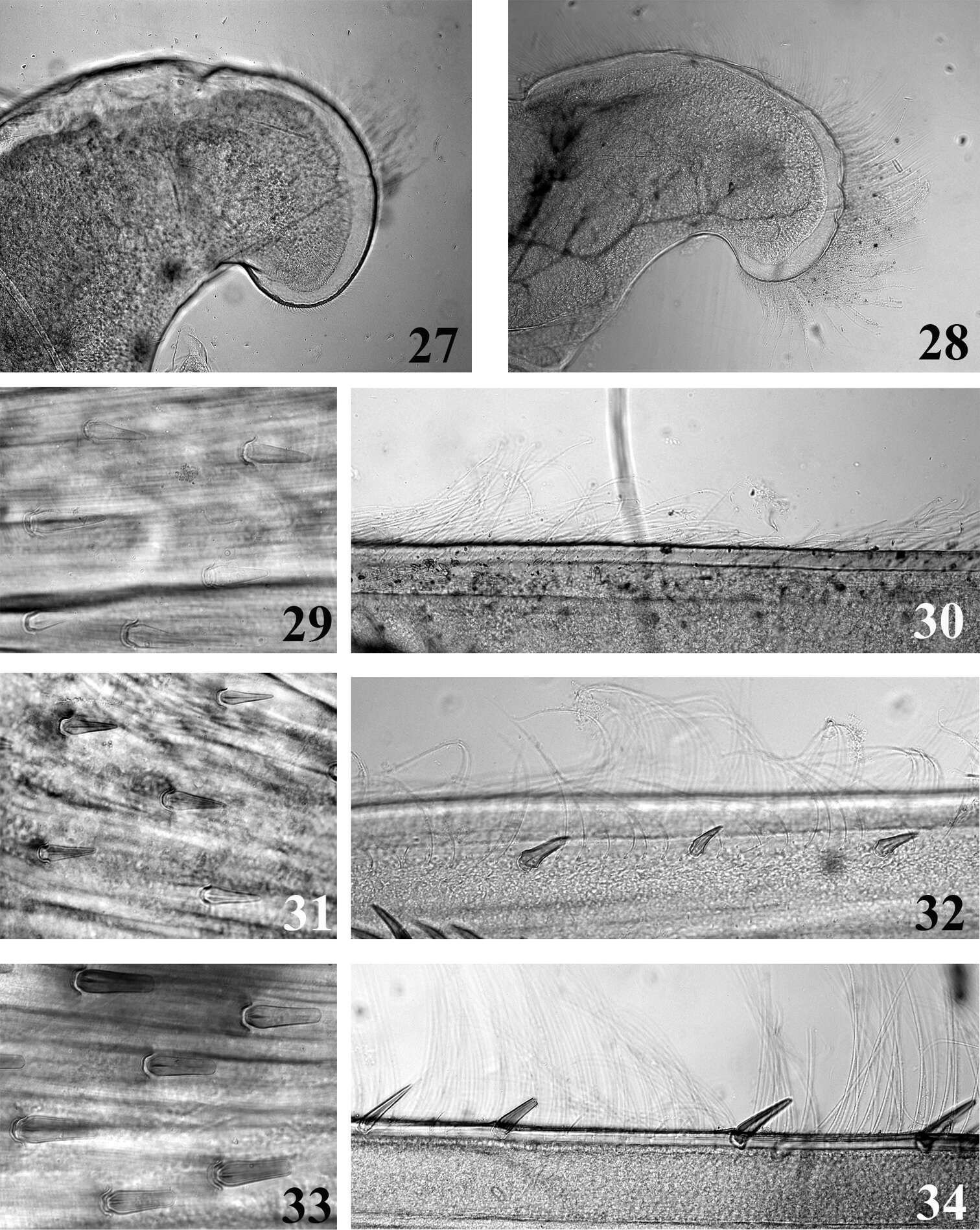
Figures 27–34.Mouthpart (27–28) and thoracic (29–34) structures of Thalerosphyrus determinatus (27, 29, 30), Thalerosphyrus sinuosus (31, 32) and Thalerosphyrus lamuriensis (28, 33, 34). 27–28 Apex of superlingua of hypopharynx 29, 31, 33 Bristles on the dorsal face of hind femur 30, 32, 34 Outer margin of hind tibia.
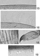
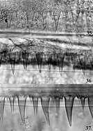
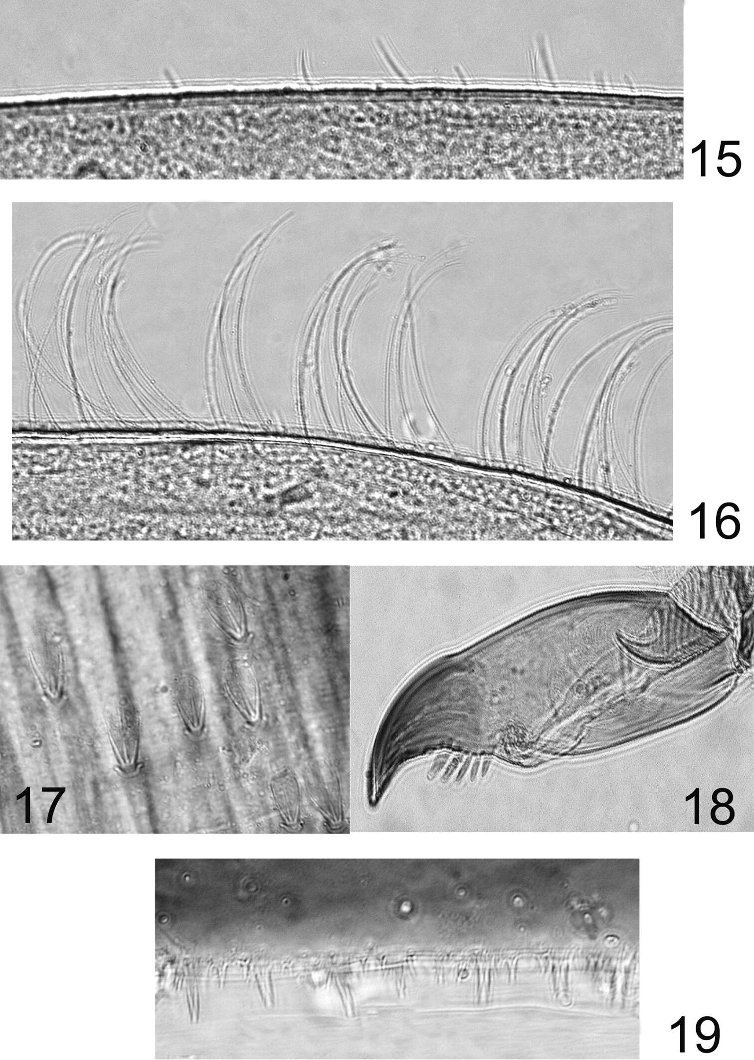
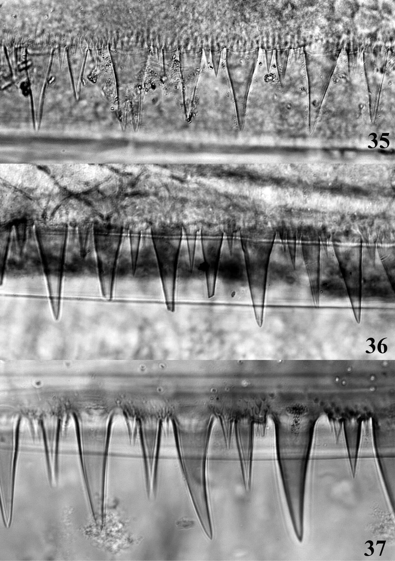
Figures 15–19.Rhithrogeniella ornata Ulmer, 1939. 15 Outer margin of the fore tibia 16 Outer margin of the hind tibia 17 Bristles on the dorsal surface of hind femur 18 Tarsal claw 19 Posterior margin of tergite V.
Figure 2.A Ventral view of abdomen of Rhithrogena siamensis Braasch & Boonsoong, 2009 B–E lamellae of gills 1 (B), 3 (C), 5 (D) and 7 (E) of Trichogenia maxillaris Braasch & Soldán, 1988 F ventral view of left maxilla of Trichogenia maxillaris Braasch & Soldán, 1988 G bristles on dorsal face of abdominal terga of Trichogenia maxillaris Braasch & Soldán, 1988 H ventral view of left maxilla of Compsoneuria langensis Braasch & Boonsoong, 2010 I-J abdominal terga (I) and tergum VII (J) of Notacanthurus baei Braasch & Boonsoong, 2009.