-
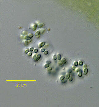
Microcystis aeruginosa (Kützing, 1833) F.T. Kützing, 1846.Collected from a freshwater aquaculture tub near Boise, Idaho December 2005. DIC
-
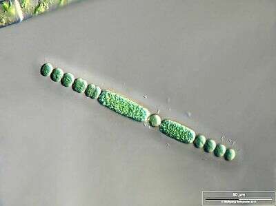
Scale bar indicates 50 µm. Sample from the pond Hegne Moor situated in the vicinity of Lake Constance. Images were taken using Zeiss Universal with Olympus C7070 CCD camera.Image under Creative Commons License V 3.0 (CC BY-NC-SA).
-
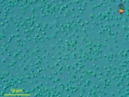
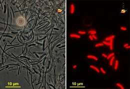
Prochlorococcus (pro-cloe-row-cock-us) marinus, a tiny globular cyanobacterium (blue-green alga). This type of organism is extremely abundant in the tropical and warm temperate regions of the open oceans, and some scientists claim it is the most abundant organism on earth. Differential interference microscopy.
data on this strain.
-
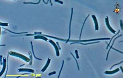
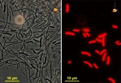
Synechococcus (sin-eck-owe-cock-us), a rod or sausage shaped cyanobacterium (blue green alga) which dominates in the upper two green layers of this microbial mat which was provided by Mike Ferris from Mushroom Spring, a thermal site in Yellowstone National Park, photograph provided by Mike Ferris and David Ward.
-
Synechococcus (sin-eck-owe-cock-us), a rod or sausage shaped cyanobacterium (blue green alga) which comes from the upper layers of a microbial mat. The cells have terminal inclusions. Other bacteria are present in the same sample. Phase contrast. Material provided by Mike Ferris from Mushroom Spring, a thermal site in Yellowstone National Park, photograph by Mike Ferris and David Patterson.
-
Synechococcus (sin-eck-owe-cock-us), a rod or sausage shaped cyanobacterium (blue green alga) which comes from the upper layers of a microbial mat. This is an aggregate of cells. Phase contrast. Material provided by Mike Ferris from Mushroom Spring, a thermal site in Yellowstone National Park, photograph by Mike Ferris and David Patterson.
-
Synechococcus (sin-eck-owe-cock-us), a rod or sausage shaped cyanobacterium (blue green alga) which comes from the upper layers of a microbial mat. This detailed cell shows the presence of deposits at the ends of the cells, as well as small aggregates within the cell. Material from Nymph Creek, a thermal site in Yellowstone National Park, photograph by Kathy Sheehan and David Patterson.
-
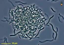
Synechococcus (sinm-eck-owe-cock-us), this pair of matched micrographs shows bacteria, mostly Synechococcus and Chloroflexus) from a mat sample. The phase contrast shot to the left shows the bacteria, the image to the right shows autofluorescence. Only the sausage-shaped Synechococcus exhibits autofluorescence. Phase contrast and fluorescence. Material provided by Mike Ferris from Mushroom Spring, a thermal site in Yellowstone National Park, photograph by Mike Ferris and David Patterson.
-
Synechococcus (sin-eck-o-cock-us) is a blue-green alga (cyanobacterium) that may have either a blue green or (as here) a red colour. Cluster of globular cells. Differential interference microscopy.
data on this strain.
-
Synechococcus (sin-eck-o-cock-us) is a blue-green alga (cyanobacterium) that may have either a blue green or (as here) a red colour. Cluster of globular cells. Differential interference microscopy.
data on this strain.
-
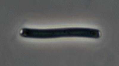
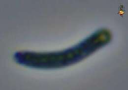
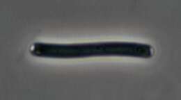
An individual Synechococcus cell.
-
These cells have been squashed slightly. They were found in water that was in placed coloured red because of the large numbers of red bacteria. This species is quite large. Differential interefernce contrast optics.
-
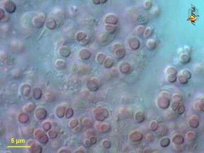
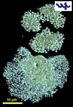
Of the genus Microcystis (Cyanobacteria, chroococales), M. wesenbergii is the easiest species to identify: the spherical cells are relatively large (5-8 µm) and are embedded in thick colorless homogeneous mucilage with clearly defined contours. It is generally less abundant in Lake Kinneret than M. aeruginosa and M. flos aquae.
-
Under conditions of nitrogen deficiency A. ovalisporum develops heterocytes (central round and empty-looking cell in this filament), in which N2 fixation by nitrogenase enzymes takes place.
-
Aphanizomenon ovalisporum (Cyanobacteria, Nostocales), showing atypical long cells in a filament grown in culture.
-
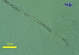
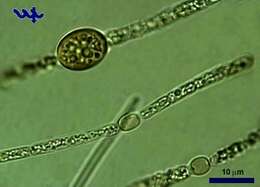
The specialized cells of Aphanizomenon ovalisporum: an akinete (large, thick-walled, egg-shaped cell in the upper filament) and a hetetocyte (egg-shaped, transparent cells in the middle and bottom filaments). The akinetes are much larger than regular cells and can be mid-filament or terminal, at times a few in a single filament, sometimes several in a row. Akinetes function as asexual resting stage and are resistant to harsh conditions. Akinete formation is induced by P-limitation. The akinetes separate from the filament and sink to the sediments, where they remain until conditions are suitable for germination.
-
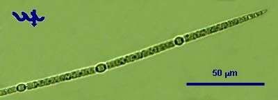
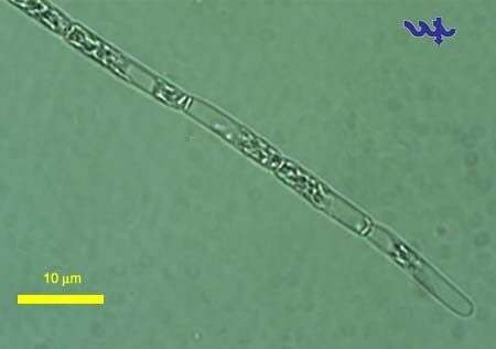
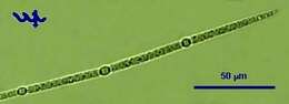
Aphanizomenon ovalisporum (Cyanobacteria, Nostocales) was the first N2-fixing filamentous cyanobacterium that ever bloomed in Lake Kinneret. It happened in 1994 when this species formed an unprecedented autumn bloom. Since then Cylindrospermosis cuspis, another N2 fixer, invaded the lake and now dominates the summer-fall phytoplankton assemblages. A. ovalisporum produces the toxin cylindrospermopsin, which makes it an undesirable species in this important source of drinking water. Aphanizomenon ovalisporum is abundant during summer and fall, being evenly distributed with depth throughout the epilimnion. The filaments are 2 â 5 µm thick and 20 â 500 µm long. Heterocytes (seen as 3 distinct oval cells dispersed along the filament) appear when N-deficient conditions prevail. Akinete formation is induced by P-deficiency. The akinetes (not seen here) are much larger than regular cells and can be mid-filament or terminal, at times a few in a single filament, sometimes several in a row. This specimen was sampled from shallow water near the Kinneret Limnological Laboratory in June 2006.
-
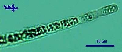
This photo shows the typical terminal cells of Aphanizomenon ovalisproum from Lake Kinneret. The filaments, 2 â 5 µm thick and 20 â 500 µm long, are terminated by an elongated hyaline cell, as seen in this picture.
-
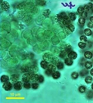
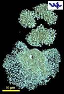
Microcystis flos-aquae together with Microcystis aeruginosa is common in Lake Kinneret throughout the year, and blooming periodically. The cells at the border and at the center of Microcystis colony looks different by chlorophyll and gas vesicles concentrations. The specimen was sampled from the shore of the lake in March 2006.
-
-
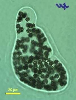
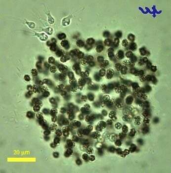
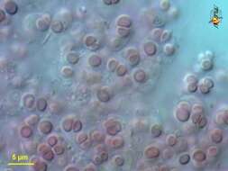
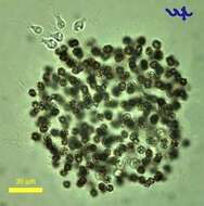
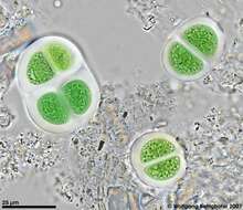
Microcystis flos-aquae (Cyanobacteria, Chroococcales) is a together with Microcystis aeruginosa are common in Lake Kinneret throughout the year, occasionally forming blooms with surface scums in winter or spring. The individual cells in a M. flos-aquae colony are 3-4 um in diameter, the colony is spherical or lens-shaped, with varying degree of spacing between cells within a colony. The dark spots clearly showing in the individual cells in this picture are due to the reflection of light from the gas vesicles. A bunch of choanoflagellates are attached at the upper left side of the colony.
-
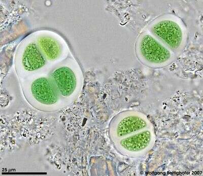
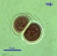
Chroococcus turgidus, (Cyanobacteria, Chroococales), from Lake Kinneret pelagic waters, April 2006, showing 2 daughter cells after division by simple binary fission â as characteristic for most Chroococcales species. This species is common in the plankton of Lake Kinneret throughout the year. Usually there are 2 â 8 cells in a colony. Clearly delimited colorless mucilaginous envelopes surround the individual cells following their contours, and the entire colony. Cell diameter: 8 â 11 µm.
-
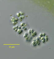
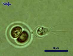
Chroococcus turgidus (Cyanobacteria, Chroococcales) is common in Lake Kinneret in recent years. In this unusual photograph a choanoflagellate is attached to its outer shell.
-
High resolution photo of Chroococcus turgidus using Planapo 63/1.4. Sample from sphagnum pond situated in the northern alpine region of Austria near Salzburg. Images were taken using Zeiss Universal with Olympus C7070 CCD camera.