-
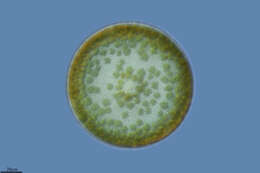
Chaetoceros diadema.Resting spores in two developing states. Scale bar indicates 50 m. Sample from North Sea near Heligoland (spring diatom bloom). Images were taken using Zeiss Universal with Olympus C7070 CCD camera.For more look at
www.protisten.de/english/gallery_main/gallery_main.htmlFor high-resolution images please ask postmaster@protisten.de.
-
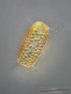
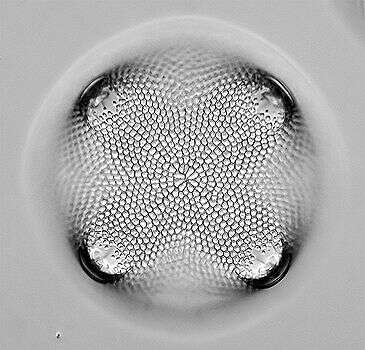
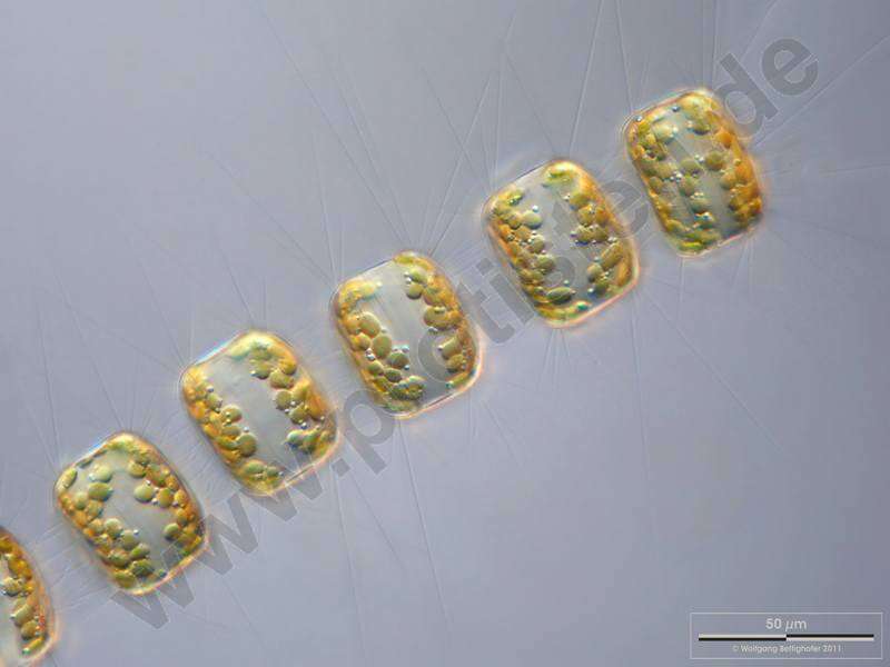
Thalassiosira punctigera.The oblique view exhibits short silicous spines, the so called occluded processes. Some chitinous spines protruding from the fultoportulae (also called strutted processes) along the dotted valve margin are also visible. Scale bar indicates 50 m. The image was built up using several photomicrographic frames with manual stacking technique. Sample from North Sea near Heligoland (spring diatom bloom). Images were taken using Zeiss Universal with Olympus C7070 CCD camera.For more look at
www.protisten.de/english/gallery_main/gallery_main.htmlFor high-resolution images please ask postmaster@protisten.de.
-
Reboredo, Galicia, Spain
-
-
-
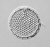
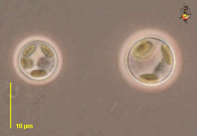
Cyclotella (sike-low-tell-a). Centric diatom, seen from valve view. Three plastid profiles are visible around the periphery of the cell. Long thin organic spines project from the cell - and are believed to have a role in flotation. The pattern of pores in the frustule is used in identification. Marine. Phase contrast.
-
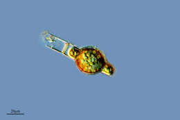
Corethron (core-eeth-ron) hystrix, centric diatom (stramenopile) with siliceous spines emerging from the border of the valves, many girdle bands (not visible here) make up the body of the cylinder. This image emphasizes the plastids. Differential interference microscopy.
data on this strain.
-
-
Balea, Galicia, Spain
-
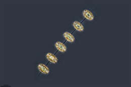
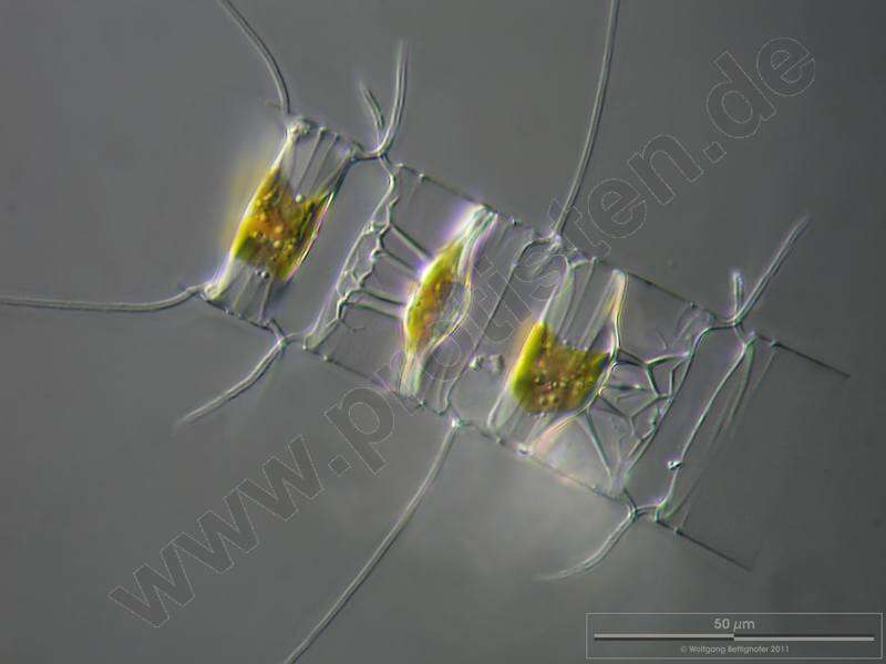
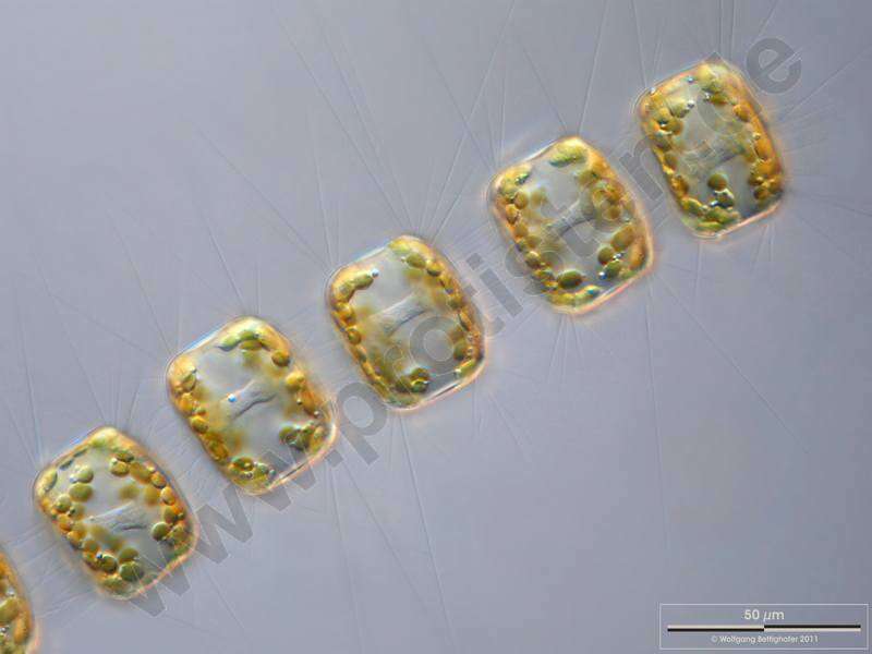
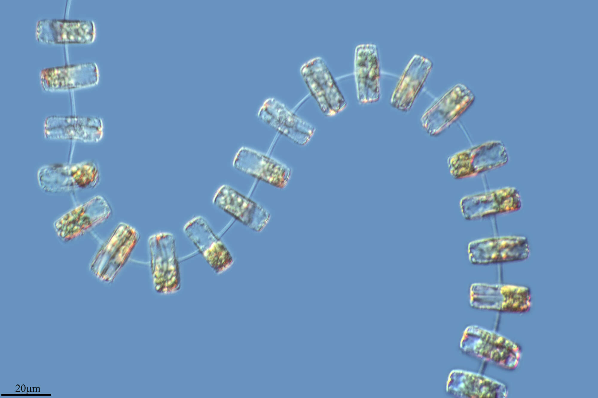
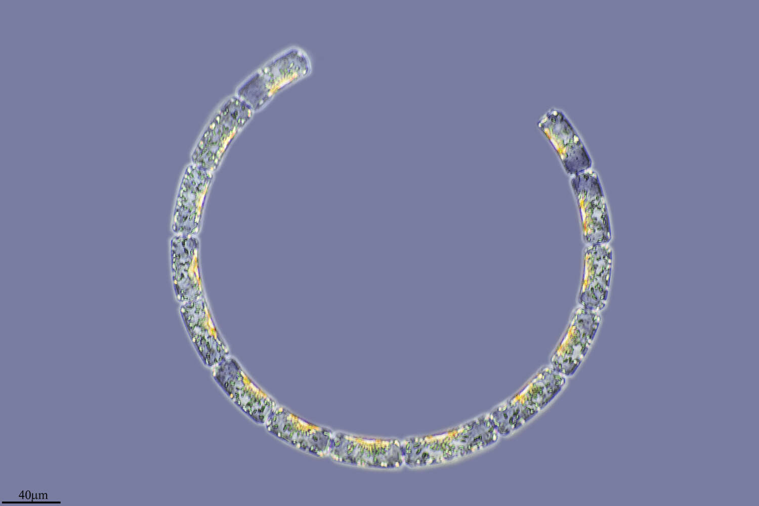
Chain of Porosira glacialis. Note that the delicate spines are chitinous. Focus on cell center showing cytoplasmic accumulation around the nucleus. Scale bar indicates 50 m. The image was built up using several photomicrographic frames with manual stacking technique. Sample from North Sea near Heligoland (spring diatom bloom). Images were taken using Zeiss Universal with Olympus C7070 CCD camera.For more look at
www.protisten.de/english/gallery_main/gallery_main.htmlFor high-resolution images please ask postmaster@protisten.de..
-
Grove, O, Galicia, Spain
-
Galende, Castile and Len, Spain
-
-
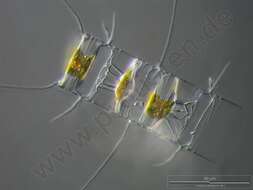
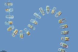
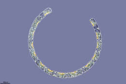
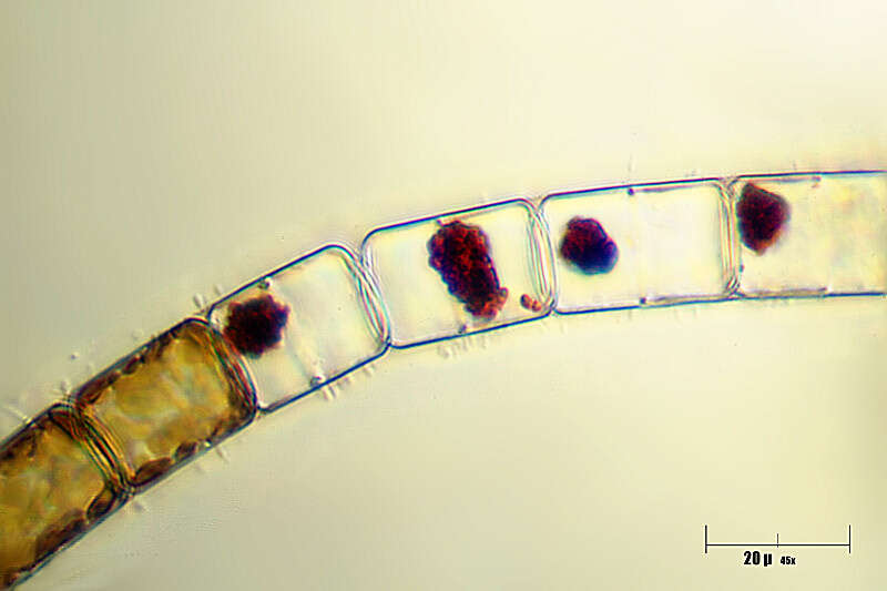
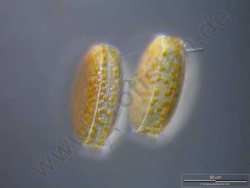
Chaetoceros diadema.Chain with vegetative cells and resting spores. Scale bar indicates 25 m. Sample from North Sea near Heligoland (spring diatom bloom). The image was built up using several photomicrographic frames with manual stacking technique. Images were taken using Zeiss Universal with Olympus C7070 CCD camera.For more look at
www.protisten.de/english/gallery_main/gallery_main.htmlFor high-resolution images please ask postmaster@protisten.de.
-
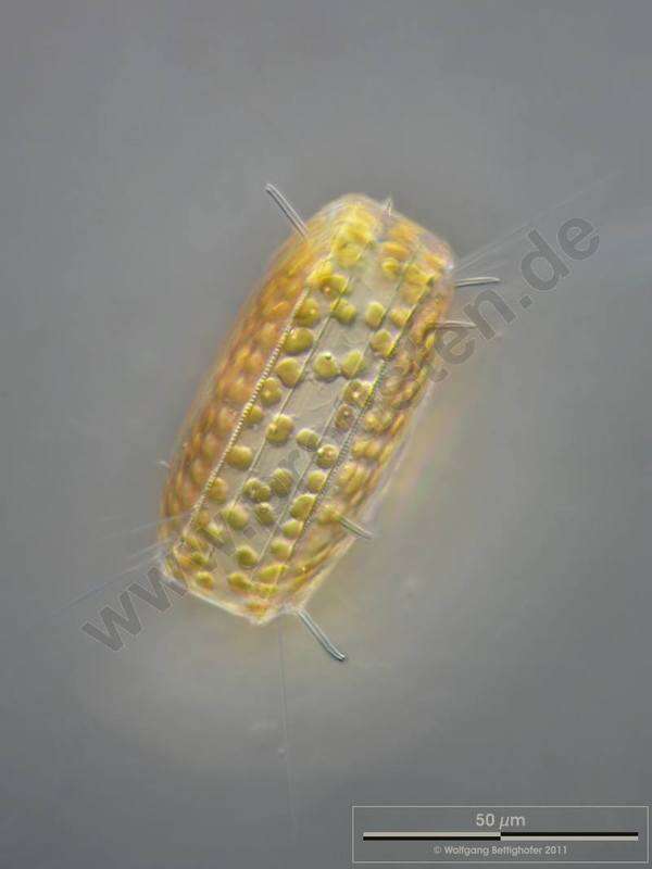
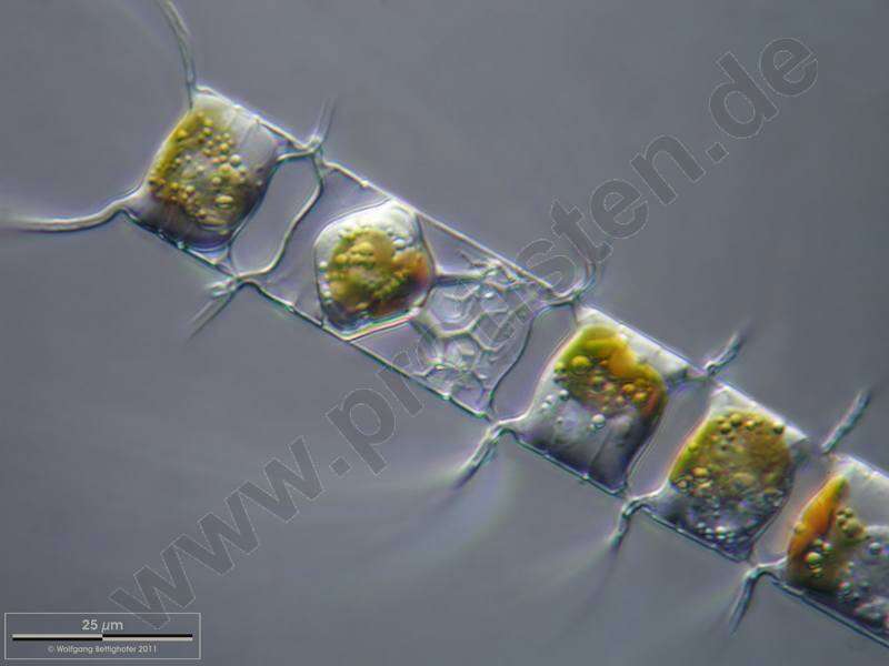
Thalassiosira punctigera.The oblique view exhibits short silicous spines, the so called occluded processes. On the lower left, lower right and central above chitinous spines are visible. Scale bar indicates 50 m. The image was built up using several photomicrographic frames with manual stacking technique. Sample from North Sea near Heligoland (spring diatom bloom). Images were taken using Zeiss Universal with Olympus C7070 CCD camera.For more look at
www.protisten.de/english/gallery_main/gallery_main.htmlFor high-resolution images please ask postmaster@protisten.de.
-
Grove, O, Galicia, Spain
-
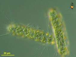
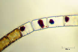
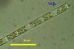
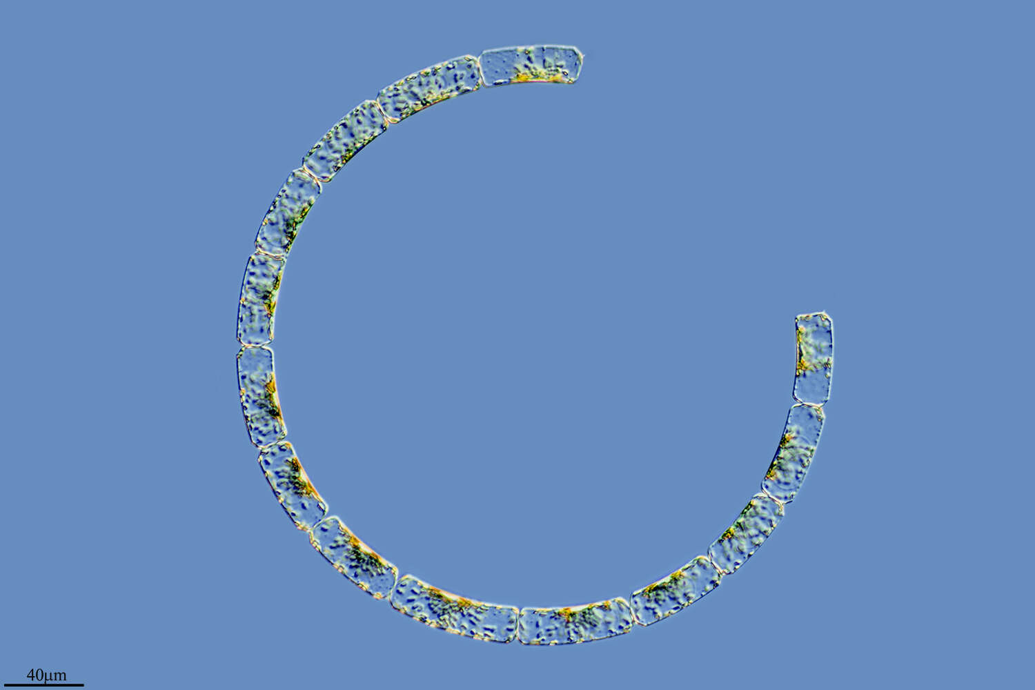
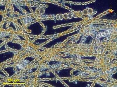
This is a typical filament of Aulacoseira (formerly Melosira) granulata (Bacillariophyta, Centrales)that was resuspended from the sediments not long before sampling. Its linking spines are of different lengths, possibly indicative of the filament having been broken at this place. In contrast, spines of equal length as in the other photo of this species are indicative of active growth. The chloroplasts here are partly compacted and do not fill the entire cell volume, indicative of the origin of this particular filament from the sediments. The other photo of this species shows actively growing, planktonic cells with chloroplasts filling the entire cell. This specimen was sampled from the shore of the lake in June 2006.
-
Cyclotella (sike-low-tell-a). Centric diatom, seen from valve view. Three plastid profiles are visible around the periphery of the cell. Long thin organic spines project from the cell - and are believed to have a role in flotation. The pattern of pores in the frustule is used in identification. Marine. Phase contrast.
-
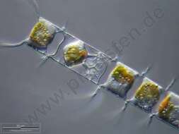
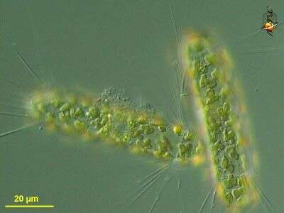
Melosira (mellow-sire-a) nummuloides, filament forming centric diatom, with multiple small plastids within the cell. Dark ground illumination. Leptosiropsis (leapt-owe-sire-op-sis) torulosa, green alga with organic wall that is produced in layers. Phase contrast microscopy.
data on this strain.
-
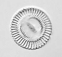
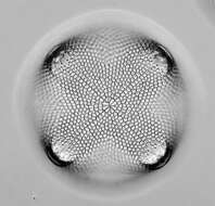
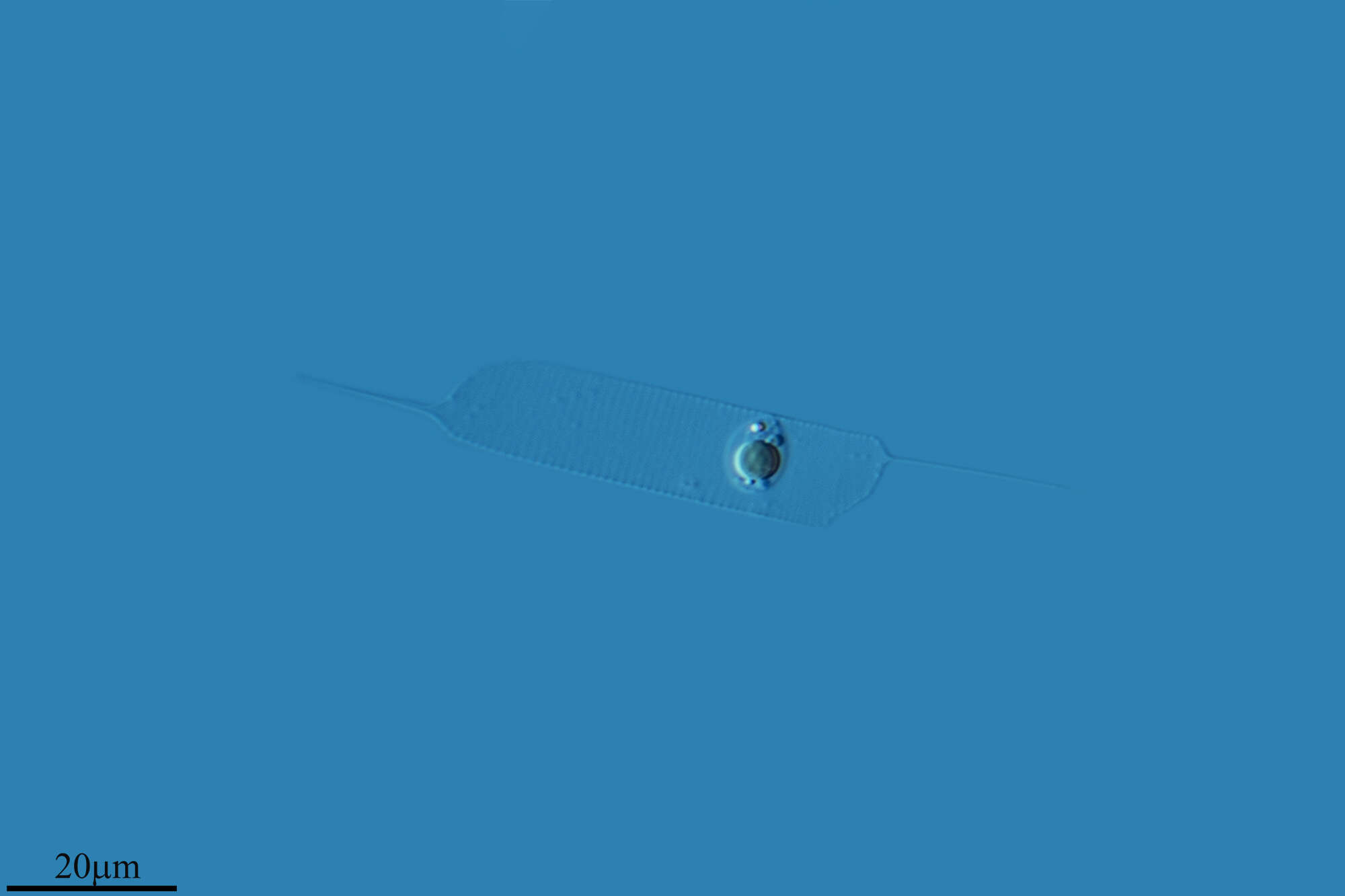
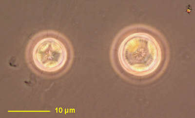
Some specimen of this centric diatom carried naviculoid ones on the valve(s). Scale bar indicates 25 µm. The image was built up using several photomicrographic frames with manual stacking technique. Sample from North Sea near Heligoland (spring diatom bloom). Images were taken using Zeiss Universal with Olympus C7070 CCD camera.
-
Balea, Galicia, Spain
-
Chain of Porosira glacialis. Note that the delicate spines are chitinous. Focus on frustule surface. Scale bar indicates 50 m. The image was built up using several photomicrographic frames with manual stacking technique. Sample from North Sea near Heligoland (spring diatom bloom). Images were taken using Zeiss Universal with Olympus C7070 CCD camera.For more look at
www.protisten.de/english/gallery_main/gallery_main.htmlFor high-resolution images please ask postmaster@protisten.de.
-
Ribadelago, Castille and Leon, Spain
-
Talamanca, Catalonia, Spain