-
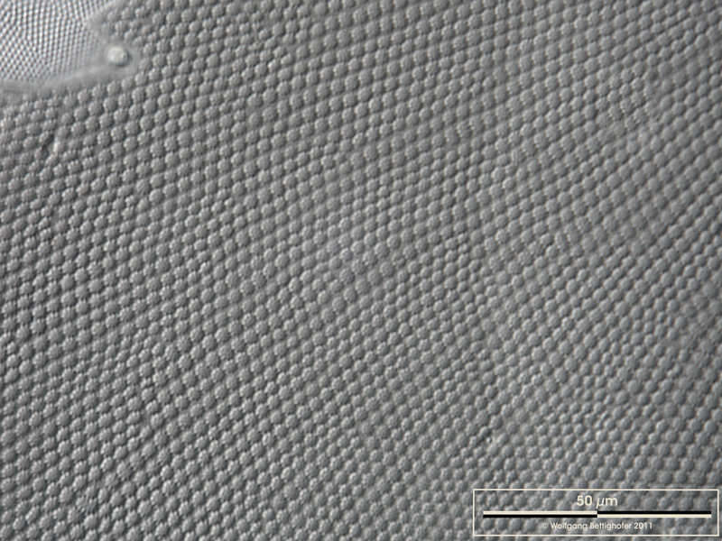
Coscinodiscus wailesii DIC closeup of valve of a living specimen. Scale bar indicates 50 µm. Sample from North Sea near Heligoland (spring diatom bloom). The image was built up using several photomicrographic frames with manual stacking technique. Images were taken using Zeiss Universal with Olympus C7070 CCD camera.Image under Creative Commons License V 3.0 (CC BY-NC-SA). Place name: North Sea around Heligoland Latitude: 54.186311 Longitude: 7.895034 Ausschnitt einer Schale, aufgenommen von einer lebenden Zelle. Multiebenen-Abbildung, manuell gestapelt. Der Messbalken markiert eine Länge von 50 µm. Probe aus der Nordsee vor Helgoland in der Zeit der Frühjahrsblüte. Mikrotechnik: Zeiss Universal, Kamera: Olympus C7070.Creative Commons License V 3.0 (CC BY-NC-SA). For permission to use of (high-resolution) images please contact postmaster@protisten.de.
-
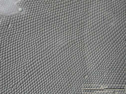
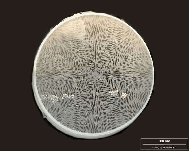
Coscinodiscus wailesii Valvar View. Scale bar indicates 100 µm. Sample from North Sea near Heligoland (spring diatom bloom). The image was built up using several photomicrographic frames with manual stacking technique. Use of SEM equipment courtesy of Lab Dr. Karl-Heinz Schäffner, Solingen, Germany. Place name: North Sea around Heligoland Latitude: 54.186311 Longitude: 7.895034 Schalenansicht. Der Messbalken markiert eine Länge von 100 µm. Probe aus der Nordsee vor Helgoland in der Zeit der Frühjahrsblüte. Die Aufnahme entstand im Labor Dr. Karl-Heinz Schäffner, Solingen.Creative Commons License V 3.0 (CC BY-NC-SA). For permission to use of (high-resolution) images please contact postmaster@protisten.de.
-
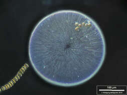
Coscinodiscus wailesii Sample from North Sea near Heligoland (spring diatom bloom). The image was built up using several photomicrographic frames with manual stacking technique. Images were taken using Leica dissecting microscope with MFT camera Olympus OM-D E-M5 II.Image under Creative Commons License V 3.0 (CC BY-NC-SA). Place name: North Sea around Heligoland Latitude: 54.186311 Longitude: 7.895034 Probe aus der Nordsee vor Helgoland in der Zeit der Frühjahrsblüte. Mikrotechnik: Leica Stereomikroskop, Kamera: Olympus OM-D E-M5 II.Creative Commons License V 3.0 (CC BY-NC-SA). For permission to use of (high-resolution) images please contact postmaster@protisten.de.
-
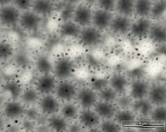
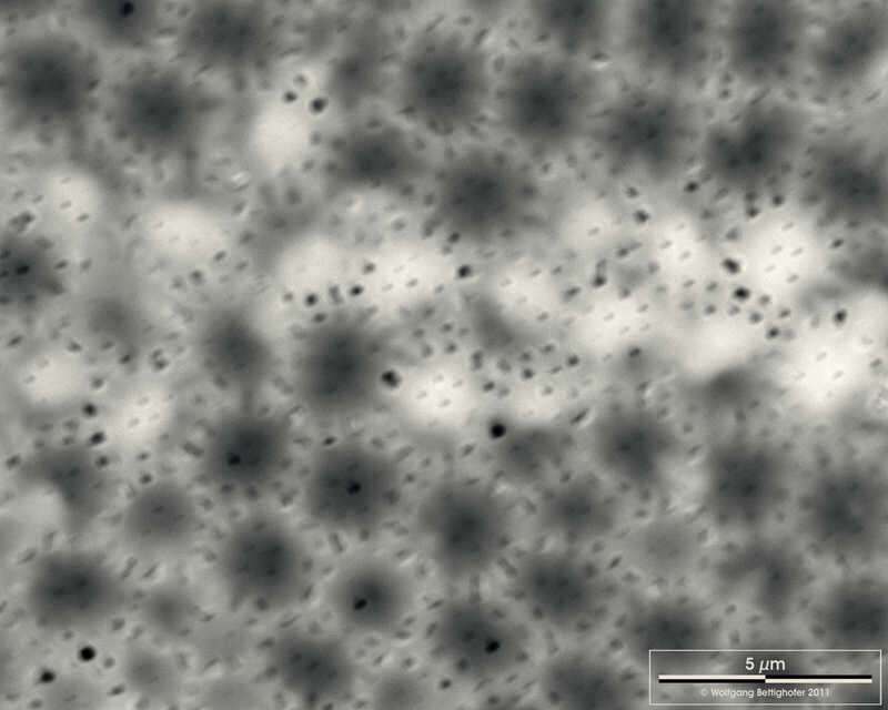
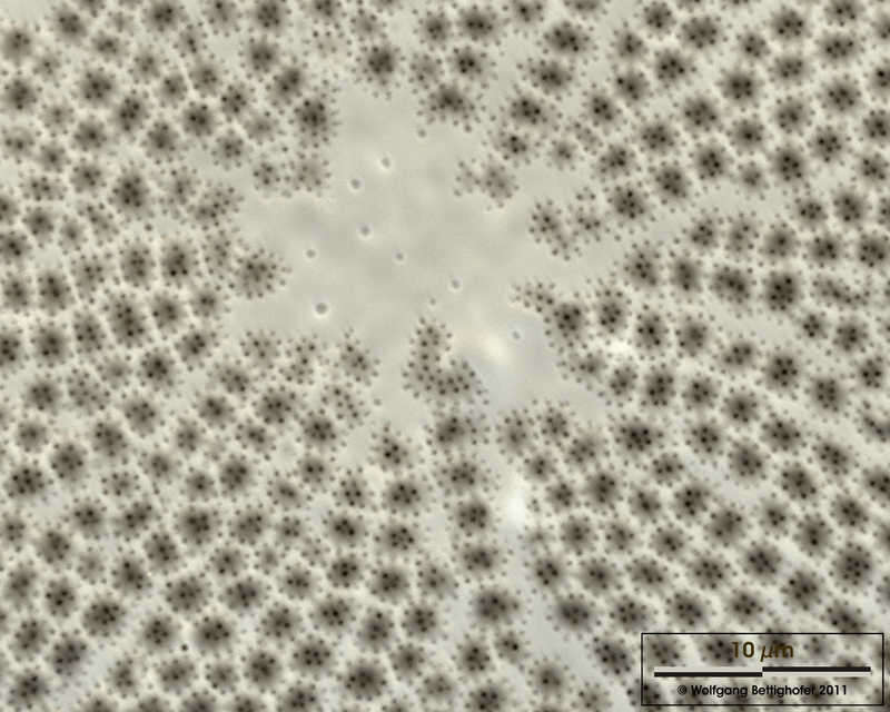
Coscinodiscus wailesii Closeup showing fine structure of valvar pores. Scale bar indicates 5 µm. Sample from North Sea near Heligoland (spring diatom bloom). Use of SEM equipment courtesy of Lab Dr. Karl-Heinz Schäffner, Solingen, Germany. Place name: North Sea around Heligoland Latitude: 54.186311 Longitude: 7.895034 Der Ausschnitt zeigt den Feinbau der Poren der Schale. Der Messbalken markiert eine Länge von 5 µm. Probe aus der Nordsee vor Helgoland in der Zeit der Frühjahrsblüte. Die Aufnahme entstand im Labor Dr. Karl-Heinz Schäffner, Solingen.Creative Commons License V 3.0 (CC BY-NC-SA). For permission to use of (high-resolution) images please contact postmaster@protisten.de.
-
Coscinodiscus wailesii Sample from North Sea near Heligoland (spring diatom bloom). The image was built up using several photomicrographic frames with manual stacking technique. Images were taken using Leica dissecting microscope with MFT camera Olympus OM-D E-M5 II.Image under Creative Commons License V 3.0 (CC BY-NC-SA). Place name: North Sea around Heligoland Latitude: 54.186311 Longitude: 7.895034 Probe aus der Nordsee vor Helgoland in der Zeit der Frühjahrsblüte. Mikrotechnik: Leica Stereomikroskop, Kamera: Olympus OM-D E-M5 II.Creative Commons License V 3.0 (CC BY-NC-SA). For permission to use of (high-resolution) images please contact postmaster@protisten.de.
-
Coscinodiscus wailesii Closeup showing hyaline central area. Scale bar indicates 10 µm. Sample from North Sea near Heligoland (spring diatom bloom). Use of SEM equipment courtesy of Lab Dr. Karl-Heinz Schäffner, Solingen, Germany. Place name: North Sea around Heligoland Latitude: 54.186311 Longitude: 7.895034 Ausschnitt aus der unperforierten Mitte der Schale. Der Messbalken markiert eine Länge von 25 µm. Probe aus der Nordsee vor Helgoland in der Zeit der Frühjahrsblüte. Die Aufnahme entstand im Labor Dr. Karl-Heinz Schäffner, Solingen.Creative Commons License V 3.0 (CC BY-NC-SA). For permission to use of (high-resolution) images please contact postmaster@protisten.de.
-
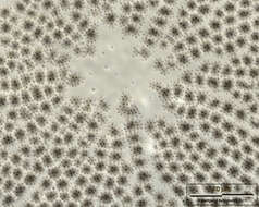
Coscinodiscus wailesii Valvar view, dark field. Scale bar indicates 100 µm. Sample from North Sea near Heligoland (spring diatom bloom). The image was built up using several photomicrographic frames with manual stacking and stitching technique. Images were taken using Zeiss Universal with Olympus C7070 CCD camera.Image under Creative Commons License V 3.0 (CC BY-NC-SA). Place name: North Sea around Heligoland Latitude: 54.186311 Longitude: 7.895034 Schale im Dunkelfeld. Multiebenen-Abbildung, manuell gestapelt. Der Messbalken markiert eine Länge von 100 µm. Probe aus der Nordsee vor Helgoland in der Zeit der Frühjahrsblüte. Mikrotechnik: Zeiss Universal, Kamera: Olympus C7070.Creative Commons License V 3.0 (CC BY-NC-SA). For permission to use of (high-resolution) images please contact postmaster@protisten.de.
-
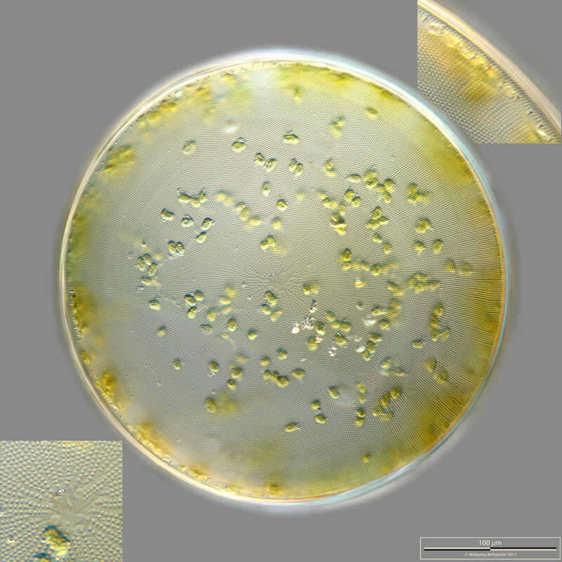
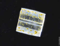
Coscinodiscus wailesii Valvar view. Insets showing marginal ring of labiate processes (upper right) and the hyaline central area (lower left). Scale bar indicates 100 µm. Sample from North Sea near Heligoland (spring diatom bloom). The image was built up using several photomicrographic frames with manual stacking and stitching technique. Images were taken using Zeiss Universal with Olympus C7070 CCD camera.Image under Creative Commons License V 3.0 (CC BY-NC-SA). Place name: North Sea around Heligoland Latitude: 54.186311 Longitude: 7.895034 Schalenansicht. Die Ausschnittsvergrößerungen zeigen den randständigen Ring der sogenannten labiate processes (oben rechts) und die unperforierte Mitte der Frustel (unten links). Multiebenen-Abbildung, manuell gestapelt. Der Messbalken markiert eine Länge von 100 µm. Probe aus der Nordsee vor Helgoland in der Zeit der Frühjahrsblüte. Mikrotechnik: Zeiss Universal, Kamera: Olympus C7070.Creative Commons License V 3.0 (CC BY-NC-SA). For permission to use of (high-resolution) images please contact postmaster@protisten.de.
-
Coscinodiscus wailesii Sample from North Sea near Heligoland (spring diatom bloom). The image was built up using several photomicrographic frames with manual stacking technique. Images were taken using Leica dissecting microscope with MFT camera Olympus OM-D E-M5 II.Image under Creative Commons License V 3.0 (CC BY-NC-SA). Place name: North Sea around Heligoland Latitude: 54.186311 Longitude: 7.895034 Probe aus der Nordsee vor Helgoland in der Zeit der Frühjahrsblüte. Mikrotechnik: Leica Stereomikroskop, Kamera: Olympus OM-D E-M5 II.Creative Commons License V 3.0 (CC BY-NC-SA). For permission to use of (high-resolution) images please contact postmaster@protisten.de.
-
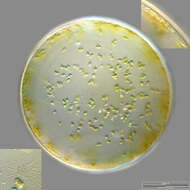
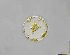
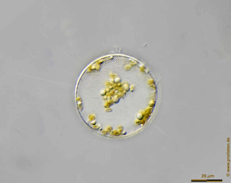
Thalassiosira spec. Scale bar indicates 25 µm. The specimen was gathered in the Kieler Förde (German Baltic Sea). Sampling date 4/2018. The image was built up using several photomicrographic frames with manual stacking technique. Images were taken using Zeiss Axioplan with Olympus OM-D M5 MKII. Image under Creative Commons License V 3.0 (CC BY-NC-SA). Place name: Baltic Sea, Kieler Förde, Kiel Fjord (Germany) Latitude: 54.3894126 Longitude: 10.1749055 Multiebenen-Abbildung, manuell gestapelt. Der Messbalken markiert eine Länge von 25 µm. Probe aus der Kieler Förde. Datum der Aufsammlung: 4/2018. Mikrotechnik: Zeiss Axioplan, Kamera: Olympus OM-D M5 MKII. Creative Commons License V 3.0 (CC BY-NC-SA). For permission to use of (high-resolution) images please contact postmaster@protisten.de.
-
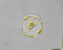
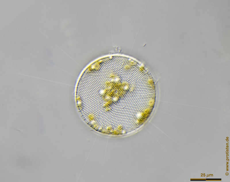
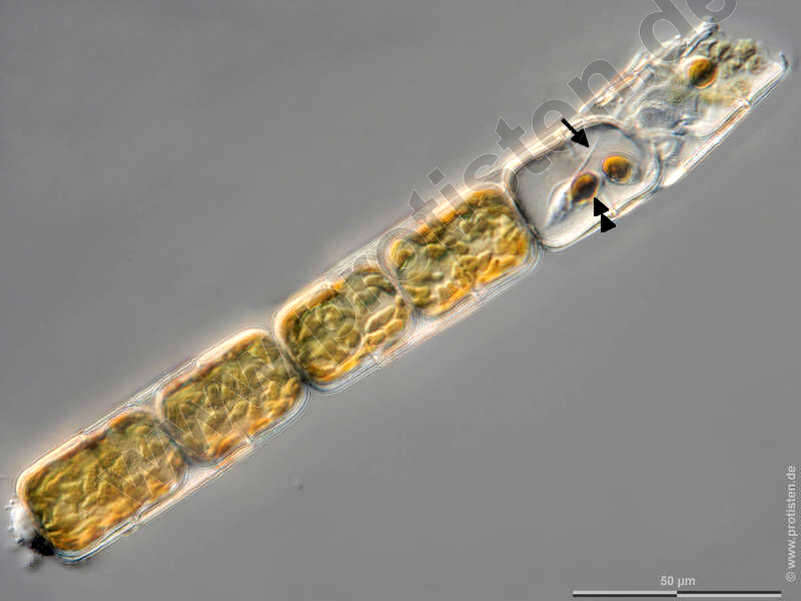
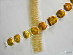
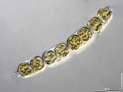
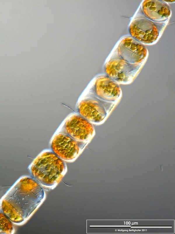
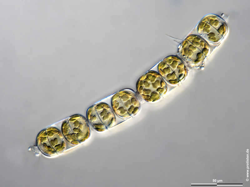
Melosira moniliformis The sexual reproduction of centric diatoms (Centrales) is characterized by oogamy. In Melosira, the spermatozoids are formed in narrower, the oogonia in broader filaments. After meiosis, four mobile spermatozoa with one flagellum develop in the male gamete mother cells (antheridia), in each of the oogonia three meiotic nuclei collaps and only one egg cell capable of fertilization remains. The images show the flagella (arrow) and a golden brown chloroplast (double arrowhead, color due to the accessory dye fucoxanthin) at the posterior end of the spermatozoids (arrowhead). After Krammer, K. and van den Hoek, C. Scale bar indicates 50 µm. The specimen was gathered in the Kieler Förde (Baltic Sea). Sampling date 1/2022. Images were taken using Zeiss Axioplan with Olympus OM-D M5 MKII. Image under Creative Commons License V 3.0 (CC BY-NC-SA). Place name: Baltic Sea, Kieler Förde, Kiel Fjord (Germany) Latitude: 54.3894126 Longitude: 10.1749055 Die sexuelle Fortpflanzung zentrischer Diatomeen (Centrales) zeichnet sich durch Oogamie aus. Bei Melosira werden die Spermien in schmaleren, die Oogonien in breiteren Zellketten gebildet. In den männlichen Gametenmutterzellen (Antheridien) entstehen nach der Meiose vier eingeißelige, bewegliche Spermatozioden, in den Oogonien sterben jeweils drei Meiose-Kerne ab und nur eine befruchtungsfähige Eizelle bleibt übrig. Die Bilder zeigen die Geißel (Pfeil) sowie einen goldbraunen Chloroplasten (Doppelpfeilkopf, Farbe aufgrund des akzessorischen Farbstoffs Fucoxanthin) am Hinterende der Spermatozoiden (Pfeilkopf).Nach Krammer, K. und van den Hoek, C. Der Messbalken markiert eine Länge von 50 µm. Probe aus der Kieler Förde. Datum der Aufsammlung: 1/2022. Mikrotechnik: Zeiss Axioplan, Kamera: Olympus OM-D M5 MKII. Creative Commons License V 3.0 (CC BY-NC-SA). For permission to use of (high-resolution) images please contact postmaster@protisten.de.
-
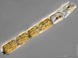
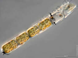
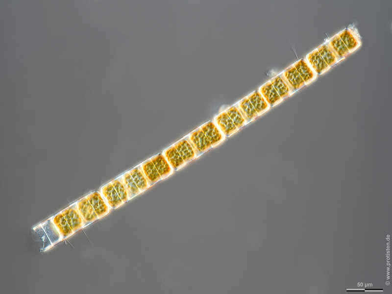
Melosira moniliformis Melosira moniliformis accompanied by Fragilaria islandica. Scale bar indicates 100 µm. The image was built up using several photomicrographic frames with manual stacking technique. Sample from North Sea near Heligoland (spring diatom bloom). Images were taken using Zeiss Universal with Olympus C7070 CCD camera.Image under Creative Commons License V 3.0 (CC BY-NC-SA). Place name: North Sea around Heligoland Latitude: 54.186311 Longitude: 7.895034 Melosira moniliformis zusammen mit Fragilaria islandica. Multiebenen-Abbildung, manuell gestapelt. Der Messbalken markiert eine Länge von 100 µm. Probe aus der Nordsee vor Helgoland in der Zeit der Frühjahrsblüte. Mikrotechnik: Zeiss Universal, Kamera: Olympus C7070.Creative Commons License V 3.0 (CC BY-NC-SA). For permission to use of (high-resolution) images please contact postmaster@protisten.de.
-
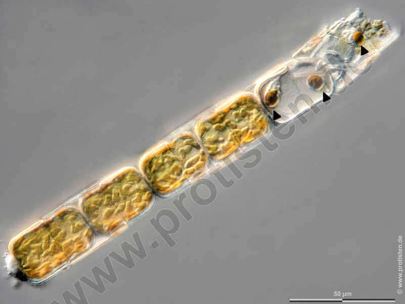
Melosira moniliformis The sexual reproduction of centric diatoms (Centrales) is characterized by oogamy. In Melosira, the spermatozoids are formed in narrower, the oogonia in broader filaments. After meiosis, four mobile spermatozoa with one flagellum develop in the male gamete mother cells (antheridia), in each of the oogonia three meiotic nuclei collaps and only one egg cell capable of fertilization remains.The images show the flagella (arrow) and a golden brown chloroplast (double arrowhead, color due to the accessory dye fucoxanthin) at the posterior end of the spermatozoids (arrowhead).After Krammer, K. and van den Hoek, C.Scale bar indicates 50 µm. The specimen was gathered in the Kieler Förde (Baltic Sea). Sampling date 1/2022. Images were taken using Zeiss Axioplan with Olympus OM-D M5 MKII. Image under Creative Commons License V 3.0 (CC BY-NC-SA). Place name: Baltic Sea, Kieler Förde, Kiel Fjord (Germany) Latitude: 54.3894126 Longitude: 10.1749055 Die sexuelle Fortpflanzung zentrischer Diatomeen (Centrales) zeichnet sich durch Oogamie aus. Bei Melosira werden die Spermien in schmaleren, die Oogonien in breiteren Zellketten gebildet. In den männlichen Gametenmutterzellen (Antheridien) entstehen nach der Meiose vier eingeißelige, bewegliche Spermatozioden, in den Oogonien sterben jeweils drei Meiose-Kerne ab und nur eine befruchtungsfähige Eizelle bleibt übrig.Die Bilder zeigen die Geißel (Pfeil) sowie einen goldbraunen Chloroplasten (Doppelpfeilkopf, Farbe aufgrund des akzessorischen Farbstoffs Fucoxanthin) am Hinterende der Spermatozoiden (Pfeilkopf).Nach Krammer, K. und van den Hoek, C.Der Messbalken markiert eine Länge von 50 µm. Probe aus der Kieler Förde. Datum der Aufsammlung: 1/2022. Mikrotechnik: Zeiss Axioplan, Kamera: Olympus OM-D M5 MKII. Creative Commons License V 3.0 (CC BY-NC-SA). For permission to use of (high-resolution) images please contact postmaster@protisten.de.
-
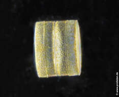
Melosira moniliformis Scale bar indicates 25 µm. The specimen was gathered in the Kieler Förde (Baltic Sea). Sampling date 1/2022. The image was built up using several photomicrographic frames with manual stacking technique. Images were taken using Zeiss Axioplan with Olympus OM-D M5 MKII. Image under Creative Commons License V 3.0 (CC BY-NC-SA). Place name: Baltic Sea, Kieler Förde, Kiel Fjord (Germany) Latitude: 54.3894126 Longitude: 10.1749055 Multiebenen-Abbildung, manuell gestapelt. Der Messbalken markiert eine Länge von 25 µm. Probe aus der Kieler Förde. Datum der Aufsammlung: 1/2022. Mikrotechnik: Zeiss Axioplan, Kamera: Olympus OM-D M5 MKII. Creative Commons License V 3.0 (CC BY-NC-SA). For permission to use of (high-resolution) images please contact postmaster@protisten.de.
-
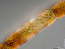
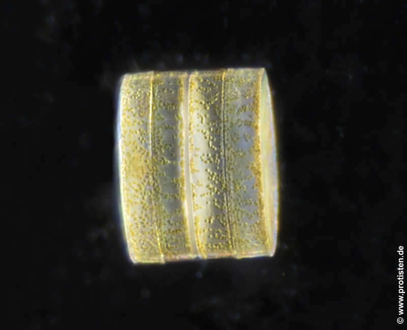
Melosira moniliformis Colony with epibiotic bacteria chains. Scale bar indicates 100 µm. The image was built up using several photomicrographic frames with manual stacking technique. Sample from North Sea near Heligoland (spring diatom bloom). Images were taken using Zeiss Universal with Olympus C7070 CCD camera.Image under Creative Commons License V 3.0 (CC BY-NC-SA). Place name: North Sea around Heligoland Latitude: 54.186311 Longitude: 7.895034 Kolonie mit aufsitzenden Bakterienketten. Multiebenen-Abbildung, manuell gestapelt. Der Messbalken markiert eine Länge von 100 µm. Probe aus der Nordsee vor Helgoland in der Zeit der Frühjahrsblüte. Mikrotechnik: Zeiss Universal, Kamera: Olympus C7070.Creative Commons License V 3.0 (CC BY-NC-SA). For permission to use of (high-resolution) images please contact postmaster@protisten.de.
-

Melosira moniliformis The sexual reproduction of centric diatoms (Centrales) is characterized by oogamy. In Melosira, the spermatozoids are formed in narrower, the oogonia in broader filaments. After meiosis, four mobile spermatozoa with one flagellum develop in the male gamete mother cells (antheridia), in each of the oogonia three meiotic nuclei collaps and only one egg cell capable of fertilization remains. The images show the flagella and a golden brown chloroplast (color due to the accessory dye fucoxanthin) at the posterior end of the spermatozoids. After Krammer, K. and van den Hoek, C.Please press the MORE button for skipping to the annotated version. Scale bar indicates 50 µm. The specimen was gathered in the Kieler Förde (Baltic Sea). Sampling date 1/2022. Images were taken using Zeiss Axioplan with Olympus OM-D M5 MKII. Image under Creative Commons License V 3.0 (CC BY-NC-SA). Place name: Baltic Sea, Kieler Förde, Kiel Fjord (Germany) Latitude: 54.3894126 Longitude: 10.1749055 Die sexuelle Fortpflanzung zentrischer Diatomeen (Centrales) zeichnet sich durch Oogamie aus. Bei Melosira werden die Spermien in schmaleren, die Oogonien in breiteren Zellketten gebildet. In den männlichen Gametenmutterzellen (Antheridien) entstehen nach der Meiose vier eingeißelige, bewegliche Spermatozioden, in den Oogonien sterben jeweils drei Meiose-Kerne ab und nur eine befruchtungsfähige Eizelle bleibt übrig. Die Bilder zeigen die Geißel sowie einen goldbraunen Chloroplasten (Farbe aufgrund des akzessorischen Farbstoffs Fucoxanthin) am Hinterende der Spermatozoiden.Nach Krammer, K. und van den Hoek, C.Bitte drücken Sie die Schaltfläche MORE, um zur kommentierten Version zu gelangen.Der Messbalken markiert eine Länge von 50 µm. Probe aus der Kieler Förde. Datum der Aufsammlung: 1/2022. Mikrotechnik: Zeiss Axioplan, Kamera: Olympus OM-D M5 MKII. Creative Commons License V 3.0 (CC BY-NC-SA). For permission to use of (high-resolution) images please contact postmaster@protisten.de.
-
Melosira moniliformis Scale bar indicates 25 µm. The specimen was gathered in the Kieler Förde (Baltic Sea). Sampling date 1/2022. The image was built up using several photomicrographic frames with manual stacking technique. Images were taken using Zeiss Axioplan with Olympus OM-D M5 MKII. Image under Creative Commons License V 3.0 (CC BY-NC-SA). Place name: Baltic Sea, Kieler Förde, Kiel Fjord (Germany) Latitude: 54.3894126 Longitude: 10.1749055 Multiebenen-Abbildung, manuell gestapelt. Der Messbalken markiert eine Länge von 25 µm. Probe aus der Kieler Förde. Datum der Aufsammlung: 1/2022. Mikrotechnik: Zeiss Axioplan, Kamera: Olympus OM-D M5 MKII. Creative Commons License V 3.0 (CC BY-NC-SA). For permission to use of (high-resolution) images please contact postmaster@protisten.de.
-
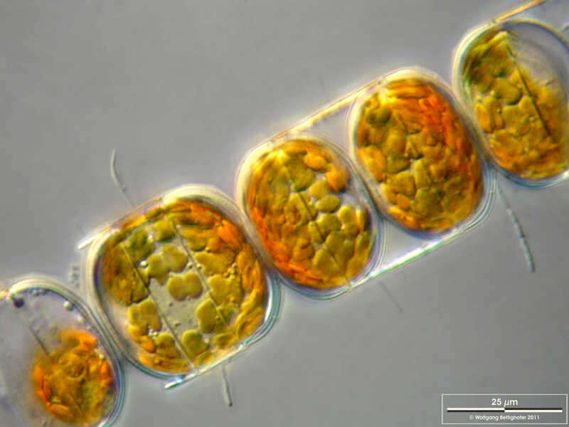
Melosira moniliformis Scale bar indicates 50 µm. The specimen was gathered in the Kieler Förde (Baltic Sea). Sampling date 1/2022. The image was built up using several photomicrographic frames with manual stacking technique. Images were taken using Zeiss Axioplan with Olympus OM-D M5 MKII. Image under Creative Commons License V 3.0 (CC BY-NC-SA). Place name: Baltic Sea, Kieler Förde, Kiel Fjord (Germany) Latitude: 54.3894126 Longitude: 10.1749055 Multiebenen-Abbildung, manuell gestapelt. Der Messbalken markiert eine Länge von 50 µm. Probe aus der Kieler Förde. Datum der Aufsammlung: 1/2022. Mikrotechnik: Zeiss Axioplan, Kamera: Olympus OM-D M5 MKII. Creative Commons License V 3.0 (CC BY-NC-SA). For permission to use of (high-resolution) images please contact postmaster@protisten.de.
-
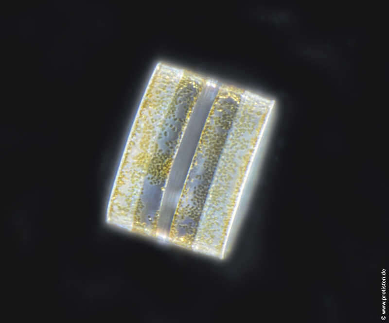
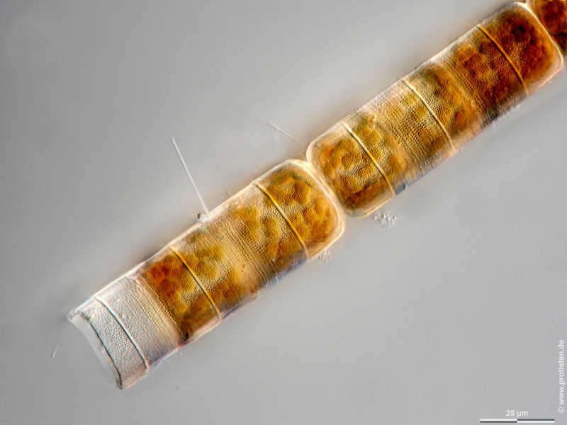
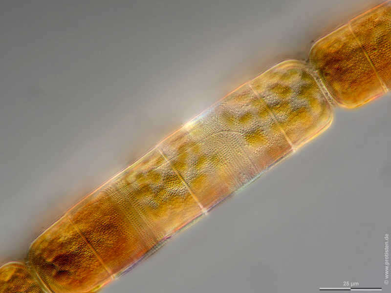
Melosira moniliformis Sample from North Sea near Heligoland (spring diatom bloom). Scale bar indicates 50 µm.The image was built up using several photomicrographic frames with manual stacking technique. Images were taken using Zeiss Axioplan with MFT camera Olympus OM-D E-M5 II.Image under Creative Commons License V 3.0 (CC BY-NC-SA). Place name: North Sea around Heligoland Latitude: 54.186311 Longitude: 7.895034 Multiebenen-Abbildung, manuell gestapelt. Der Messbalken markiert eine Länge von 50 µm. Probe aus der Nordsee vor Helgoland in der Zeit der Frühjahrsblüte. Mikrotechnik: Zeiss Axioplan Kamera: Olympus OM-D E-M5 II.Creative Commons License V 3.0 (CC BY-NC-SA). For permission to use of (high-resolution) images please contact postmaster@protisten.de.
-
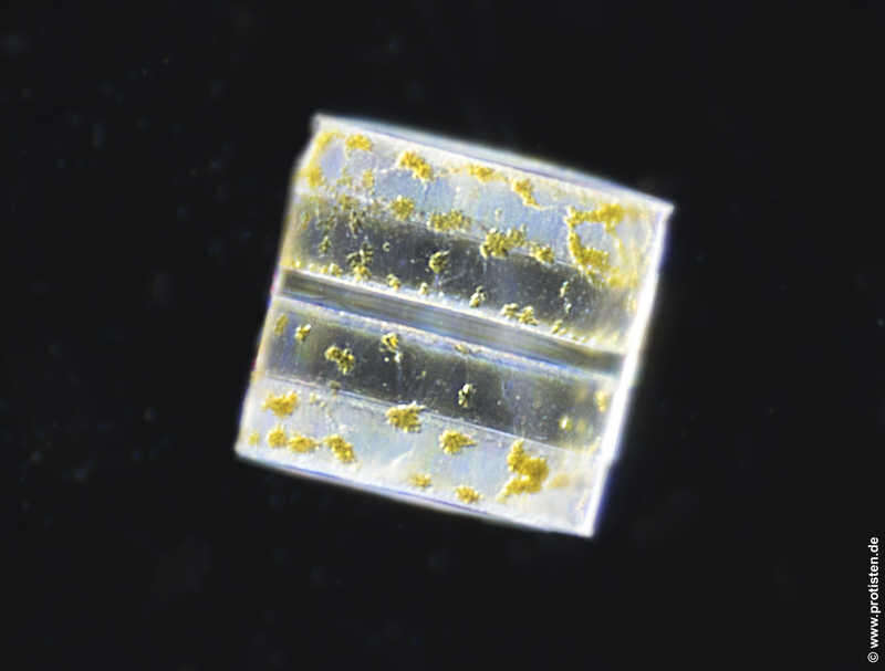
Melosira moniliformis Colony with epibiotic bacteria chains. Scale bar indicates 25 µm. The image was built up using several photomicrographic frames with manual stacking technique. Sample from North Sea near Heligoland (spring diatom bloom). Images were taken using Zeiss Universal with Olympus C7070 CCD camera.Image under Creative Commons License V 3.0 (CC BY-NC-SA). Place name: North Sea around Heligoland Latitude: 54.186311 Longitude: 7.895034 Kolonie mit aufsitzenden Bakterienketten. Multiebenen-Abbildung, manuell gestapelt. Der Messbalken markiert eine Länge von 25 µm. Probe aus der Nordsee vor Helgoland in der Zeit der Frühjahrsblüte. Mikrotechnik: Zeiss Universal, Kamera: Olympus C7070.Creative Commons License V 3.0 (CC BY-NC-SA). For permission to use of (high-resolution) images please contact postmaster@protisten.de.
-
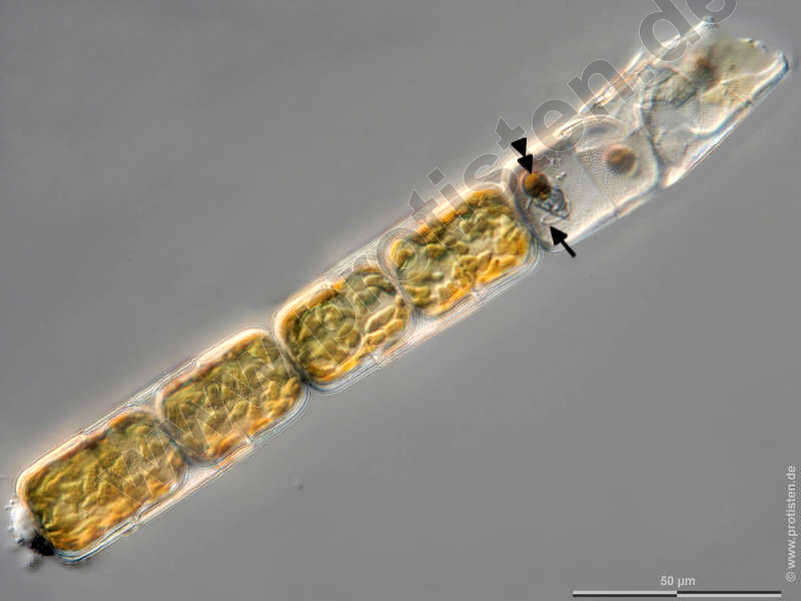
Melosira moniliformis The sexual reproduction of centric diatoms (Centrales) is characterized by oogamy. In Melosira, the spermatozoids are formed in narrower, the oogonia in broader filaments. After meiosis, four mobile spermatozoa with one flagellum develop in the male gamete mother cells (antheridia), in each of the oogonia three meiotic nuclei collaps and only one egg cell capable of fertilization remains. The images show the flagella (arrow) and a golden brown chloroplast (double arrowhead, color due to the accessory dye fucoxanthin) at the posterior end of the spermatozoids (arrowhead). After Krammer, K. and van den Hoek, C. Scale bar indicates 50 µm. The specimen was gathered in the Kieler Förde (Baltic Sea). Sampling date 1/2022. Images were taken using Zeiss Axioplan with Olympus OM-D M5 MKII. Image under Creative Commons License V 3.0 (CC BY-NC-SA). Place name: Baltic Sea, Kieler Förde, Kiel Fjord (Germany) Latitude: 54.3894126 Longitude: 10.1749055 Die sexuelle Fortpflanzung zentrischer Diatomeen (Centrales) zeichnet sich durch Oogamie aus. Bei Melosira werden die Spermien in schmaleren, die Oogonien in breiteren Zellketten gebildet. In den männlichen Gametenmutterzellen (Antheridien) entstehen nach der Meiose vier eingeißelige, bewegliche Spermatozioden, in den Oogonien sterben jeweils drei Meiose-Kerne ab und nur eine befruchtungsfähige Eizelle bleibt übrig. Die Bilder zeigen die Geißel (Pfeil) sowie einen goldbraunen Chloroplasten (Doppelpfeilkopf, Farbe aufgrund des akzessorischen Farbstoffs Fucoxanthin) am Hinterende der Spermatozoiden (Pfeilkopf).Nach Krammer, K. und van den Hoek, C. Der Messbalken markiert eine Länge von 50 µm. Probe aus der Kieler Förde. Datum der Aufsammlung: 1/2022. Mikrotechnik: Zeiss Axioplan, Kamera: Olympus OM-D M5 MKII. Creative Commons License V 3.0 (CC BY-NC-SA). For permission to use of (high-resolution) images please contact postmaster@protisten.de.