-
Fig 1: Gonyaulax polygramma Schematic diagram (ventral view) redrawn from Tomas et al. 1997.
-
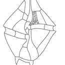
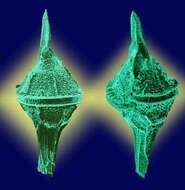
Two cells, two faces.
-
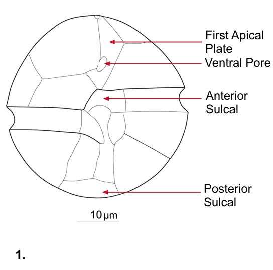
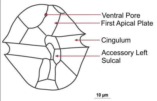
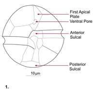
Fig 1: Alexandrium ostenfeldii Schematic drawing of a cell showing plate patterns on ventral side of cell
-
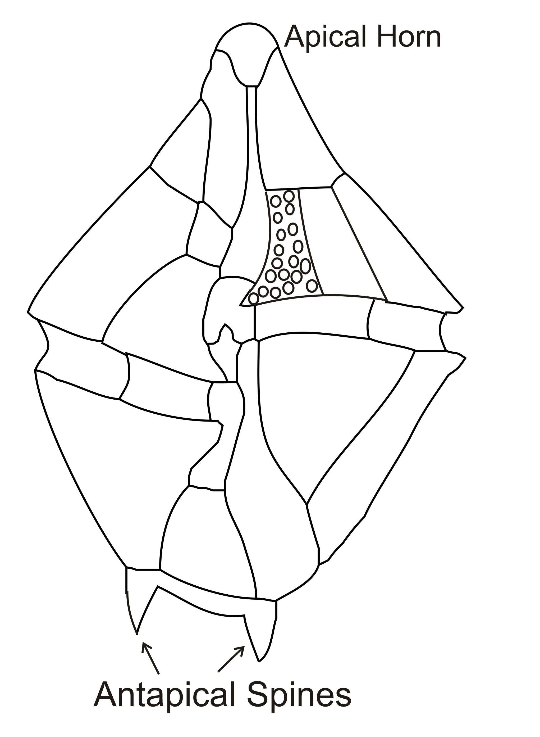
G. spinifera cells are slightly longer than broad. The epictheca has convex sides and a small epical horn. The hypotheca has a 2-4 antapical spines. The sulcus extends almoust the whole length of the cell. The cingulum is deeply excavated and displaced by 2 or more widths. G. spinifera is sometimes confused with Gonyaulax digitale.
-
Uit: www.nies.go.jp/biology/ mcc/strainlist_a.htm
Ecomare
Alexandrium; Alexandrium.
-
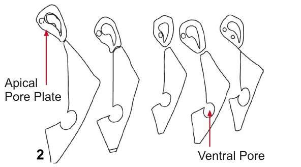
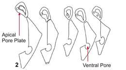
Fig 2: Alexandrium ostenfeldii Schematic drawing of a cell showing the morphological variation in APC and 1'
-
Cells are elongate with a short apical horn and two distinct winged antapical spines. The left spine is longer than the right one. The epitheca is slightly larger than the hypotheca and the girdle is offset by 1-2 girdle widths.
-
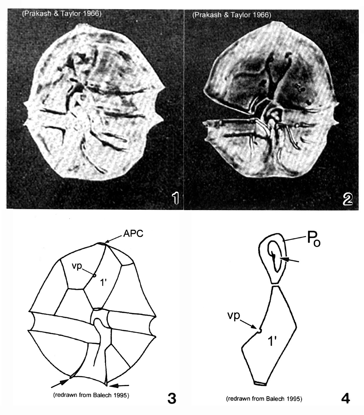
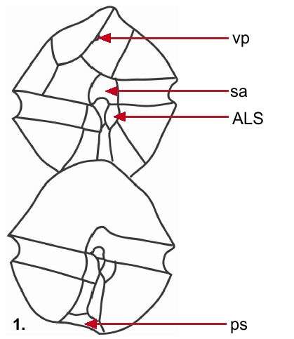
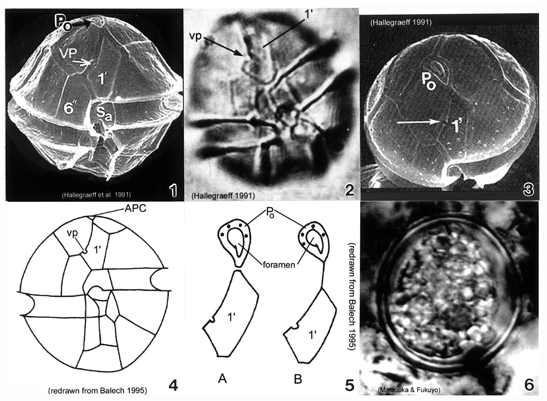
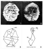
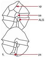
Plate 1. Alexandrium acatenella. Figs. 1-2. LM: ventral view of empty thecae. Cell small to medium, longer than wide, angular to round. Conical epitheca with shoulders; larger than hypotheca. Figs. 3-4. Line drawings. Fig. 3. Ventral view: 1' plate bears ventral pore (vp). Hypotheca with two antapical spines (arrows). Fig. 4. Po comes in direct contact with 1' plate. APC: comma-shaped foramen (arrow).
-
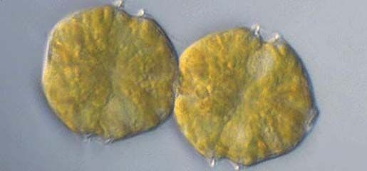
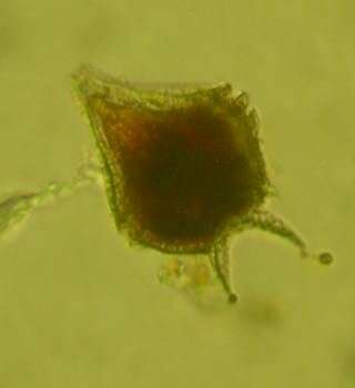
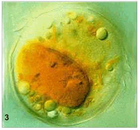
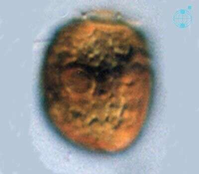
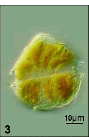
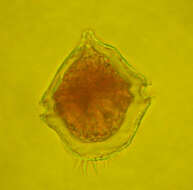
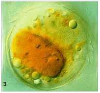
Fig 3: Alexandrium ostenfeldii whole cell with food vacuole
-
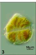
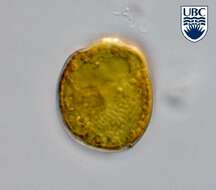
Adenoides spec., a so far undescribed taxon. Left lateral view, mid cell focus, note the granula reserve material in the cell.
-
Fig 1: Schematic diagram of Alexandrium affine, showing ventral (top) and dorsal (bottom) views.
-
Left lateral view, mid cell focus. Note the centrally located nucleus and storage material (granules).
-
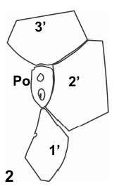
Fig 2: Schematic drawing of the Po and surrounding apical plates
-
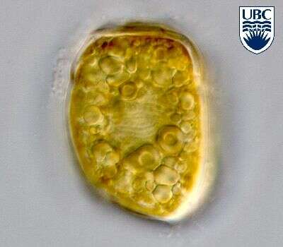
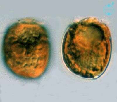
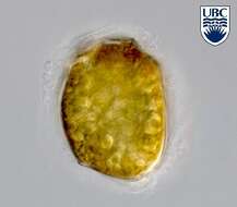
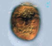
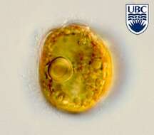
Adenoides (add-en-oi-dees) eludens (Herdman) Balech 1956. The image on the left shows the left lateral view of a cell, with yellow-brown plastids with a ring -like pyrenoid. The small epicone is almost not visible. The image on the right shows a mid-focus plane through a cell, with a pusule near the anterior end and the nucleus near the posterior end.
-
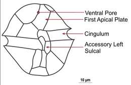
Plate 3. Alexandrium minutum. Fig. 1. SEM: ventral view. Cell small and ellipsoidal. Epitheca conical, larger than hypotheca. Hypotheca short and wide; antapex obliquely flattened. Intercalary bands present. Cingulum deep, lipped; displaced 1X its width. Sulcus shallow (sa=anterior sulcal plate). Apical pore plate (Po) in direct contact with 1' plate. Fig. 2. LM: ventral view. Ventral pore (vp) present on 1' plate. Fig. 3. SEM: apical view. Po large, narrow and oval; indirectly connected to 1' plate. Vp present (arrow). Figs. 4-5. Line drawing. Fig. 4. Ventral view. 1' plate slender and rhomboidal. Fig. 5. Po connection to 1' plate: a. direct; b. indirect via thin suture. Fig. 6. LM: cyst circular in apical view.
-
Fig 1: Alexandrium tamarense Schematic drawing of a cell showing plate patterns on ventral side of cell
-
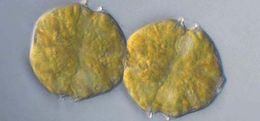
Adenoides (add-en-oi-dees) eludens (Herdman) Balech 1956. The image shows the left lateral view of a cell, with yellow-brown plastids with a ring-like pyrenoid. The small epicone is almost not visible.
-
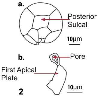
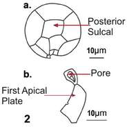
Fig 2: Alexandrium tamarense Schematic drawing of a cell in posterior view and b. apical pore coplex with 1'.
-
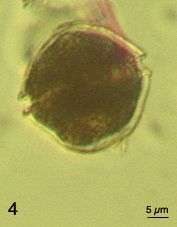
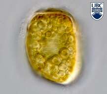
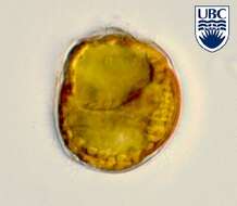
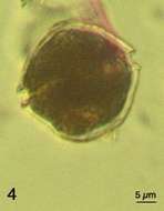
Adenoides eludens (Herdman) Balech 1956 is shown here from its right lateral cell side. Note the large pusule in the upper part of the cell and the nucleus in the lower part.
-
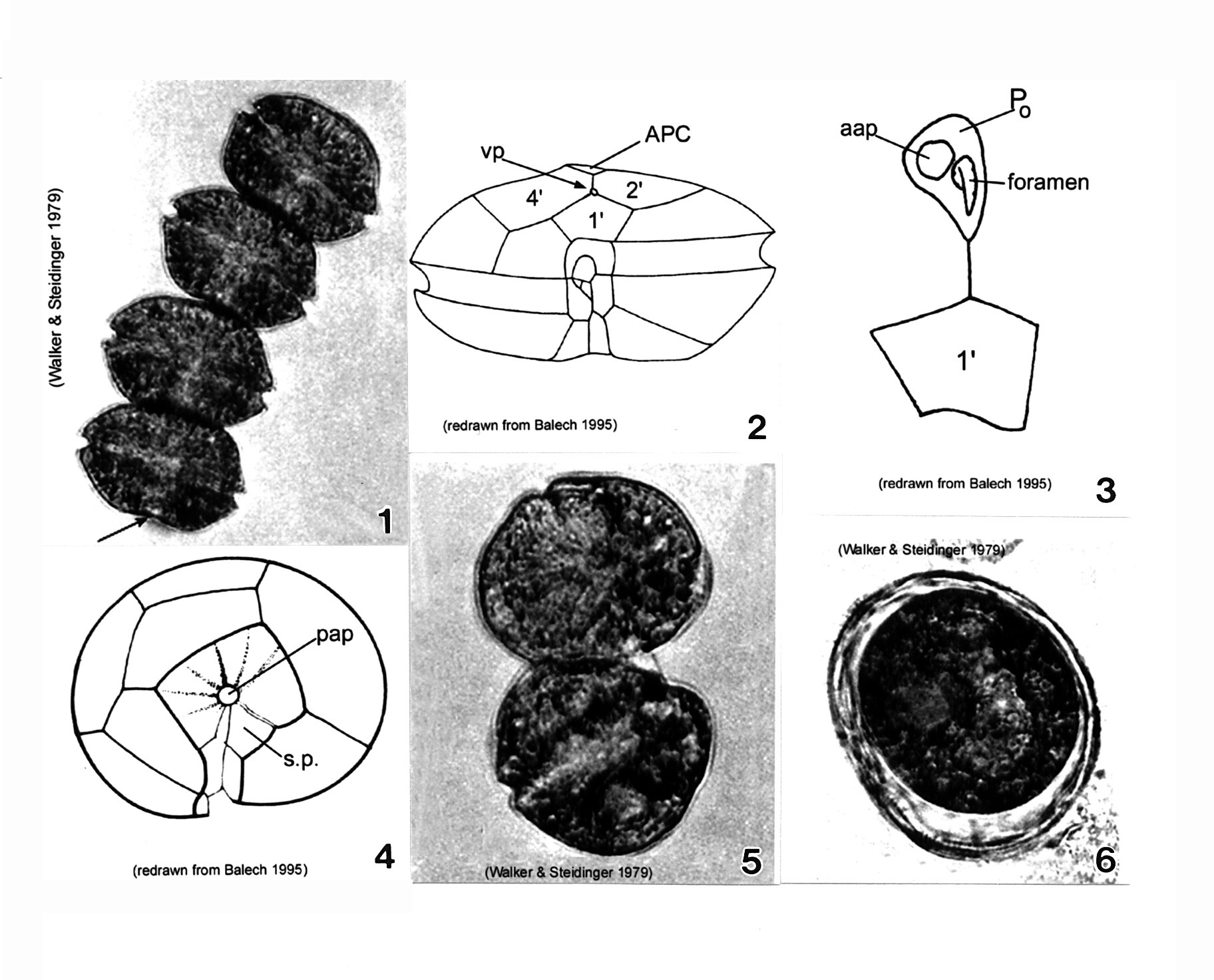
Plate 4. Alexandrium monilatum. Fig. 1. LM: four-cell chain. Cells large, wider than long, flattened anterio-posteriorly. Antapex slightly concave (arrow). Figs. 2-4. Line drawings. Fig. 2. Ventral pore (vp) depicted (Florida specimens) at anterior margin of 1' plate where it comes in contact with plates 2' and 4'. Cingulum (C) deeply excavated, wide, descending; displaced one time its width. Fig. 3. Apical pore plate (Po) does not come in contact with 1' plate. Anterior attachment pore (aap) large, round and dorsally situated in the APC. Foramen comma-shaped. Fig. 4. Antapical view: posterior sulcal plate (sp) large, rhomboid and concave with radial markings. Posterior attachment pore (pap) large and centrally located. Figs. 5-6. LM. Fig. 5. Two isogamous gametes fusing at oblique angles. Fig. 6. Mature resting cysts: dark and round, with a triple layered wall.
-
Fig 3: Alexandrium tamarense Live cell in dorsal view
-
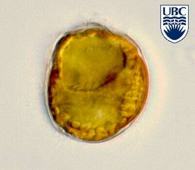
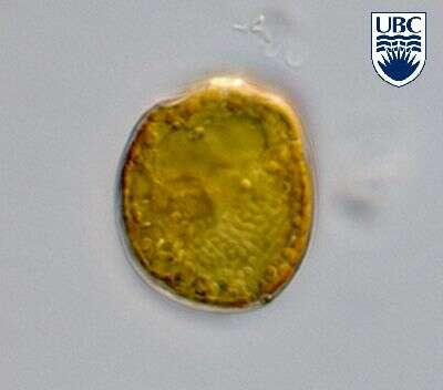
Right lateral view. Note the ring-like structure. It is a starch sheath around the pyrenoid, a structure of the plastid.
-
Fig 4: Alexandrium tamarense Lugol's preserved cell
-
Mid cell focus showing the nucleus in the lower part of the cell and the pusule in the upper half.