-
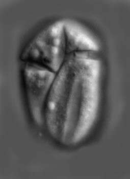
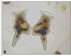
Fig 4: Dinophysis caudata Image of a Lugol's preserved cell in lateral view
-
This is Amphidinium cf. glabrum in that it looks like but is not fully identical with the usual concept of this species.
-
Durinskia baltica theca observed in marine muds and sandy sediments in the vicinity of Broome, Western Australia in September 2003. This image was taken using phase contrast optics. This work was supported by the Australian Biological Resources Study.
-
Togula jolla Flo Jorgensen, Murray et Daugbjerg 2004
-
Prorocentrum emarginatum, an empty valve, observed in marine muds and sandy sediments in the vicinity of Broome, Western Australia in September 2003. This image was taken using phase contrast optics. This work was supported by the Australian Biological Resources Study.
-
Fig 6: Dinophysis caudata Image of a pair of Lugol's preserved cells in lateral view cells in lateral view
-
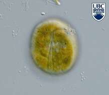
Amphidinium (am-fee-din-ee-um) pellucidum Herdman 1922. The image shows a cell in ventral view. The cell is colourless (contains no plastids). The cingulum is near the anterior end of the cell and is slightly descending. There is an apical groove present.
-
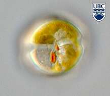
Durinskia baltica, showing its stigma, observed in marine muds and sandy sediments in the vicinity of Broome, Western Australia in September 2003. This image was taken using differential interference contrast optics. This work was supported by the Australian Biological Resources Study.
-
Togula jolla Flo Jorgensen, Murray et Daugbjerg 2004
-
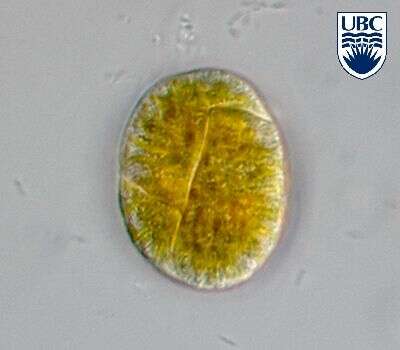
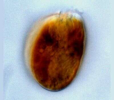
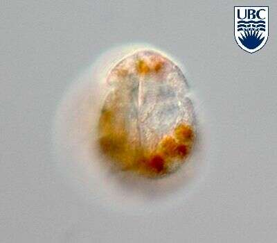
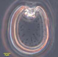
Prorocentrum (pro-row-sent-rum) lima (Ehrenberg) Dodge 1975. The image shows one of the two valves of a cell. The cingulum is not visible. The plastids are yellow-brown, and surround a large circular pyrenoid in the centre of the cell.
-
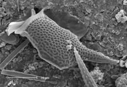
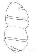
Fig 7: SEM of Dinophysis caudata
-
Amphidinium pellucidum
-
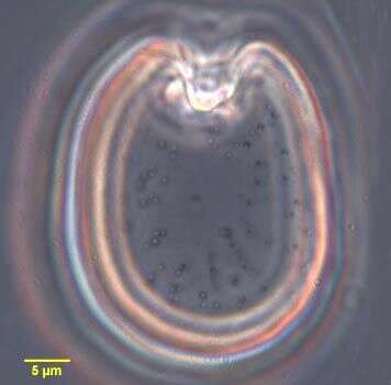
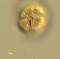
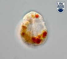
Durinskia baltica (Levander 1892) Carty et Cox 1986. This phototrophic species is also known from the plankton. Cell in ventral view, note the bright red stigma in the sulcal area.
-
Togula jolla Flo Jorgensen, Murray et Daugbjerg 2004
-
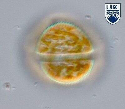
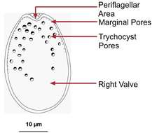
Prorocentrum lima valves are oval, 40 - 45 microns long, 27-33 microns wide , length to width ratio 1.3 - 1.5. Valve surface smooth. Valve pores scattered except in the centre, 58 - 80 per cell, approximately 0.3 microns diameter, round, oval to slightly kidney shaped. Marginal pores around periphery of cell, 51-74 per cell, round to oval approximately 0.3 microns diameter. Intercalary band smooth. Apical area consists of a small triangular indentation. A small flange (apical collar) is present. Plastids large, orange-brown. A pyrenoid, 7 - 8 microns diameter, is situated centrally. Nucleus in the posterior of the valve.
-
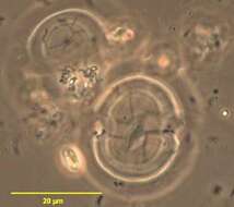
Fig 1: Cochlodinium polykrikoides Schematic diagram (ventral view) redrawn from Tomas et al. 1997.
-
Amphidinium pellucidum
-
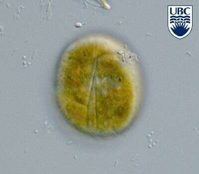
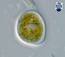
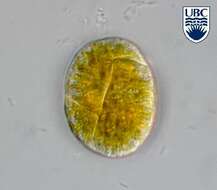
Durinskia baltica (Levander 1892) Carty et Cox 1986. Dorsal view showing the cingulum (transverse furrow) and golden-brown chloroplasts.
-
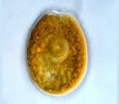
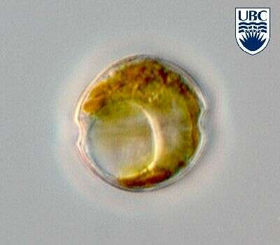
Prorocentrum (pro-row-sent-rum) mexicanum Tafall 1942. The image shows the valve of a cell. The plastid is yellow-brown. There is a small apical spine present. The cingulum is not visible.
-
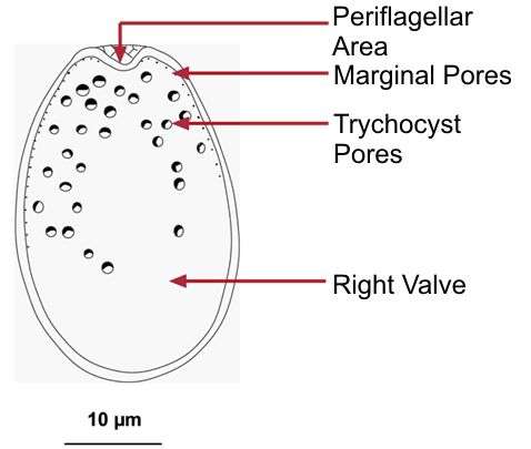
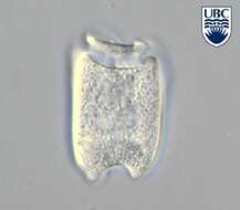
Fig 1: Schematic drawing of Prorocentrum lima
-
Amphidinium pellucidum
-
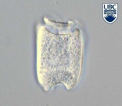
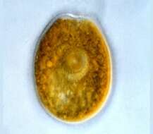
Durinskia baltica (Levander 1892) Carty et Cox 1986. Mid cell focus showing the large pusule.
-
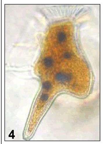
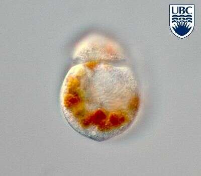
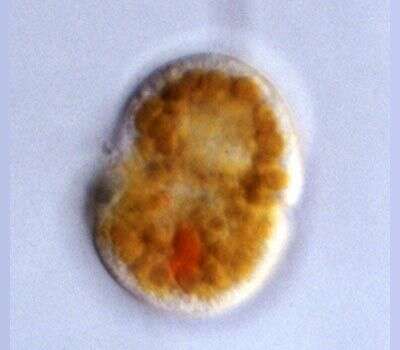
Spiniferodinium (spin-if-err-o-din-ee-um) galeiforme Horiguchi & Chihara 1987. The image shows a mid-focal plane through a cell. The cingulum is in the middle of the cell. The nucleus is in the anterior end of the cell. There are many yellow-brown plastids present.
-
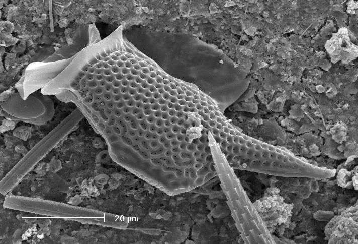
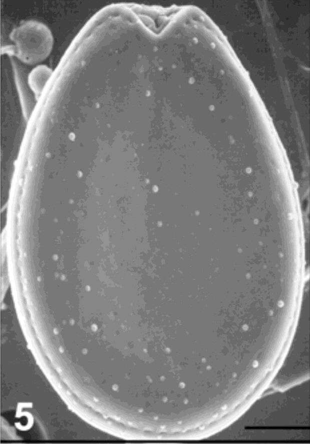
Fig 5:Prorocentrum lima SEM image of cell in right valve view