-
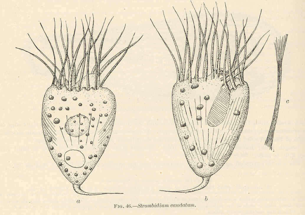
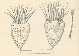
Strombidium caudalum.
-
Algue brune, Alaria fistulosa, Postels et Ruprecht.
-
Pheophycees (Algues brunes) Fucacees, Halidrys siliquosa (L.) Lyngh..
-
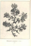
Pheophycees (Algues brunes) Sphacelariees, Halopteris scoparia (L.) Sauv..
-
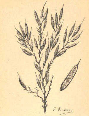
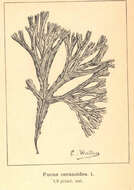
Fucus ceranoides.
-
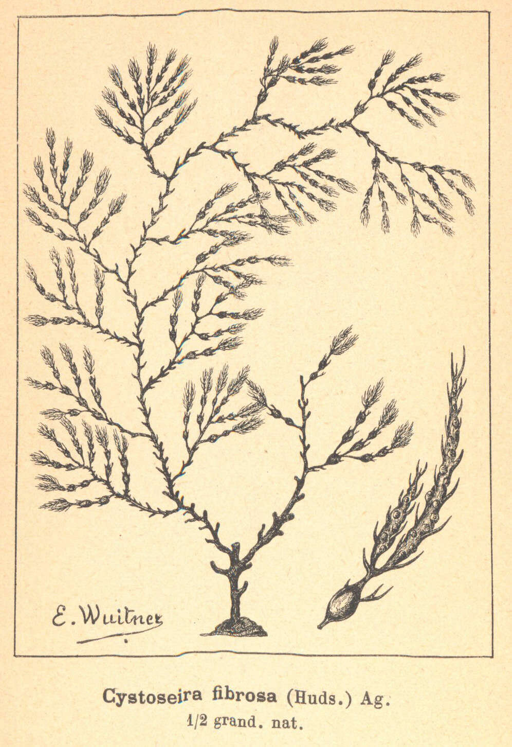
Pheophycees (Algues brunes) Fucacees, Cystoseira fibrosa (Huds.) Ag..
-
Portion of Leaf of Alaria Fistulosa, Showing Midrib.
-
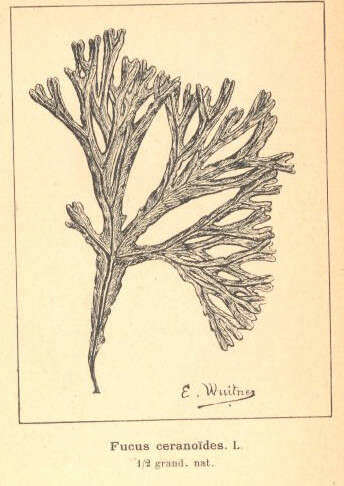
Pheophycees (Algues brunes) Fucacees, Fucus ceranoides. L..
-
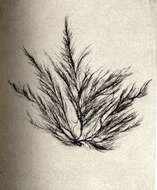
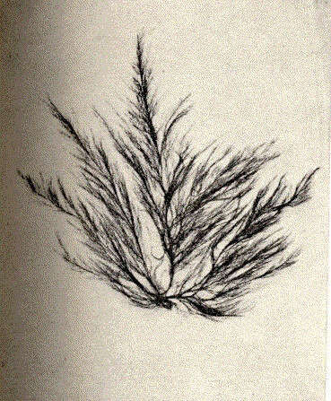
Ectocarpus littoralis.
-
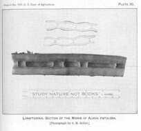
Longitudinal Section of the Midrib of Alaria Fistulosa.
-
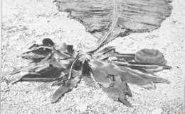
Spore Leaves of Alaria fistulosa. [There were about 220 on this plant.]
-
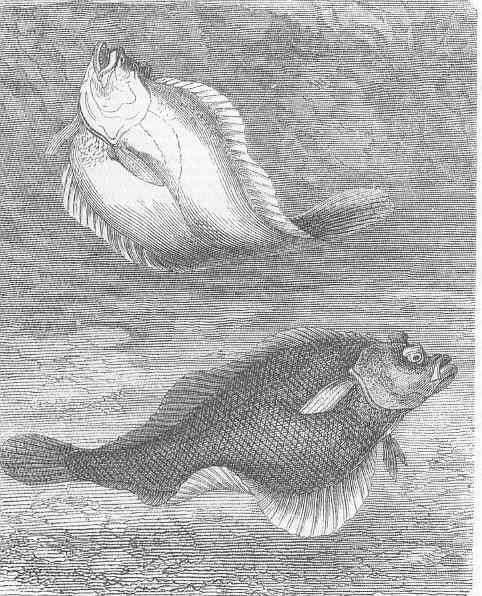
Dab (platessa Imanda).
-
-
Public Domain, U.S. Government Work 2011 Barry H. Rosen Courtesy of life.nbii.gov
NBII images
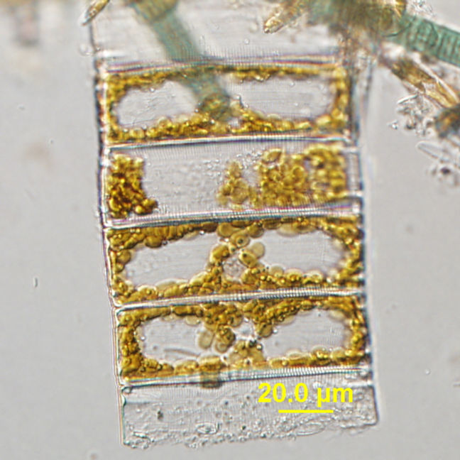
Category hierarchy: Microorganisms | AlgaeDescription: Eunotia chain micrograph. Sample was collected from Silver Springs, Florida.Capture device: Olympus DP71Locality: Latitude: 2.859009900000000e+001; Longitude: -8.119031699999999e+001
-
Public Domain, U.S. Government Work 2011 Barry H. Rosen Courtesy of life.nbii.gov
NBII images
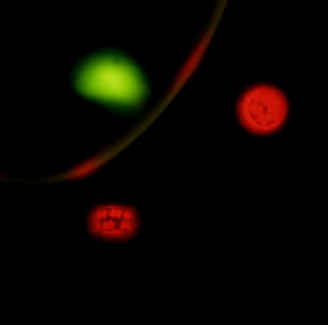
Category hierarchy: Microorganisms | AlgaeDescription: Discoid chromatophores and the nucleus easily observed with epifluorescent illumination. Sample was collected from Raccoon River, Iowa.Capture device: DP71Capture details: 400x with microscopeOriginal date: 20091002|||112306Locality: Latitude: 2.859009900000000e+001; Longitude: -8.119031699999999e+001
-
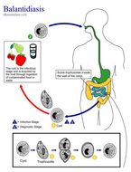
This illustration depicts the life cycle of Balantidium coli, the causal agent of Balantidiasis.Created: 2002
-
Note the developmental tetrad configuration of these Babesia sp. trophozoites, which resemble P. falciparum.Created: 1973
-
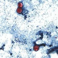
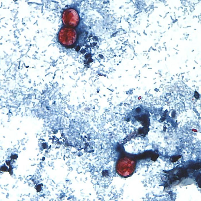
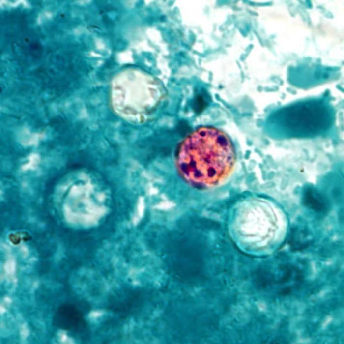
This photomicrograph of a fresh stool sample, which had been prepared using a 10% formalin solution, and stained with safranin, revealed the presence of three uniformly stained Cyclospora cayetanensis oocysts in the field of view.Created:
-
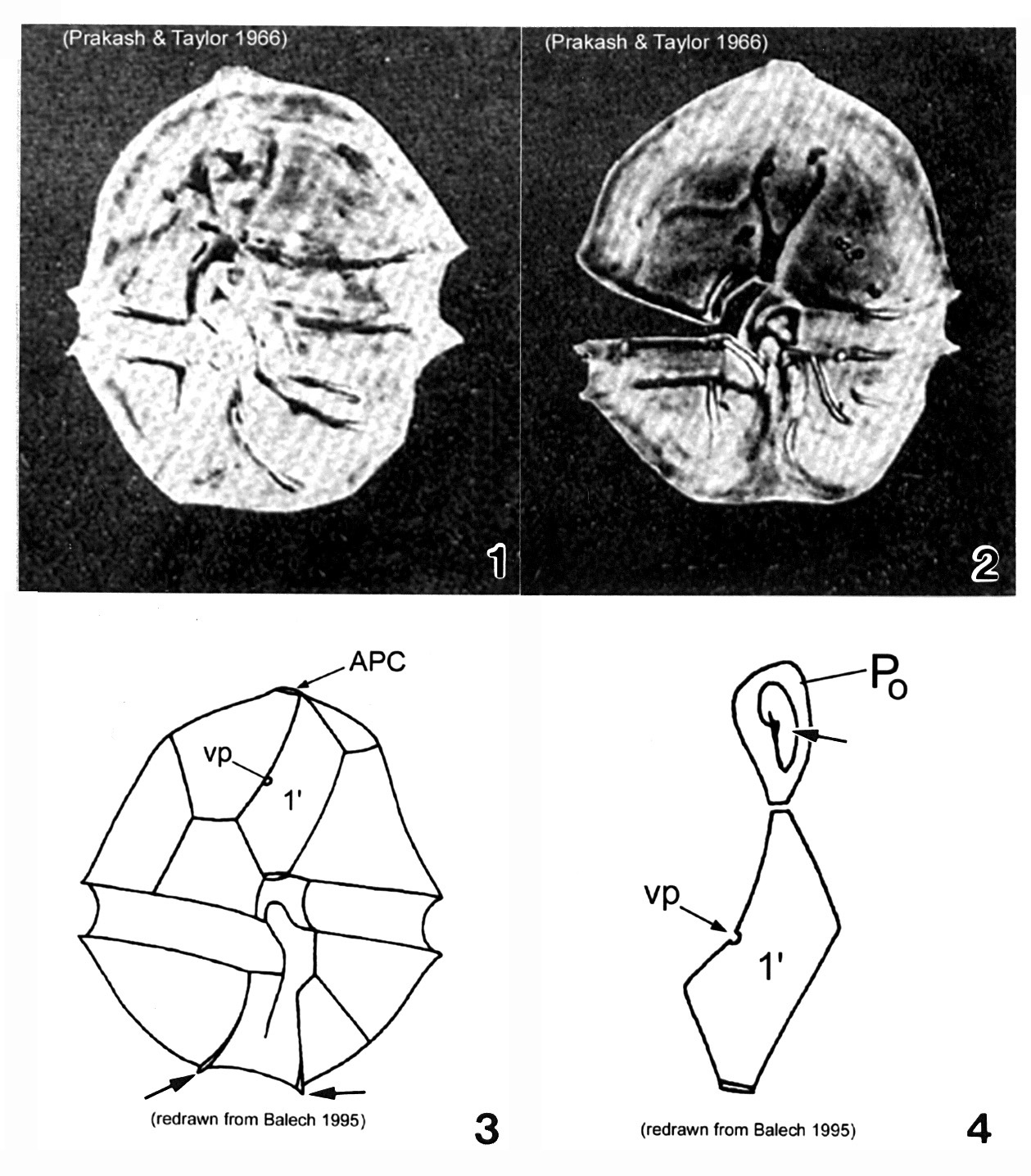
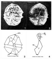
Plate 1. Alexandrium acatenella. Figs. 1-2. LM: ventral view of empty thecae. Cell small to medium, longer than wide, angular to round. Conical epitheca with shoulders; larger than hypotheca. Figs. 3-4. Line drawings. Fig. 3. Ventral view: 1' plate bears ventral pore (vp). Hypotheca with two antapical spines (arrows). Fig. 4. Po comes in direct contact with 1' plate. APC: comma-shaped foramen (arrow).
-
Public Domain, U.S. Government Work 2011 Barry H. Rosen Courtesy of life.nbii.gov
NBII images
Category hierarchy: Microorganisms | MicrofloraDescription: Discoid chromatophores and the nucleus easily observed with normal illumination. Specimen was collected from Raccoon River, Iowa.Capture device: DP71Original date: 20091002|||113054Locality: Latitude: 2.859009900000000e+001; Longitude: -8.119031699999999e+001
-
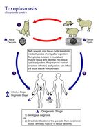
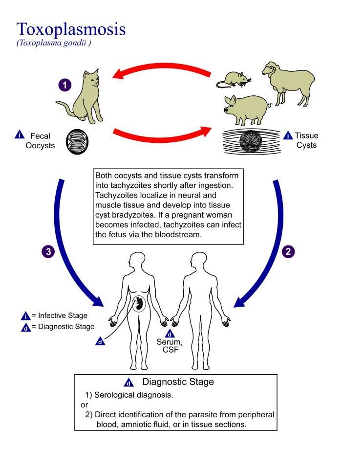
This is an illustration of the life cycle of Toxoplasma gondii, the causal agent of Toxoplasmosis.Created: 2002
-
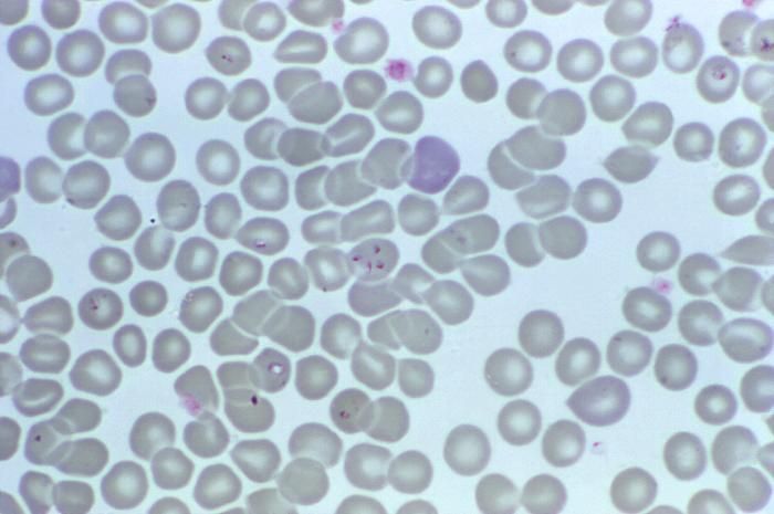
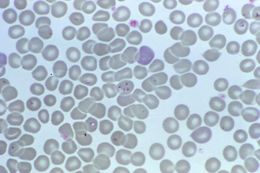
Magnified 1000X, this photomicrograph revealed the presence of what were determined to be numbers of intraerythrocytic Babesia sp. ring-form parasites.Created: 1974
-
This photomicrograph of a fresh stool sample, which had been prepared using a 10% formalin solution, and stained with modified acid-fast stain, revealed the presence of four Cyclospora cayetanensis oocysts in the field of view. Compared to wet mount preparations, the oocysts are less perfectly round and have a wrinkled appearance due to this method of fixation. Most importantly, the staining is variable among the four oocysts.Created:
-
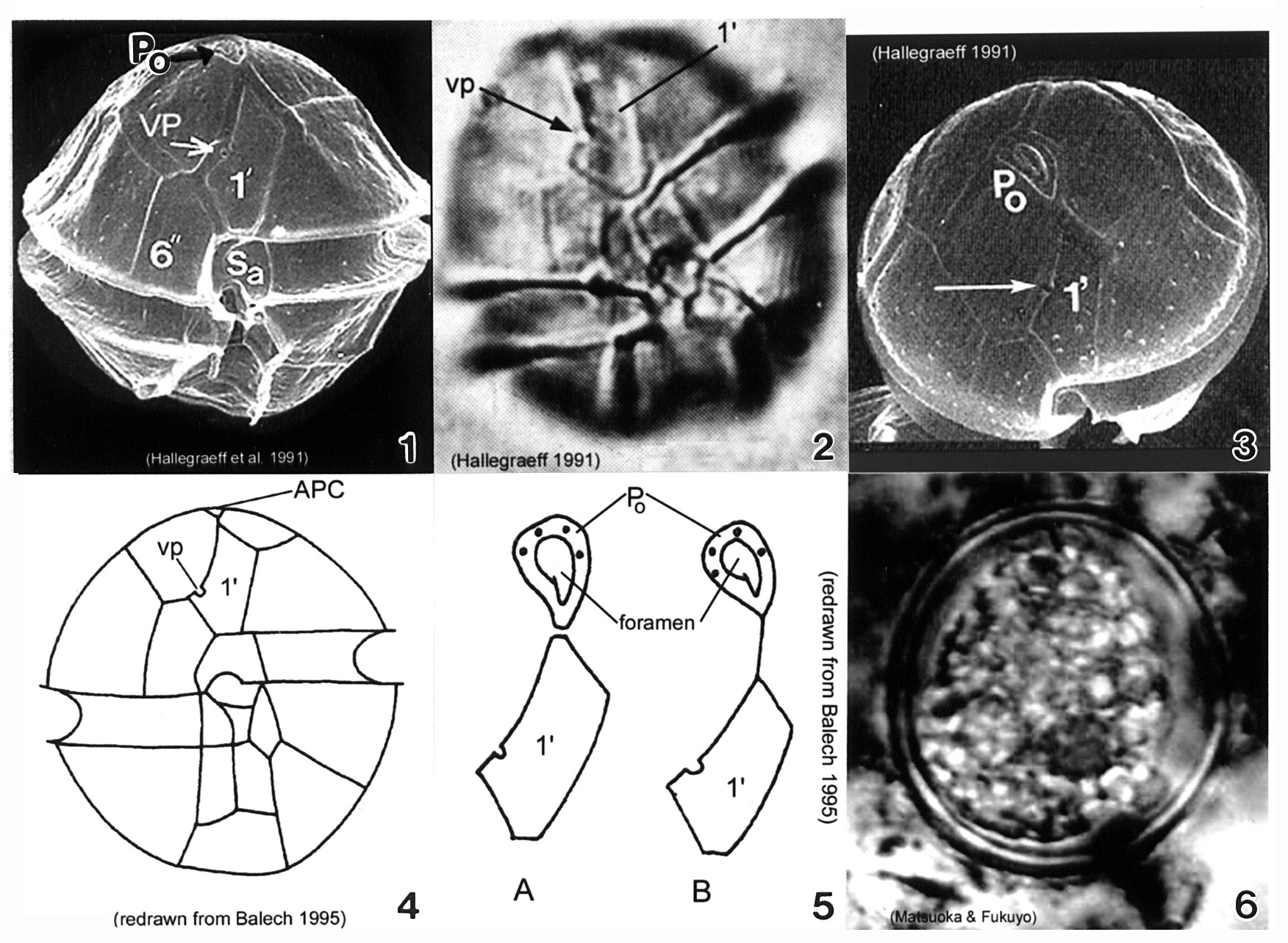
Plate 3. Alexandrium minutum. Fig. 1. SEM: ventral view. Cell small and ellipsoidal. Epitheca conical, larger than hypotheca. Hypotheca short and wide; antapex obliquely flattened. Intercalary bands present. Cingulum deep, lipped; displaced 1X its width. Sulcus shallow (sa=anterior sulcal plate). Apical pore plate (Po) in direct contact with 1' plate. Fig. 2. LM: ventral view. Ventral pore (vp) present on 1' plate. Fig. 3. SEM: apical view. Po large, narrow and oval; indirectly connected to 1' plate. Vp present (arrow). Figs. 4-5. Line drawing. Fig. 4. Ventral view. 1' plate slender and rhomboidal. Fig. 5. Po connection to 1' plate: a. direct; b. indirect via thin suture. Fig. 6. LM: cyst circular in apical view.


