-
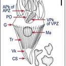
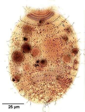
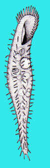
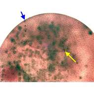
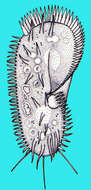
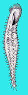
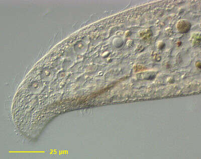
Dorsal infraciliature of the large nassulid ciliate Obertrumia aurea (Ehrenberg, 1833; Foissner 1987). Synonym of Nassula aurea. Obertrumia is distinguished by it's bipartite "hypostomial frange", linear arrays of ciliary tufts. A sigmoid ventrolateral frange runs anteriorly and to the left. The dorsal termination of this part is seen as a vertical arrangement of rectangular collections of kinetids (blue arrow). It terminates at the left end of the horizontal line of ciliary tufts on the dorsal surface (yellow arrow). The uniform longitudinal somatic kineties are well seen here. In vivo the cell appears brightly colored (orange, green or violet) due to multiple food vacuoles containing ingested cyanobacteria. Numerous small mucocysts give the cortex a roughly granular appearance. O. aurea feeds mainly on cyanobacteria. Silver carbonate stain (see Foissner, W.Europ. J. Protistol.27,313-330;1991). Collected from a freshwater pond near Idaho City, Idaho Septmeber 2004. Brightfield optics.
-
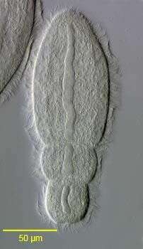
Group portrait of the endocommensal astomatid ciliate, Anoplophrya maupasi(Cépède,1910) in the digestive tract of the oligochaete, Aelosoma hemprichi (see description of characteristics in: Lom, J., 1956. Arch. Protistenk., 101: 286). Collected from a freshwater pond near Boise, Idaho. June 2005. DIC.
-
-
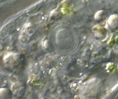
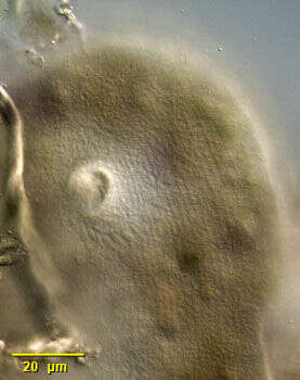
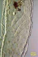
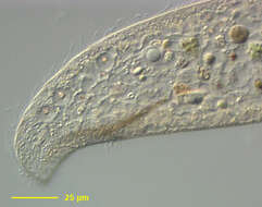
Detail of one of the two macronuclei of Loxodes striatus with its adherent micronucleus. There are two spheroid macronuclei, one anterior and one in the mid body, each with an adherent micronucleus. The arrangement of the macronuclei and the pellicular striations distinguish L. striatus from the similar L. vorax and L. rostrum. Many food vacuoles are seen. Found in polysaprobic habitats. Feeds on cyanobacteria, algae, flagellates and other ciliates. From organically enriched freshwater pond sediment near Boise, Idaho. DIC optics.
-
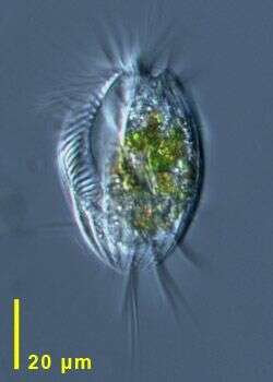
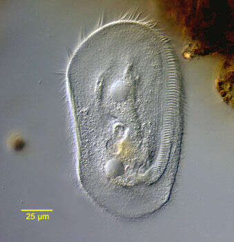
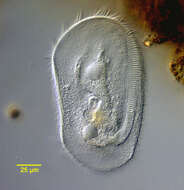
This Euplotes was in Sample 2 taken adjacent to Tvärminne Zoological Station, coastal, algal material 3rd April 2012. A good sharp picture, best images not too large. Usable.
-
-
-
-
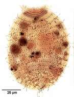
The identity of this organism is unconfirmed. This image is focussed just below the ventral surface. There seem to be 2 rows of marginal cirri - a distinguished feature for Holosticha, and there is a collection of transverse cirri at the back. The inner region of the Adoral Zone of Membranelles that arcs around the front of the cell is quite evident - but is larger than is usual for the genus Holosticha. The relationships between Holosticha and close relatives are subject to debate. Nomarski optics.
-
-
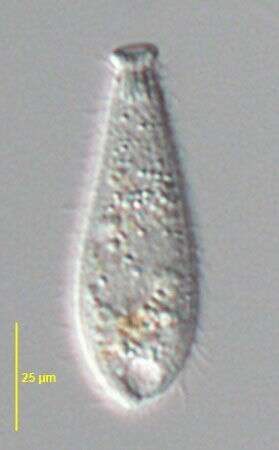
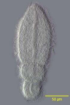
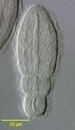
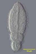
Fully contracted Enchelydium fusidens KAHL, 1930. This population is about 25% smaller than the one described by Kahl.The important morphological featues (oral bulge, extrusomomes, macronucleus) match those of E. fusidens. DIC.
-
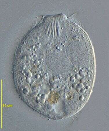
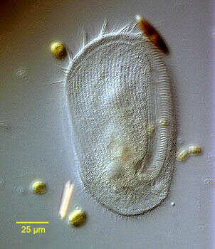
Portrait of Obertrumia aurea(Ehrenberg, 1833; Foissner 1987). Widely distributed nassulid ciliate with well developed cytopharyngeal basket or cyrtos (viewer's upper left). Fed organisms may contain brightly colored orange, blue and purple food vacuoles. O. aurea has a bipartite hypostomial frange (not seen here). From a freshwater pond near Boise, Idaho. Oblique illumination This image was taken by William Bourland. He now uses a Zeiss Axioskop 2 with a Spot Insight CCD camera (Diagnostic Instruments).
-
View of the bandform macronucleus of the endocommensal astomatid ciliate, Anoplophrya maupasi (Cépède,1910) from the digestive tract of the oligochaete,Aelosoma hemprichi (see description of characteristics in: Lom, J., Arch. Protistenk.,101:p.286,1956).There are two smaller opisthes seen here. Collected from a freshwater pond near Boise, Idaho. June 2005. DIC.
-
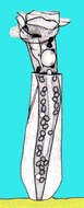
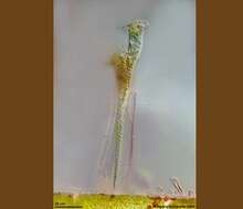
Thuricola (thurr-ick-cola) folliculata and 3 swarmers on the bottom of the lorica. The transparent lorica is equiped with a valve which closes the aperture as cell retracts. This specimen shows endosymbiotic algae. This specimen was collected in freshwater ponds near Konstanz, Germany. Differential interference contrast.
-
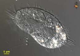
Detail of Loxodes striatus, a medium-size karyorelict ciliate. Loxodes striatus is colorless to slightly brown. The cell body is elongate, rounded anteriorly and posteriorly and highly laterally compressed. The anterior is bent ventrally forming a short beak-like rostrum. Very flexible. Somatic ciliature on the left side is restricted to a marginal kinety. On the right surface there are regular longitudinal kineties. In this species there are longitudinal pellicular striations on the left surface. The slit shaped cytostome is located in a ventral concavity posterior to the rostrum. A thin cone of dark fibrils forms a primitive cytopharynx at the posterior end of the cytostome (seen here). There are two spheroid macronuclei, one anterior and one in the mid body, each with an adherent micronucleus (both seen well in this image). The arrangement of the macronuclei and the pellicular striations distinguish L. striatus from the similar L. vorax and L. rostrum. Refractile concretions of barium sulfate occupy several Müller's vesicles on the dorsal side. These probably act as statoreceptors, orienting the organism in the gravitational field. There are also subcortical pigment granules, which may have chemo- and phototactic functions. Both the vesicles and pigment granules are seen here. Found in polysaprobic habitats. Feeds on cyanobacteria, algae, flagellates and other ciliates. From organically enriched freshwater pond sediment near Boise, Idaho. DIC optics.
-
-
-
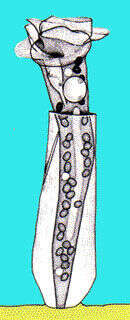
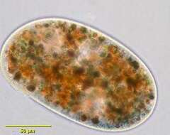
Portrait of Vasicola ciliata (Tatem, 1869), a metacystid ciliate that produces a thin, transparent pseudochitinous vase-like lorica with shallow transverse corrugations (seen in this image). In the lorica the cell body assumes a more globular shape. The circular cytostome is located in the center of the truncate anterior end. It opens into a cytopharynx supported by indistinct trichites. There are three concentric ciliary rings around the cytostome, the innermost with a single ciliary row, the middle with a double ciliary row and a third ring of four ciliary rows. The uniform longitudinal ciliary rows are formed of dikinetids kinetosomes the alignment which gives the appearance of transverse ciliary bands (paratenes). There is a sparse tuft of long caudal cilia (only one long eccentric posterior cilium is seen in the similar genus, Metacystis and another metacystid, Pelatractus, lacks caudal cilia). Multiple food vacuoles are visible in the cytoplasm. The central spherical macronucleus is not seen in this image. The base of the lorica is partly filled with the expelled contents of defecation vacuoles. A single peripheral contractile vacuole is located in the posterior third of the cell. A large, clear terminal vacuole may occur but is not seen in this image. The cell often vacates the lorica when disturbed. Vasicola ciliata is sapropelic and feeds on sulfur bacteria. Collected from stagnant freshwater pond sediment near Boise,Idaho;43°19'07.45"N 115°27'31.99"W, elev.4712 ft.;October2005. Phase contrast.
-
-
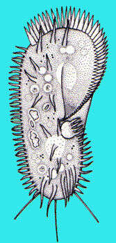
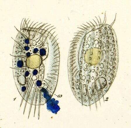
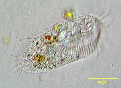
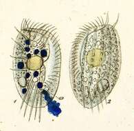
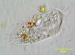
Stylonychia, a widely distributed hypotrich ciliate. The dorsoventrally flattened body is elongate and broadly rounded anteriorly, narrowing posteriorly. The adoral zone of membranelles is strongly developed and rests on an anteriorly protruding collar. The two rows of marginal cirri are slightly out of the focal plane in this image. They do not meet posteriorly. The three characteristic caudal cirri are seen here. There are short dorsal cilia (not seen here). Two ellipsoid macronuclei are visible in this image. One of two small spherical micronuclei is seen at the inferior margin of the posterior macronucleus. This individual has been feeding on diatoms and green algae. From freshwater pond near Boise, Idaho. Brightfield illumination.
-
partially contracted Enchelydium fusidens KAHL, 1930. This population is about 25% smaller than the one described by Kahl.The important morphological featues (oral bulge, extrusomomes, macronucleus) match those of E. fusidens. DIC.
-
Detail of the large Nnassulid ciliate Obertrumia aurea (Ehrenberg, 1833; Foissner 1987). Synonym of Nassula aurea. Obertrumia is distinguished by it's bipartite "hypostomial frange", linear arrays of ciliary tufts. A sigmoid ventrolateral frange (seen most clearly posterior to the cytostome in this image) runs anteriorly and to the left and is separated from a horizontal line of ciliary tufts on the dorsal surface. The cell appears brightly colored (orange, green or violet) due to multiple food vacuoles containing ingested cyanobacteria. Numerous small mucocysts give the cortex a roughly granular appearance. O. aurea feeds mainly on cyanobacteria. Collected from a freshwater pond near Boise, Idaho November 2003. DIC optics.
-
Surface view of the endocommensal astomatid ciliate, Anoplophrya maupasi(Cépède,1910) from the digestive tract of the oligochaete,Aelosoma hemprichi (see description of characteristics in: Lom, J., Arch.Protistenk., 101:286, 1956). Collected from a freshwater pond near Boise, Idaho. June 2005. DIC.
-
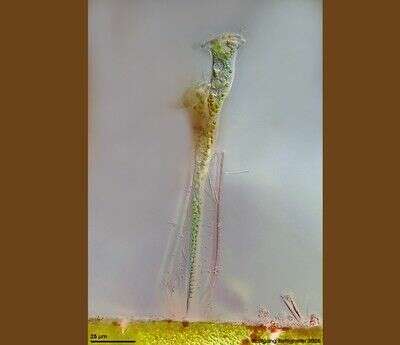
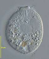
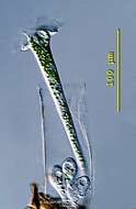
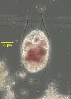
This sessile peritrich ciliat builds chitinous lorica with flap valve. The species houses symbiontic chlorellae. Multi layer image (DOF) shows ciliat and whole lorica with flap valve and epibiontic bacteria. Scale bar indicates 25 µm.See ZIP archive for more. Collected from littoral region (stand of Phragmites) of a rain storage reservoir in Kiel (Schleswig-Holstein, Germany). Images were taken using Zeiss Universal with Olympus C7070 CCD camera.