-
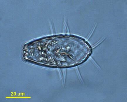
Metromonas grandis Larsen and Patterson, 1990. Cell outline is leaf shaped or slightly roundish. Cells are 5 to 11 microns long (mostly 7 to 10 microns), 4 to 10 microns wide, about 2 microns deep and dorso-ventrally flattened. One side of the cell appears folded. The cells have two flagella, a long flagellum is 1.2 to 2.5 times the length of the cell and trails behind the cell when gliding. There is a short inactive flagellum, less than 2 microns long, which inserts to the right of the major flagellum and is always present. The cells attach to the substratum with the longer flagellum and move with a nodding action - like a pendulum. The nucleus is near the flagellar insertion.
-
-
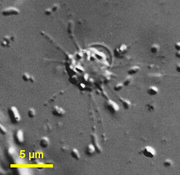
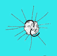
Massisteria (mass-hysteria) is a cercomonad flagellate, with one species (M. marina ), measuring 2.5 to 6.5 microns, dorso-ventrally flattened irregular body. Cells produce delicate pseudopodia with extrusomes, which extend radially from the cell and normally adhere to the substrate. Two short curved flagella arise from the dorsal side of the cell and are relatively inactive in trophic cells, and are very hard to see. Cells may withdraw pseudopodia, and swim using the flagella. Phase contrast.
-
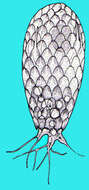
Euglypha (you-gly-fa) a shelled amoeba with filose pseudopodia (although this picture is only of the test) and with the test covered in small overlapping scales and a terminal aperture.Differential interference contrast.
-
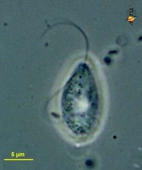
Metromonas (met-row-moan-ass) simplex (Griessmann, 1913) Larsen and Patterson, 1990. Cells are obovate, 3 to 8 microns long (mostly 4 to 7 microns), 2 to 6 microns wide and dorso-ventrally flattened, and have smooth pellicle. The abflagellar margin of the cell is thicker than the (posterior) margin. Two flagella of very unequal length arise from the posterior part of the cell. The major flagellum is always present, is about 1.5 to 3.0 times the length of the cell and may be attached to the substrate. The short inactive flagellum is about 1 microns long and inserts to the right of the major flagellum. It may be difficult to see. The cells normally attach to the substratum and swing from side to side like a pendulum and the cells may also glide with the cell body in front of the flagellum. More common than M. grandis.
-
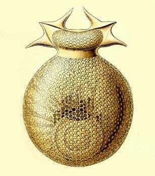
Challengeria murrayi.
-
Massisteria (mass-hysteria) is a cercomonad flagellate, with one species (M. marina ), measuring 2.5 to 6.5 microns, dorso-ventrally flattened irregular body. Cells produce delicate pseudopodia with extrusomes, which extend radially from the cell and normally adhere to the substrate. This image shows the two short curved flagella which arise from the dorsal side of the cell. The flagella are relatively inactive in trophic cells, and are very hard to see. Cells may withdraw pseudopodia, and swim using the flagella. Differential interference contrast.
-
-
Metromonas simplex (Griessmann, 1913) Larsen and Patterson, 1990. Cells are obovate, 3 to 8 microns long (mostly 4 to 7 microns), 2 to 6 microns wide and dorso-ventrally flattened, and have smooth pellicle. The margin of the cell that is away from the flagella is thicker than the (posterior) margin. Two flagella of very unequal length arise from the posterior part of the cell. The major flagellum is always present, is about 1.5 to 3.0 times the length of the cell and may be attached to the substrate. The short inactive flagellum is about 1 microns long and inserts to the right of the major flagellum. It may be difficult to see. The cells normally attach to the substratum and swing from side to side like a pendulum and the cells may also glide with the cell body in front of the flagellum.
-
-
Cedar Swamp, Woods Hole, Massachusetts, USA. Photoed by Hwan Su Yoon.
-
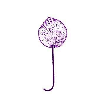
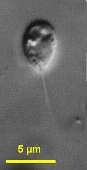
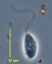
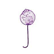
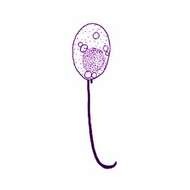
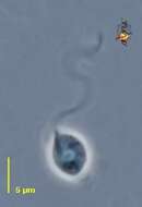
This is a long exposure phase contrast micrograph of Metromonas simplex in situ. This is one of my all time favourites as it so effectively captures the movements of this distinctive predator. Attaching by the crook of the long flagellum, the cell swings actively in an arc - increasing the probability of encountering another protist - its food.
-
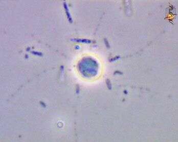
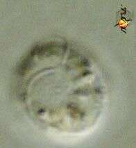
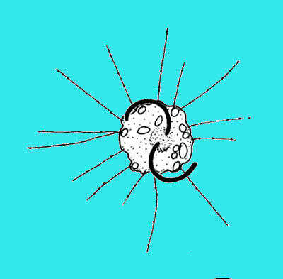
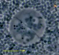
Massisteria (mass-hysteria) marina Larsen and Patterson, 1990. Cells are 3 to 6.5 microns and dorso-ventrally flattened irregular body. The cells produce delicate pseudopodia with extrusomes, which extend radially from the cell and normally adhere to the substrate. Two short curved flagella arise from the dorsal side of the cell and are relatively inactive. Rarely observed.
-
Samples from Sediment at Cedar swamps, Woods Hole, Massachusatts. Photographed by Hwan Su Yoon.
-
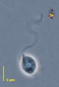
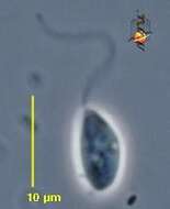
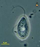
Proleptomonas (pro-lep-toe-moan-ass) a ellipsoidal heterotrophic flagellate of uncertain affinities, one apical flagellum emerging from the front of the cell, nucleus usually located anteriorly. Reported usually from soils. Phase contrast.
-
Massisteria marina Larsen and Patterson, 1990. Cells are 3 to 6.5 microns and dorso-ventrally flattened irregular body. The cells produce delicate pseudopodia with extrusomes, which extend radially from the cell and normally adhere to the substrate. Two short curved flagella arise from the dorsal side of the cell and are relatively inactive.
-
Samples from Sediment at Cedar swamps, Woods Hole, Massachusatts. Photographed by Hwan Su Yoon.
-
Proleptomonas (pro-lep-toe-moan-ass) a ellipsoidal heterotrophic flagellate of uncertain affinities, one apical flagellum emerging from the front of the cell, nucleus usually located anteriorly. Reported usually from soils. Phase contrast.
-
Cholamonas (coal-a-moan-ass), cercomonad flagellate described early in the third millenium, with two subapically inserting flagella. Phase contrast micrograph.
-
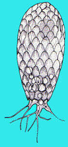
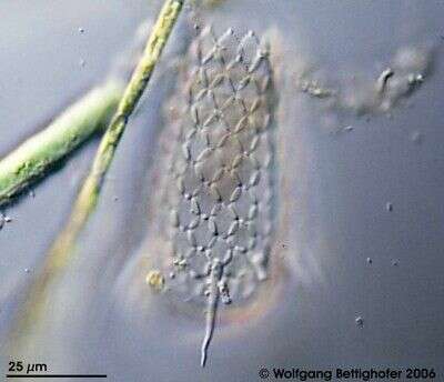
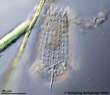
Members of testate amoebae group Euglyphidae form siliceous plates for constructing shell. Sample collection by Martin Kreutz from Simmelried near Konstanz(Baden-Wuerttemberg, Germany). This image was taken using Zeiss Universal with Olympus C7070 CCD camera.
-
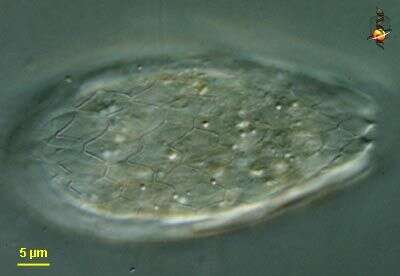
Proleptomonas (pro-lep-toe-moan-ass) a ellipsoidal heterotrophic flagellate of uncertain affinities, one apical flagellum emerging from the front of the cell, nucleus usually located anteriorly. Reported usually from soils. Bilaterally symmetrical cyst. Phase contrast.
-
Gymnochlora (jim-know-clore-a), one of the amoeboid algae but like other members of the chlorarachniophytes the plastids result from a secondary endosymbiotic event involving a eukaryotic green alga, a nucleomorph is present. With bright green plastids. Differential interference microscopy.
data on this strain.
-
Apertural plates of Euglypha cristata Leidy, 1874. Found in a soil sample from Pyhä-Luosto National Park, Finland. DIC.
-
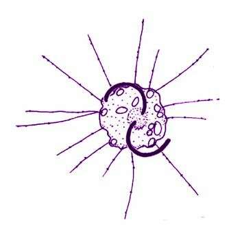
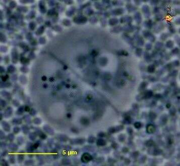
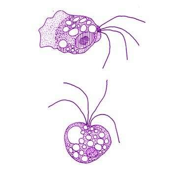
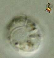
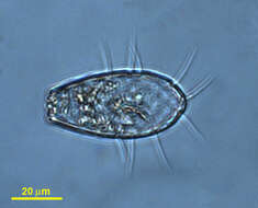
Quadricilia rotundata (Skuja, 1948) V+rs, 1992. Cells are 5-9 x 10-15 microns, globular cells 10-20 microns in diameter. Cell globular, or nearly so, with 4 (-8) unequal, smooth, acronematic flagella inserted anteriorly in a shallow depression. The flagella are 1-3 times the diameter of the cell body. Many thin, branched pseudopodia may be produced from any point of the cell surface. Cytoplasm sometimes highly vesiculate, nucleus central or in the cell anterior.