-
Bloomfield Track
-
Hong Kong Wetland Park
-
Diana M. P. Galassi, Paola De Laurentiis, Barbara Fiasca
Zookeys
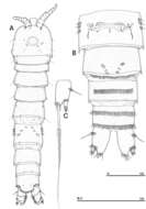
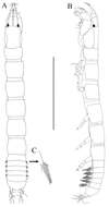
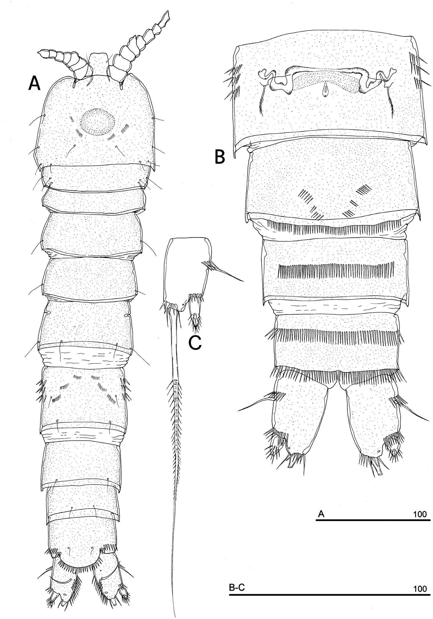
Figure 10.Phyllognathopus inexspectatus sp. n. (♀). A habitus, dorsal view B abdomen, ventral view C caudal ramus, ventral view (scale bars in μm).
-
Kristine N. White, James Davis Reimer
Zookeys
Figure 3.Leucothoe amamiensis sp. n., holotype male, 5.9 mm, RUMF-ZC-1654.
-
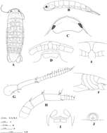
Eduardo Suárez-Morales, Humberto Camisotti, Alberto Martín
Zookeys
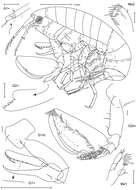
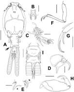
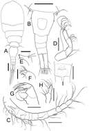
Figure 1.Caligus evelynae sp.n., adult female from Venezuela: A habitus, dorsal view B sternal furca, ventral view C antennule D antenna E postantennal process (b) and maxillule (a) F maxilla G detail of calamus and canna H maxilliped I genital complex and abdomen, ventral view. Scale bars: A, I=0.5 mm, B–F, H =0.1 mm, G=0.05 mm.
-
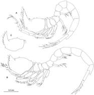
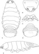
Figure 2.Ithyleucon sorbei gen. et sp. n. A preadult female holotype (ICMU12101901), whole animal in lateral view B preadult male paratype (ICMU12101904) C carapace of immature male paratype (ICMU12101906).
-
Tomislav Karanovic, Mark J. Grygier, Wonchoel Lee
Zookeys
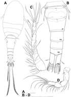
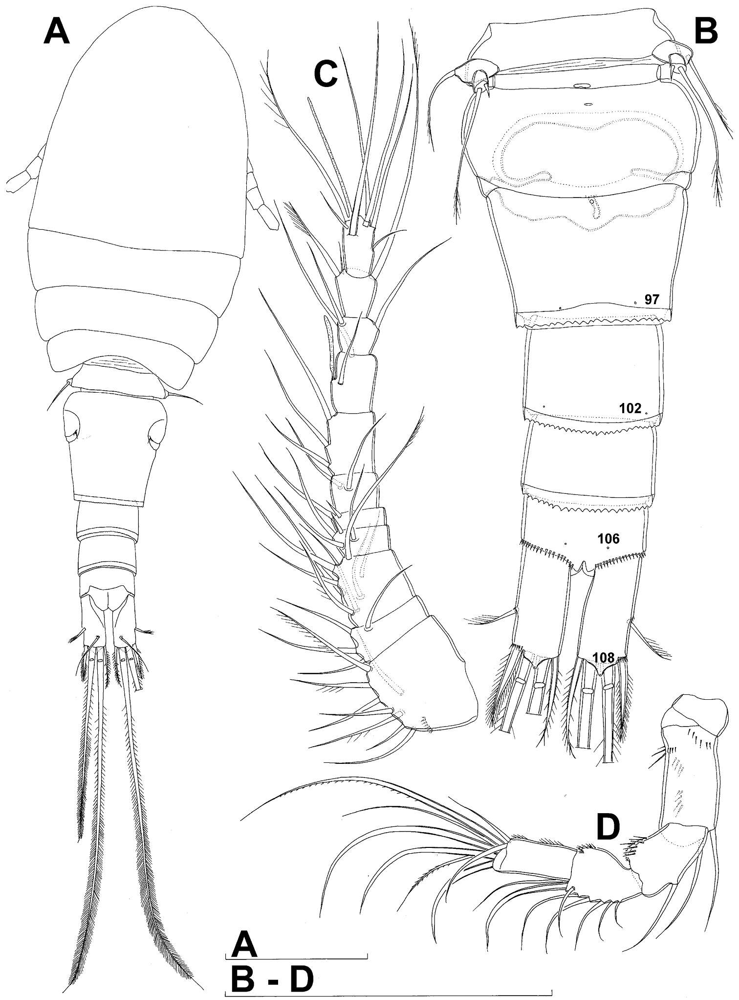
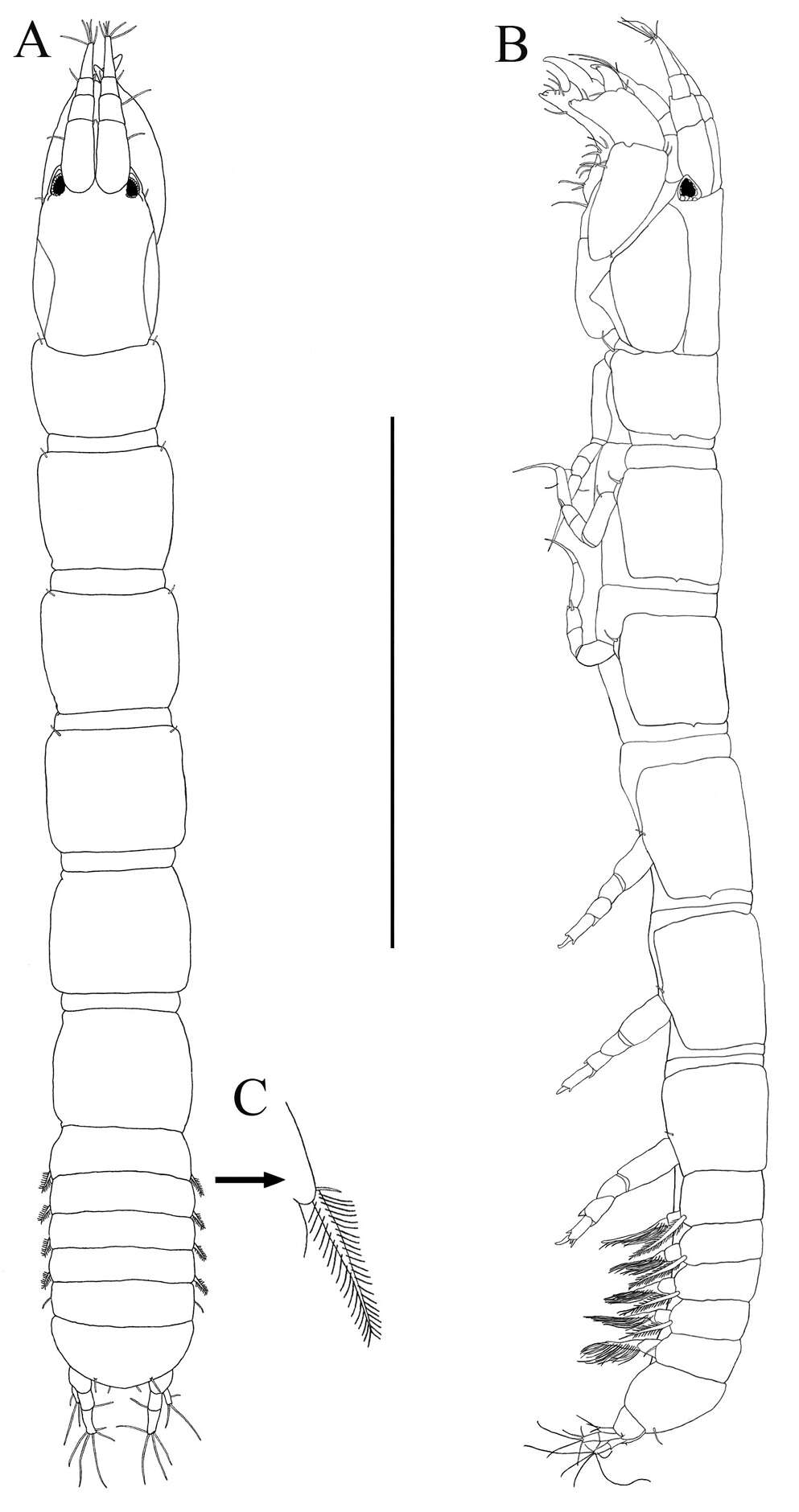
Figure 1.Diacyclops ishidai sp. n., holotype female: A habitus, dorsal view B urosome, ventral view C antennula, dorsal view D antenna, dorsal view. Arabic numerals numbering sensilla and pores consecutively from anterior to posterior end of body, and from dorsal to ventral side (excluding appendages). Scale bars 100 μm.
-
Nancy F. Mercado-Salas, Eduardo Suárez-Morales, Alejandro M. Maeda-Martínez, Marcelo Silva-Briano
Zookeys
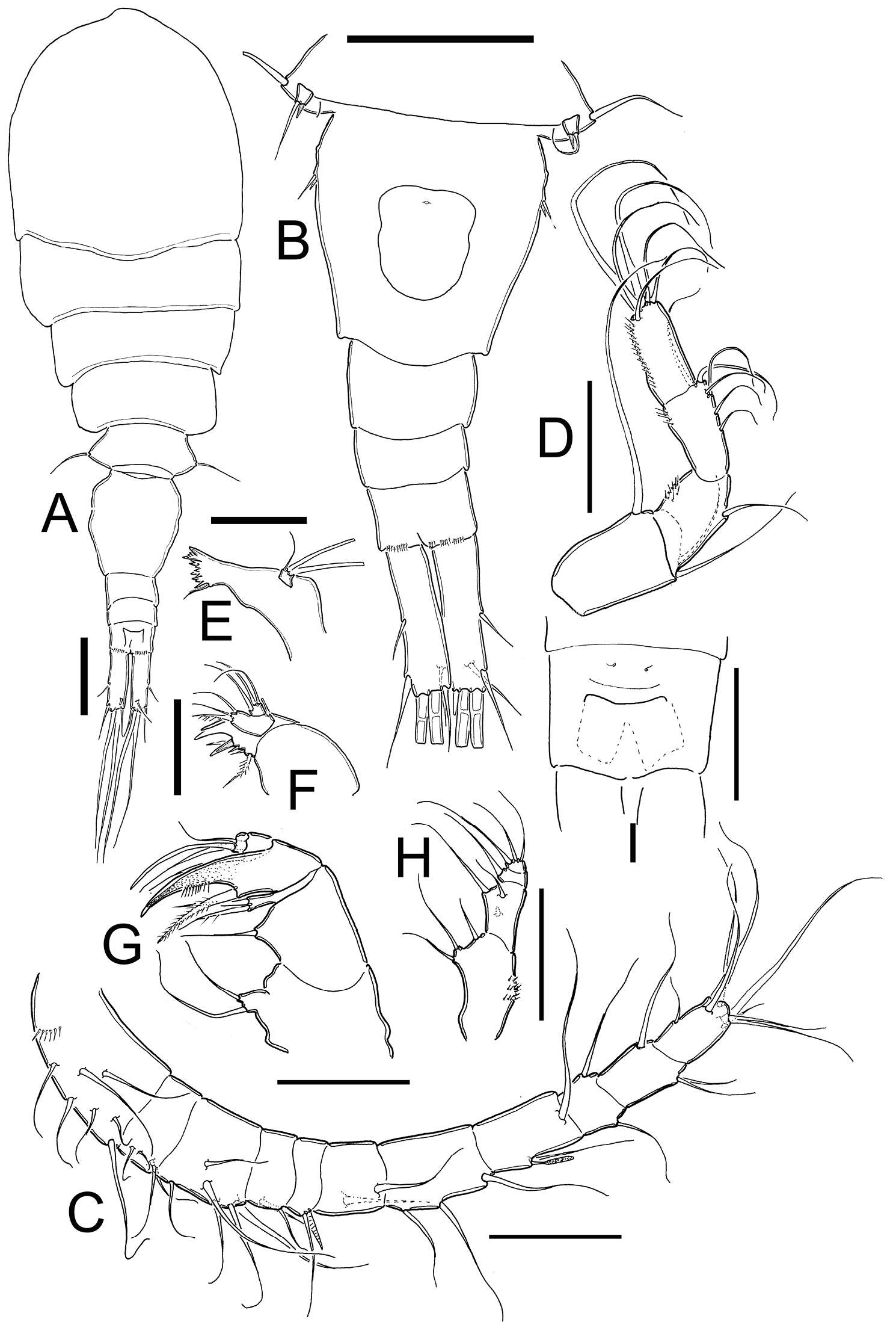
Figure 1.Metacyclops deserticus sp. n., female holotype from Coahuila, Mexico. A habitus, dorsal view B urosome, ventral view C antennule D antenna E mandible F maxillule G maxilla H maxilliped I anal operculum. Scales bars A–B= 100µm; C–I= 50 µm.
-
Eduardo Suárez-Morales, Jani Jarquín-González
Zookeys
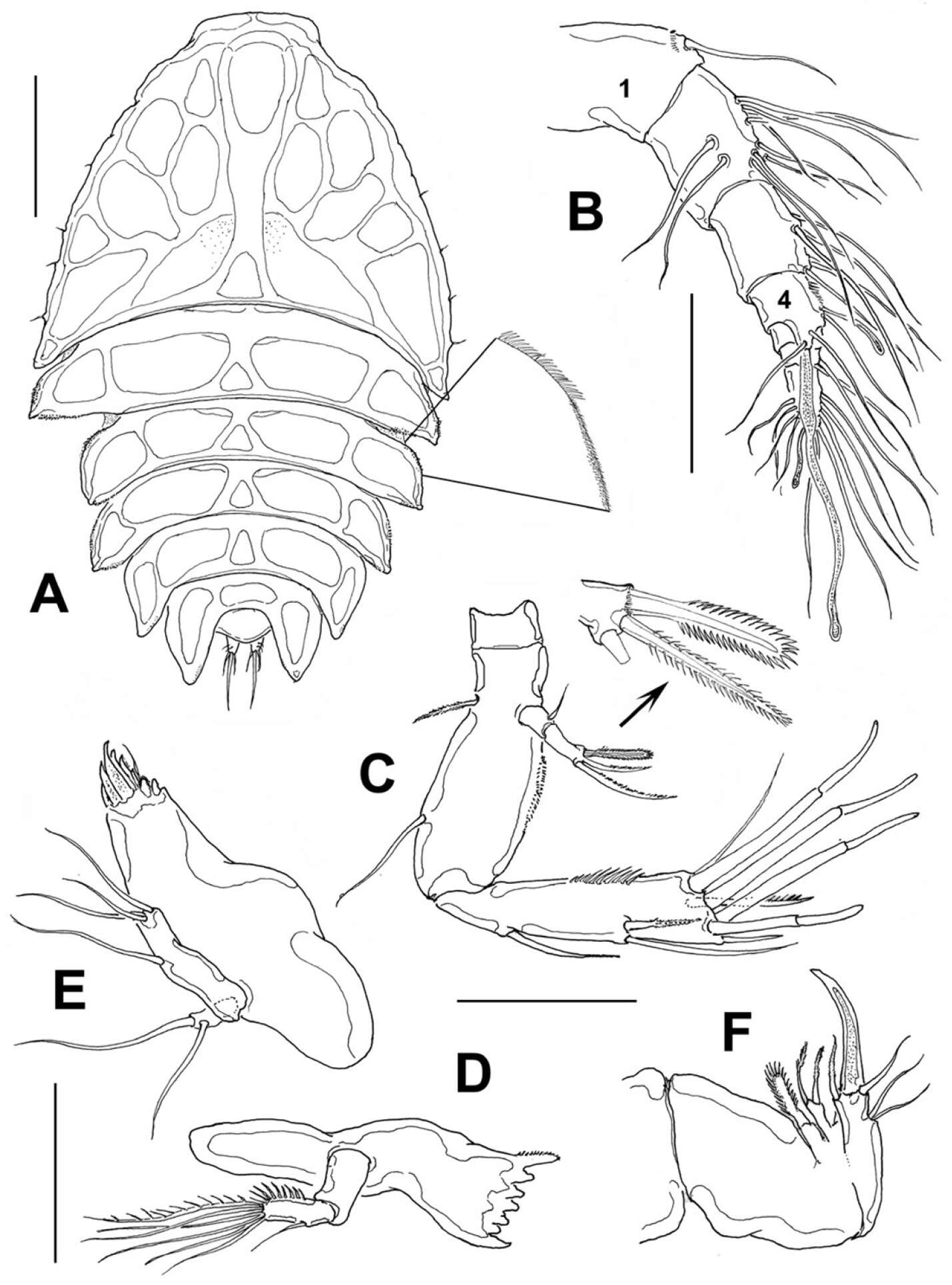
Figure 2.Peltidium nayarit sp. n., from Playa Careyeros, Nayarit, Mexican Pacific. A adult female, habitus, dorsal view, showing detail of ornamentation of epimeral processes of cephalothorax B antennule C antenna D mandible E maxillule F maxilla. Scales bars: A= 250 μm, B–F=100 μm.
-
Terue C. Kihara, Carlos E. F. Rocha
Zookeys
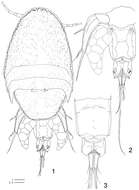
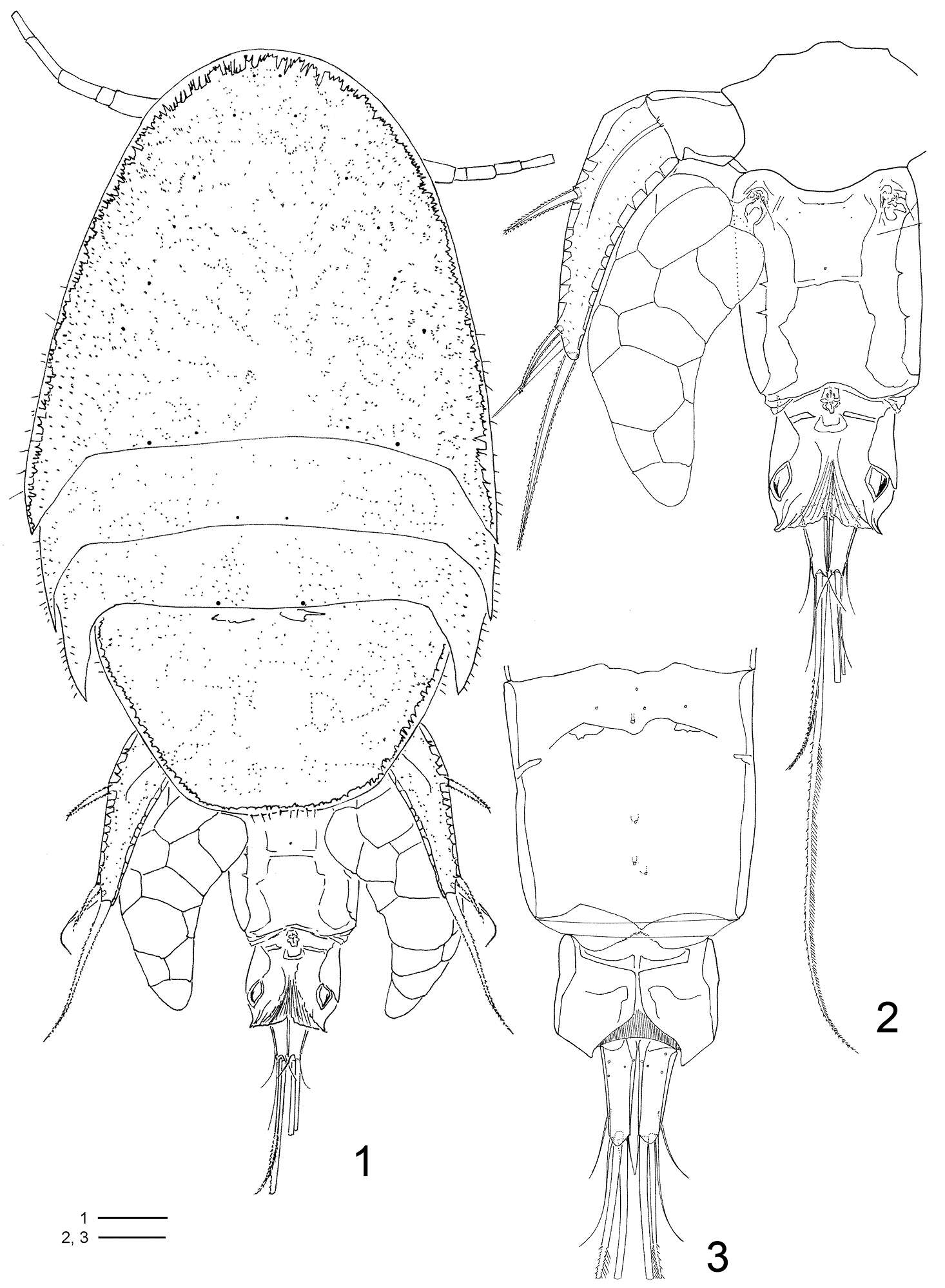
Figures 1–3.Clausidium rodriguesi sp. n. Female: 1 habitus, dorsal 2 urosome, dorsal 3 urosome lacking somite bearing P5, ventral. Scale bars: 1 = 100 μm; 2, 3 = 50 μm.
-
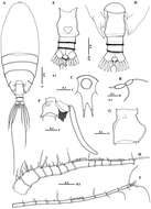
Mohsen M. El-Sherbiny, Ali M. Al-Aidaroos
Zookeys
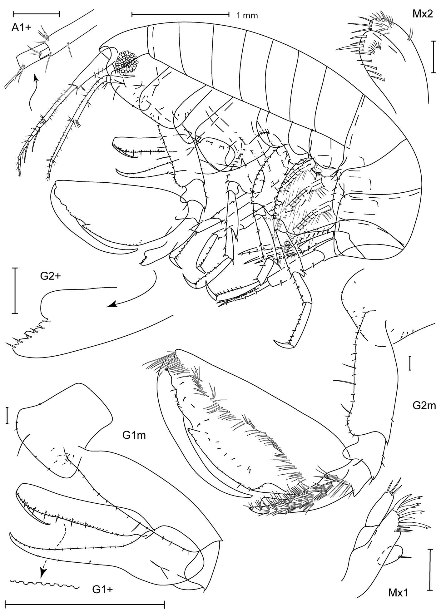
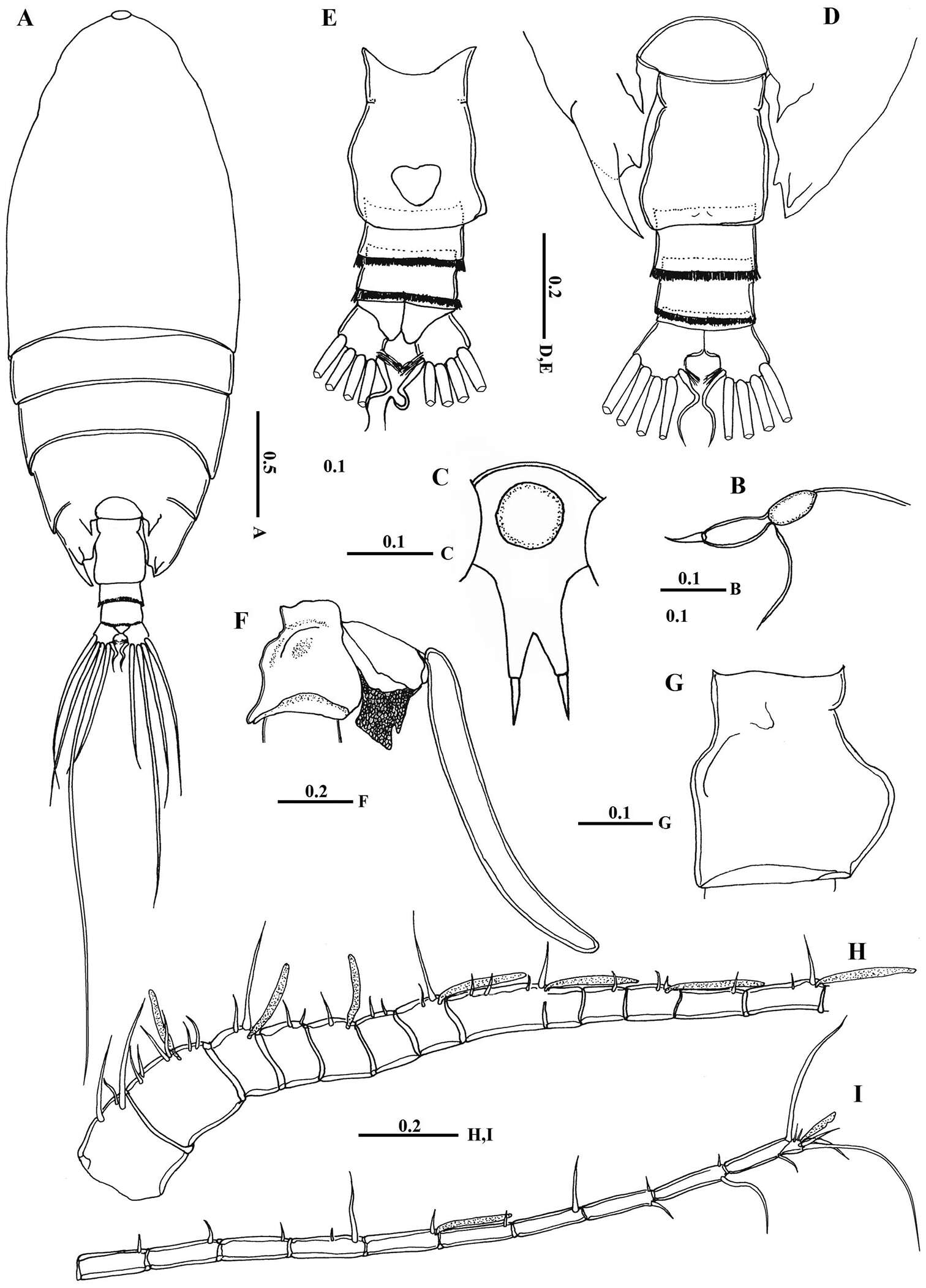
Figure 2.Macandrewella cochinensis female from the northern Red Sea. A habitus, dorsal view B rostrum, lateral view C rostrum, ventral view D posterior prosome and urosome, dorsal view E urosome, ventral view F genital double-somite with spermatophore, lateral view (right) G genital double-somite, lateral view (right) H–I antennules. All scale bars in mm.
-
Young-Hyo Kim, Ed A. Hendrycks
Zookeys
Figure 1.Socarnes tongyeongensis sp. n., female, 8.8 mm, Gyeongpo, Pungwha-ri, Sanyang-eup, Tongyeong-si, Korea.
-
Eknarin Rodcharoen, Niel L. Bruce, Pornsilp Pholpunthin
Zookeys
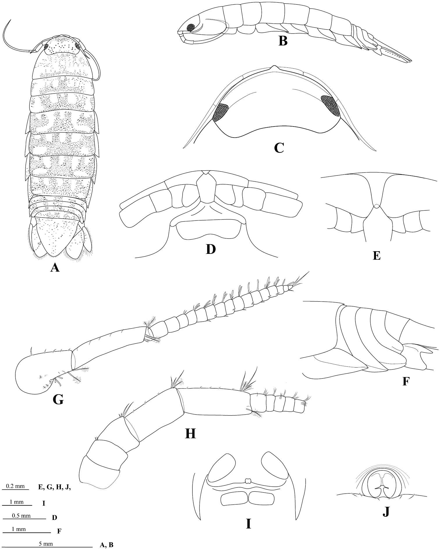
Figure 2.Cirolana songkhla sp. n., male holotype (PSUZC-CR0281-01) (13.7 mm) (A–F), male paratype (PSUZC-CR0281-2) (11.2 mm) (G–H), male paratype (PSUZC-CR0281-2) (13.8 mm) (I–J). A dorsal view B lateral view C head, dorsal view D frons E detail of frontal lamina F pleon G antennule H antennal peduncle I antero-ventral view of penial opening J ventral view of penial opening.
-
Shigenori Karasawa, Kenshi Goto
Zookeys
Figure 1.Burmoniscus kitadaitoensis, male, holotype, TOYA-Cr 14899. A, B Pleopod 1 endopodite C pleopod 1 exopodite D pleopod 2 endopodite E pleopod 2 exopodite F genital papilla. Scale bars: A, C–E 200 μm, B 50 μm.
-
Andrés G. Morales-Núñez, Richard W. Heard
Zookeys
Figure 2.Paratanais rosadi sp. n., holotype female: A dorsal view B lateral view C enlargement of articulated setulate seta on pleonite-1. Scale bar A–B 1.0 mm.
-
Kerry A. Hadfield, Niel L. Bruce, Nico J. Smit
Zookeys
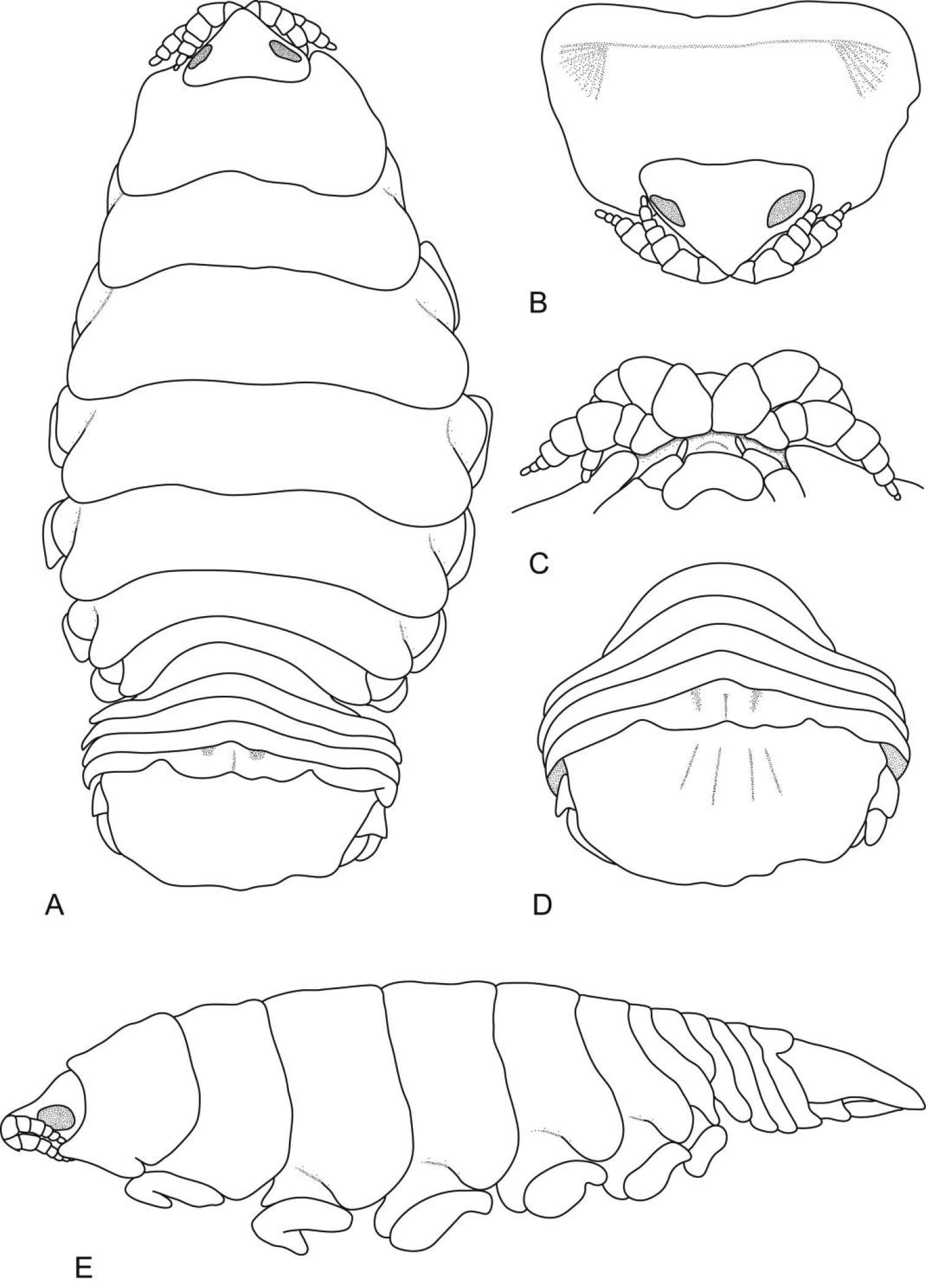
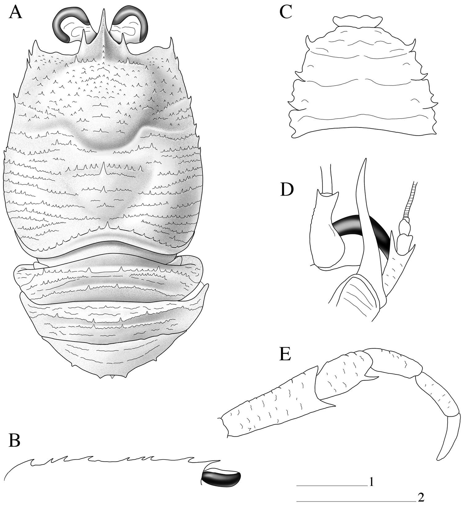
Figure 1.Ceratothoa africanae sp. n. female holotype (29 mm) (SAM A45937): A dorsal view B antero-dorsal view of pereonite 1 and cephalon C ventral view of cephalon D dorsal view of pleotelson E lateral view.
-
Patricia Cabezas, Enrique Macpherson
Zookeys
Figure 1.Paramunida haigae sp. n. male holotype, 16.6 mm (LACM–CR1973-3312). Christmas (Kiritimati) Island. A carapace and abdomen, dorsal view B carapace, lateral profile C sternum D left antennule and antenna, ventral view E right maxilliped 3, lateral view. Scale: 5 mm (scale 1 for A–C, E; scale 2 for D).
-
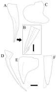
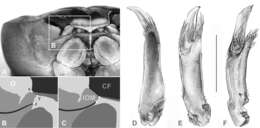
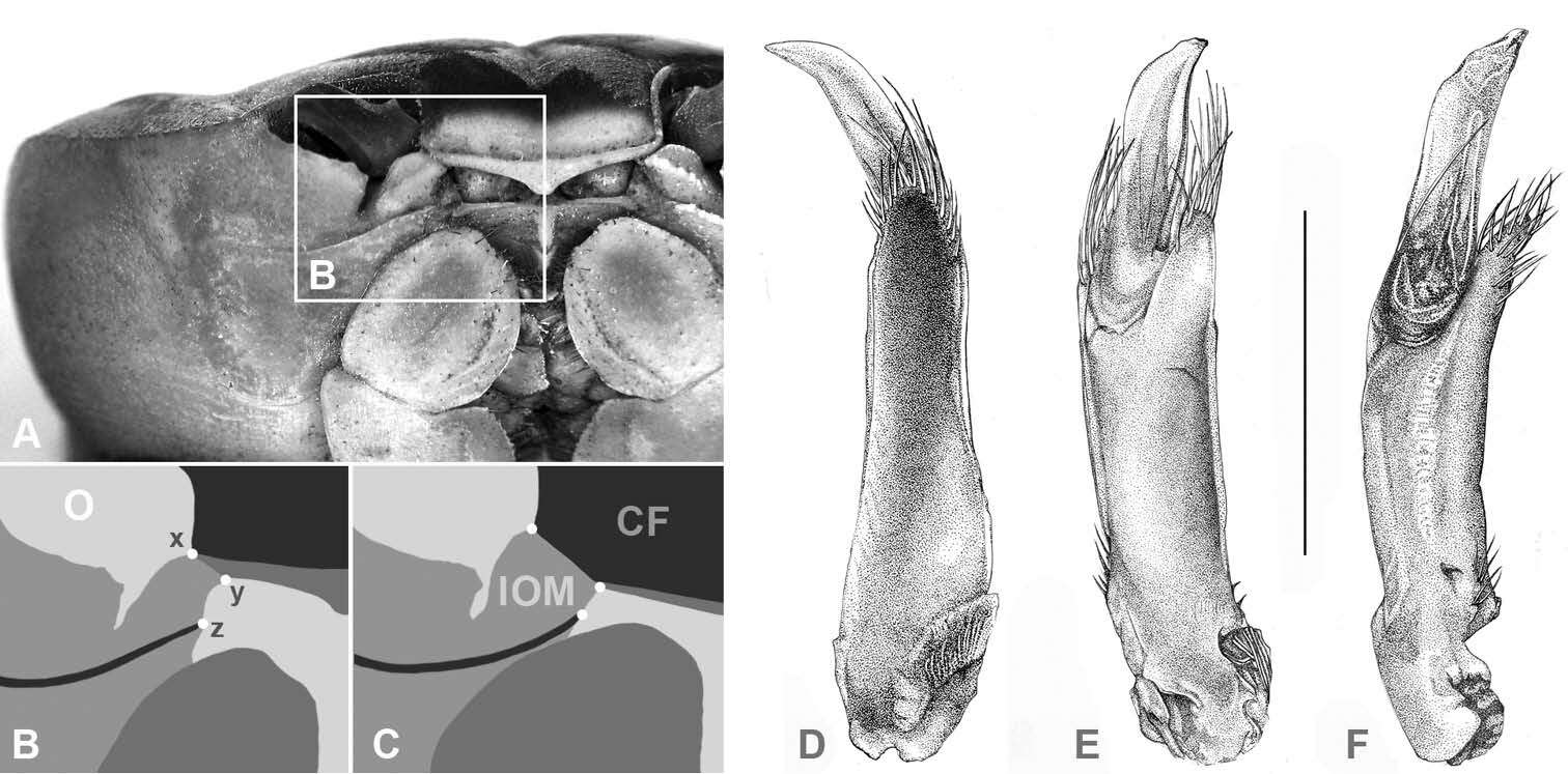
Figure 2.(CF) Carapace front; (O) orbit; (IOM) mesial lobe of infraorbital margin; (x) widest width of CF; (z) mesial end of suborbital crista; (x–y) width of IOM at point of contact with CF; (y–z) shortest distance between CF and mesial end of suborbital crista; A, B Atlantic Gecarcinus lateralis (Freminville 1835), male, carapace width (CW) 31 mm, Costa Rica, Puerto Viejo C Gecarcinus nobilii sp. n., holotype, male, CW 31 mm, Ecuador, Punta Galera (LACM CR 1968-477). First male gonopod: Gecarcinus nobilii sp. n., holotype: D mesial view E lateral view F Pacific Gecarcinus lateralis (sensu Türkay 1973), CW 31 mm, Costa Rica, Hermosa Beach, lateral view; Scale bar = 5 mm.
-
Figure 2.Mesocletodes elmari sp. n., adult female, paratype 2. CLSM photograph of a Congo-red stained specimen, lateral view. Scale bar: 100 µm
-
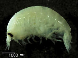
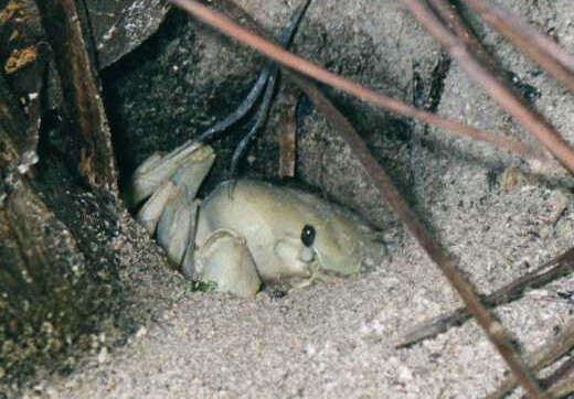
Kent Island, Chesapeake Bay, Maryland, USA
-
Subject: Live Animal | Type: Photo | Life Stages And Gender: Adult/Sexually Mature
-
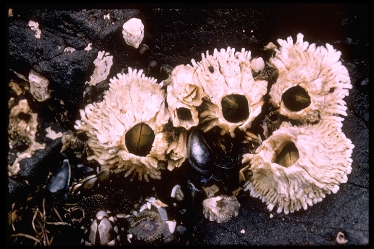
1999 California Academy of Sciences
CalPhotos
-
-
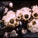
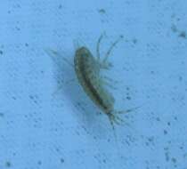
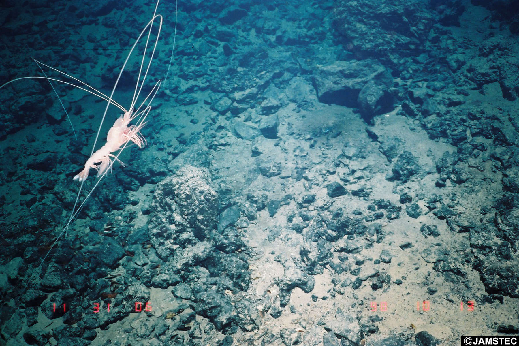
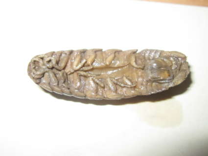
Description: Isopoda Aega sp., possibly Aega bicarinata Leach, 1818.
This specimen was caught using a specimen collector. This particular family of Isopoda, the Aegidae, are temporary parasites on marine fishes. They engorge themselves with food (presumably blood), from their hosts, then dislodge and sit on the bottom to digest their meal.
The specimen seen here is about 5 cm long from head to tail end. Item Type: Image Title: Aega sp. Copyright: Serpent project Species: Aega sp. Behaviour: Probably a temporary parasite, feeding on blood of fish host. The anterior first 3 pairs of pereiopods are hooked to hold on to the host whereas the rest of the pereipods are 'walking legs'. Site: Arctic -- Barents -- Norway Site Description: Seafloor Depth (m): 310 Latitude: 71 deg 00' 00" N Longitude: 21 deg 00' 00" E Project Partners: Oceaneering ROV: Magnum 142 Deposited By: Dr K Kroeger Deposited On: 16 November 2011