-
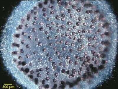
A colonial spumellarian radiolarian, image and identification by Dave Caron. This is an example of one of the four types of large amoebae which is common in the marine water column.
-
Individual isolated from the Hamble estuary, southern England. Image courtesy of Dr. Elisabeth Alve, University of Oslo. Citation: Alve, E. and Murray, J.W. Ecology and taphonomy of benthic foraminifera in a temperate mesotidal inlet. Journal of Foraminiferal Research 24:18-27.
-
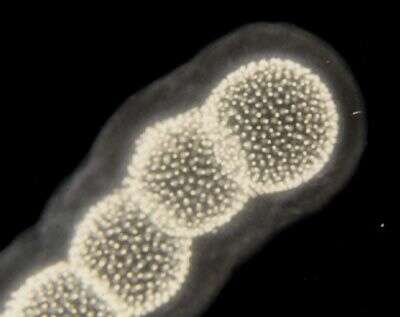
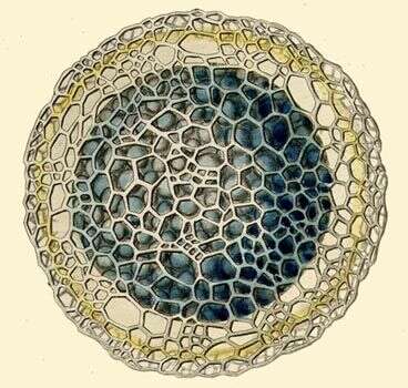
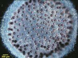
A colonial spumellarian radiolarian (Collosphaera sp.) composed of numerous central capsules (purple porous spheres) connected to one another by cytoplasmic strands and enclosed in a clear gelatinous sheath secreted by the radiolarian cytoplasm.
-
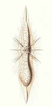
A light microscopic view of a living nassellarian radiolarian (Eucyrtidium acuminata) showing the reddish pigmented cytoplasm within the siliceous, conical shell.
-
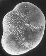
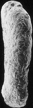
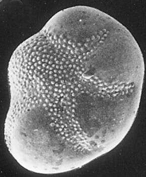
Bulimina inflata.
-
The outer surface of the test is much smoother on this specimen than in most members of its species. Image courtesy of Elisabeth Alve, University of Oslo. Originally published in J. Foram. Res. 16: 261-284; used with permission.
-
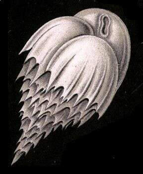
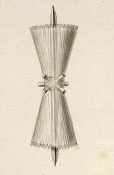
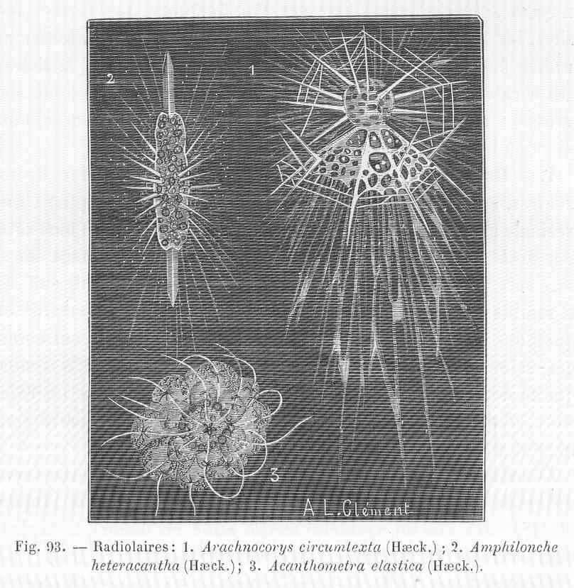
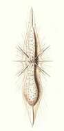
Haeckel says: The central capsule contains numerous spherical nuclei and is enclosed by the hyaline calymma, which forms conical sheaths around the spines.
-
-
-
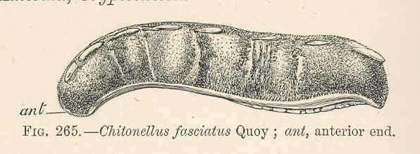
Chitonellus fasciatus Quoy; ant, anterior end.
-
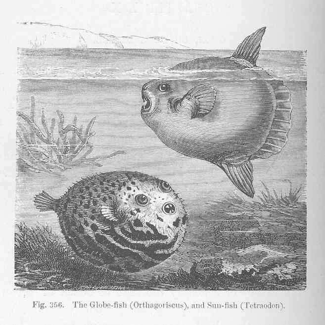
Globe-fish (Orthagoiscus), and Sun-fish (Tetraodon).
-
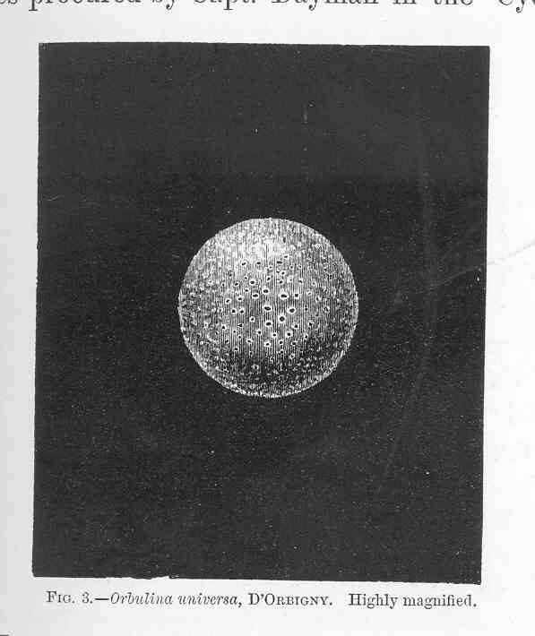
Orbulina universa, d'Orbigny.
-
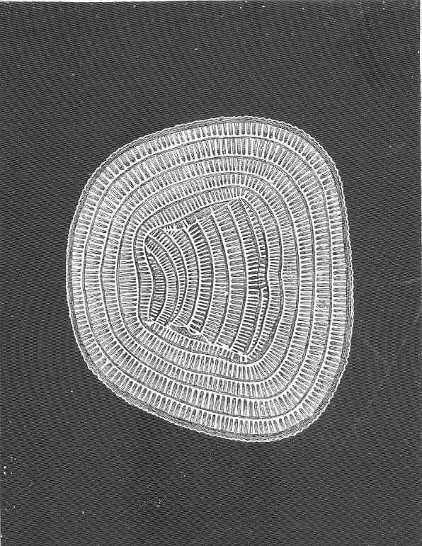
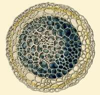
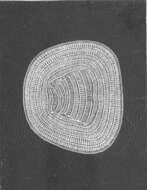
Orbitolites tenuissimus, Carpenter MSS..
-
Radiolaires. 1, Arachnocorys circumtexta (Haeck.); 2, Amphilonche heteracantha (Haeck.)
-
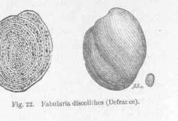
Fabularia discolithes (Defraroe).
-
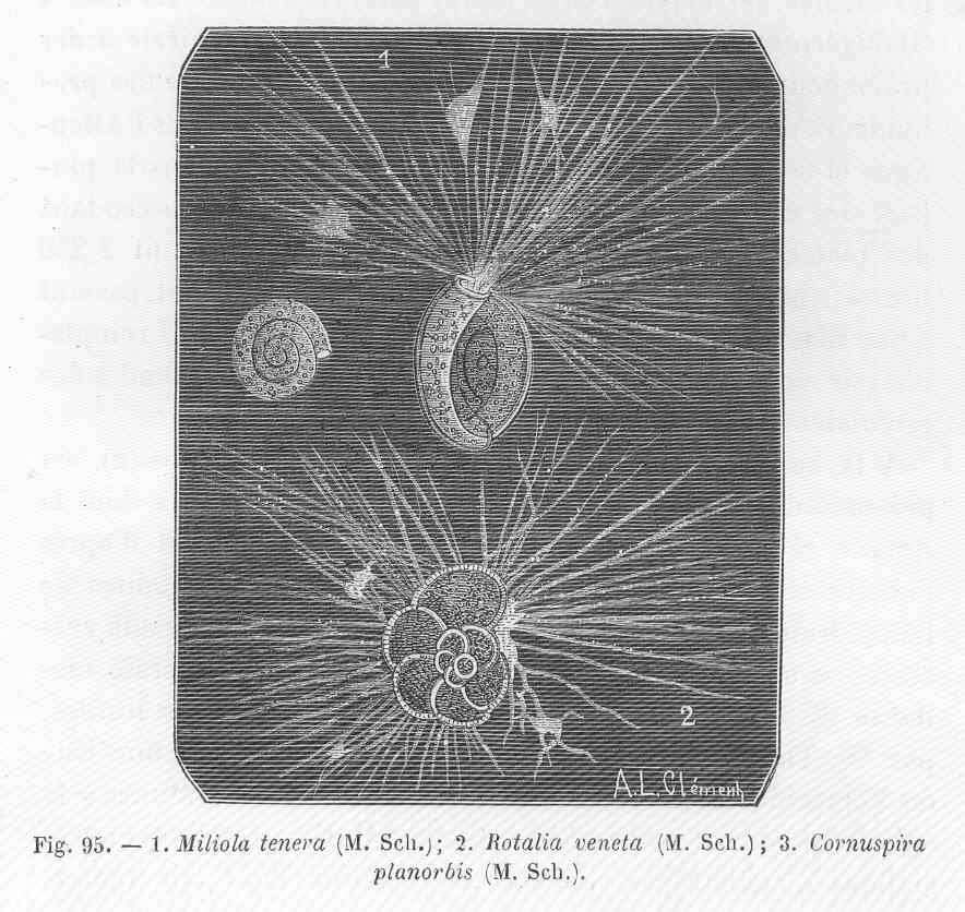
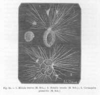
Miliola tenera (M.Sch.)-1, Rotalia veneta (M. Sch.)-2, Cornuspira planorbis (M.Sch).-3.
-
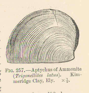
Aptychus of Ammonite (Trigonellites latus), Cameridge Clay, Ely.
-
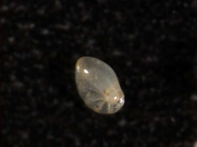
Extreme macro: take a good binocular microscope, a good camera and just focus your microfossils ;)
-
SEM image of the foraminiferan Planorbulina acervalis
-
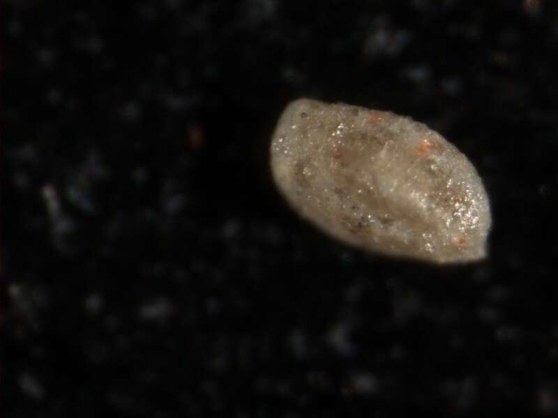
Extreme macro: take a good binocular microscope, a good camera and just focus your microfossils ;)
-
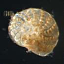
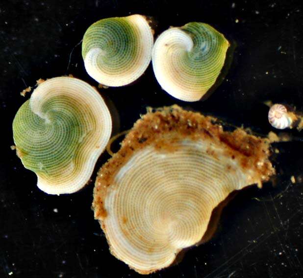
Live individuals of the foraminiferans Archaias angulatus and one specimen of Coscinospira (=Peneroplis) antillarum from Belize
-
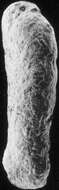
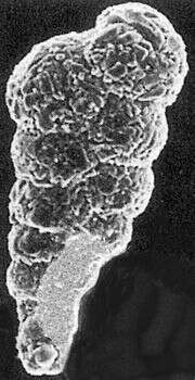
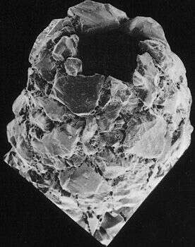
Foraminiferans living in polluted environments often show alterations in the morphology of their tests. This individual, isolated from a site in Norway which is contaminated with heavy metals, exhibits reduced chamber size in some of its chambers (notice that the test is not evenly rounded.) Image courtesy of Dr. Elisabeth Alve, University of Oslo. Citation: Alve, E. Benthic foraminifera reflecting pollution. Journal of Foraminiferal Research 21:1-19.
-
A closer view of the aperture. Aperture morphology is one of the important diagnostic characteristics for foram identification. Image courtesy of Elisabeth Alve, University of Oslo. Originally published in J. Foram. Res. 16: 261-284; used with permission.
-
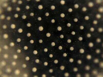
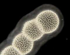
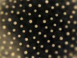
Sphaerozoum (sphere-owe-zoo-um), detail of the surface of a colony, in which many individual organisms can be seen. In the centre of each of the bright regions is the capsule. This is an example of one of the four types of large amoebae which is common in the marine water column. Dark ground image by Dave Caron.