-
Canella Radea, Aristeidis Parmakelis, Vassilis Papadogiannis, Despoina Charou, Kostas A. Triantis
Zookeys
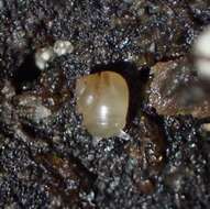
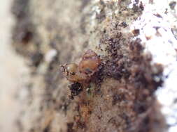
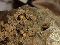
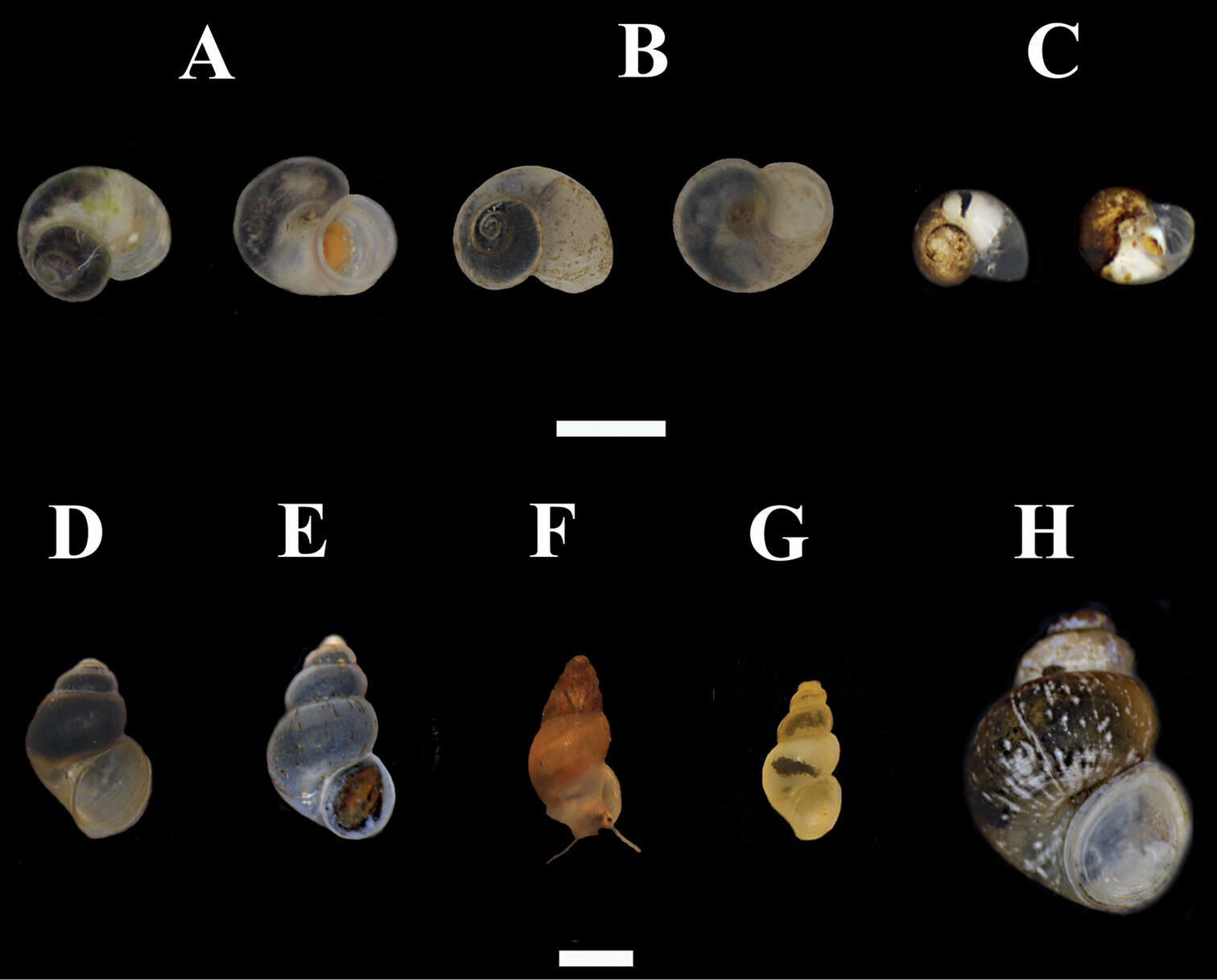
Figure 3.Hydrobioids collectedduring the survey in mainland and insular Greece. A Daphniola exigua (dorsal and ventral view) B Isimerope semele (dorsal and ventral view, Megali Vrysi) C Pseudoislamia balcanica (dorsal and ventral view, Ag. Sophia) D Pseudamnicola pieperi (Olympos) E Radomaniola cf. curta (spring of Louros river) F Radomaniola cf. curta (Ag. Sophia spring) G Trichonia trichonica H Pseudobithynia eubooensis. Scale bar 1 mm.
-
Robert Hershler, Hsiu-Ping Liu, Corbin Bradford
Zookeys
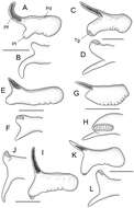
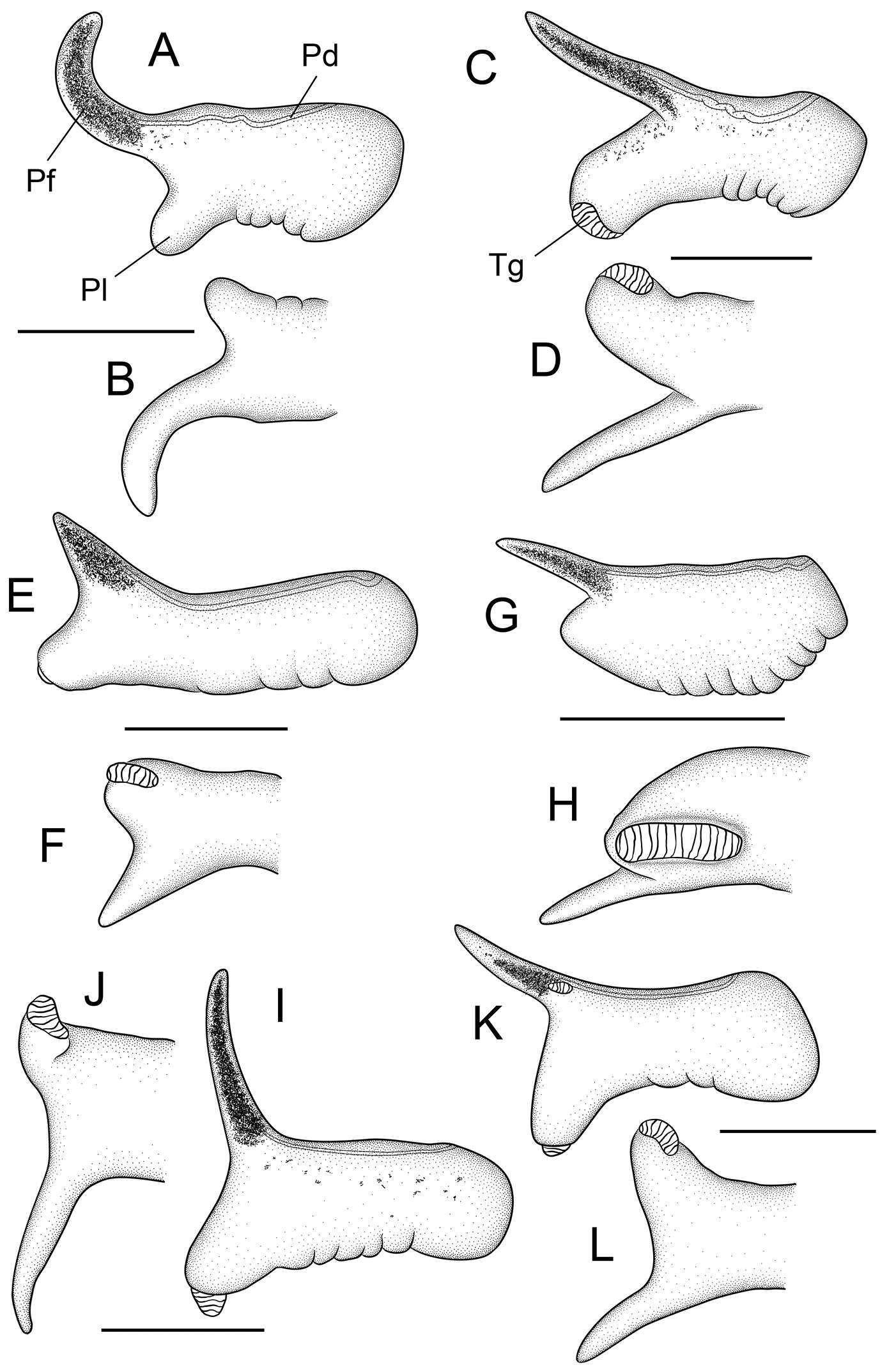
Figure 4.Penes (dorsal, ventral surfaces). A, B Pyrgulopsis licina sp. n., USNM 850346 C, D Pyrgulopsis perforata sp. n., BellMNH 20891 E, F Pyrgulopsis sanchezi sp. n., USNM 883361 G, H Pyrgulopsis micrococcus, BellMNH 20663 I, J Pyrgulopsis turbatrix, USNM 860699 K, L Pyrgulopsis turbatrix, USNM 883373. Scale bars A–C 250 µm; D–L 500 µm. Pd penial duct Pf penial filament Pl penial lobe Tg terminal gland.
-
Robert Hershler, Hsiu-Ping Liu, Corbin Bradford
Zookeys
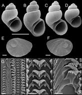
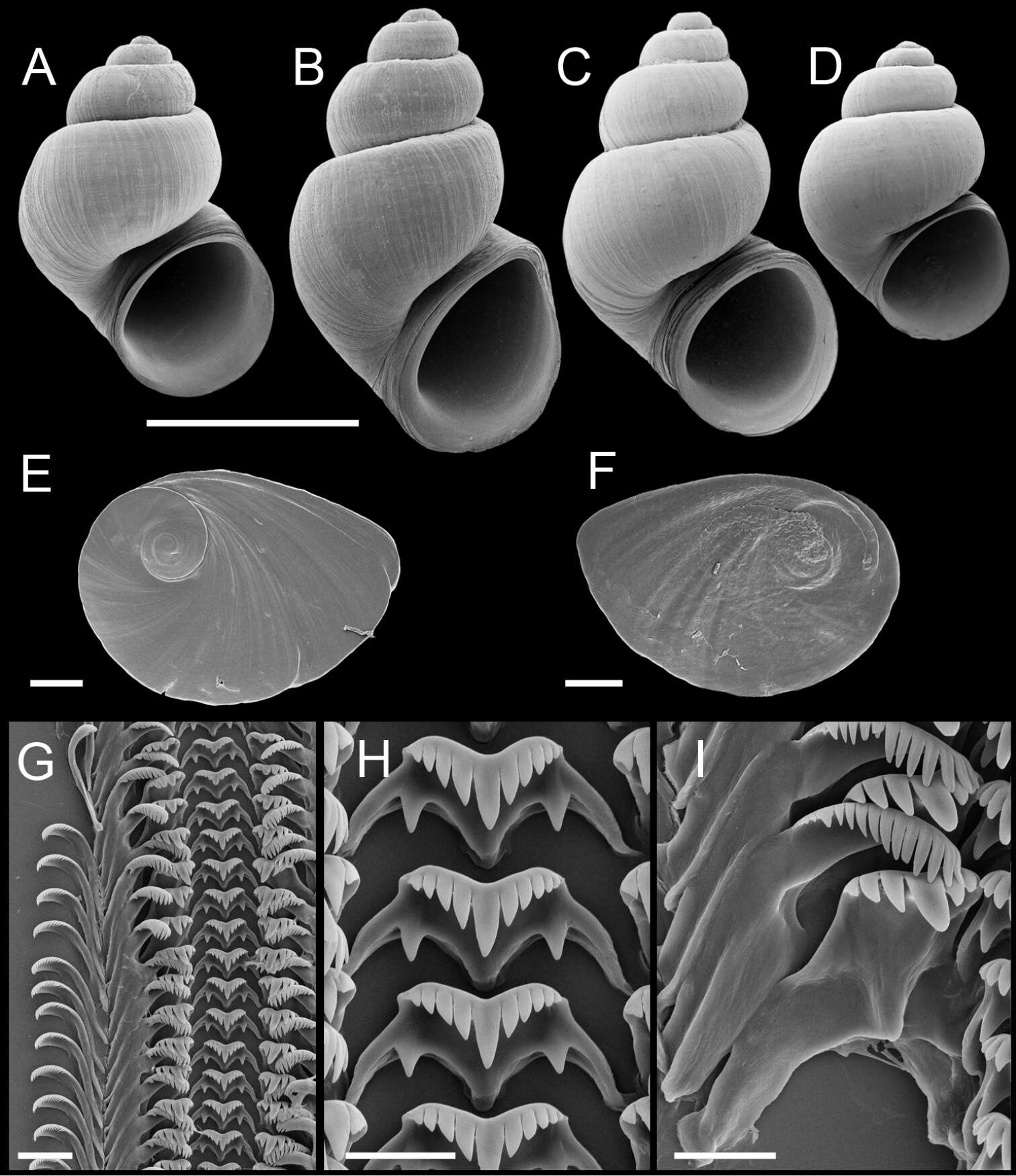
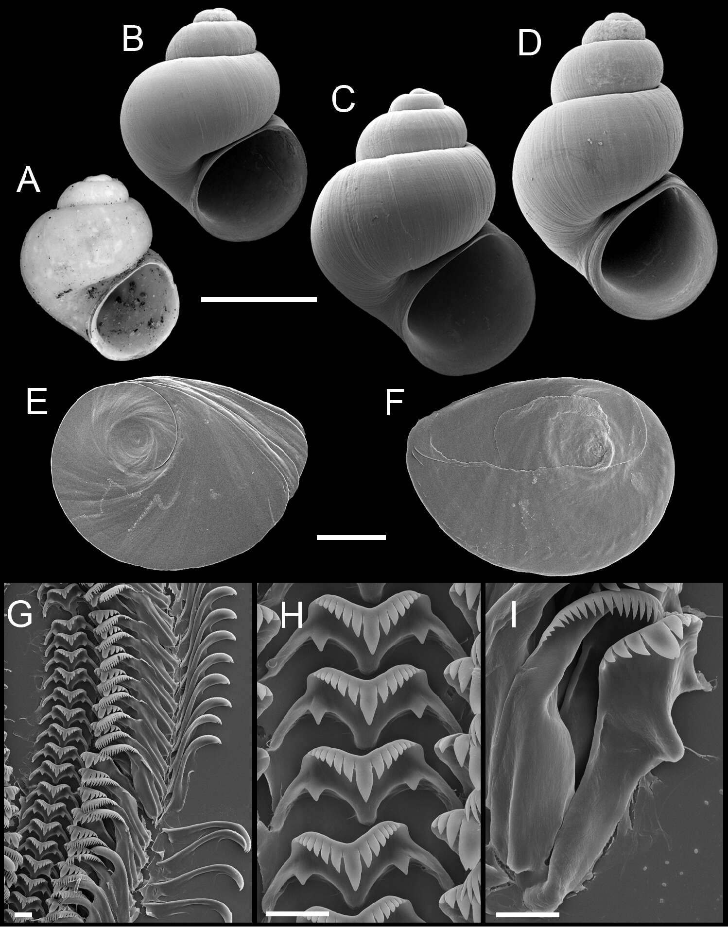
Figure 7.Shells, opercula and radula, Pyrgulopsis sanchezi sp. n. A Holotype, USNM 850333 B–D Shells, USNM 853505, USNM 1204755, USNM 853501 E, F Opercula (outer, inner sides), USNM 883361 G Portion of radular ribbon, USNM 883361 H Central teeth, USNM 883361 I Lateral and inner marginal teeth, USNM 883361. Scale bars A–D 1.0 mm; E, F 100 µm; G 20 µm; H, I 10 µm.
-
Robert Hershler, Hsiu-Ping Liu, Corbin Bradford
Zookeys
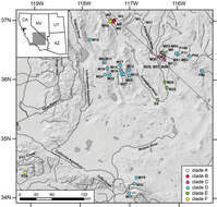
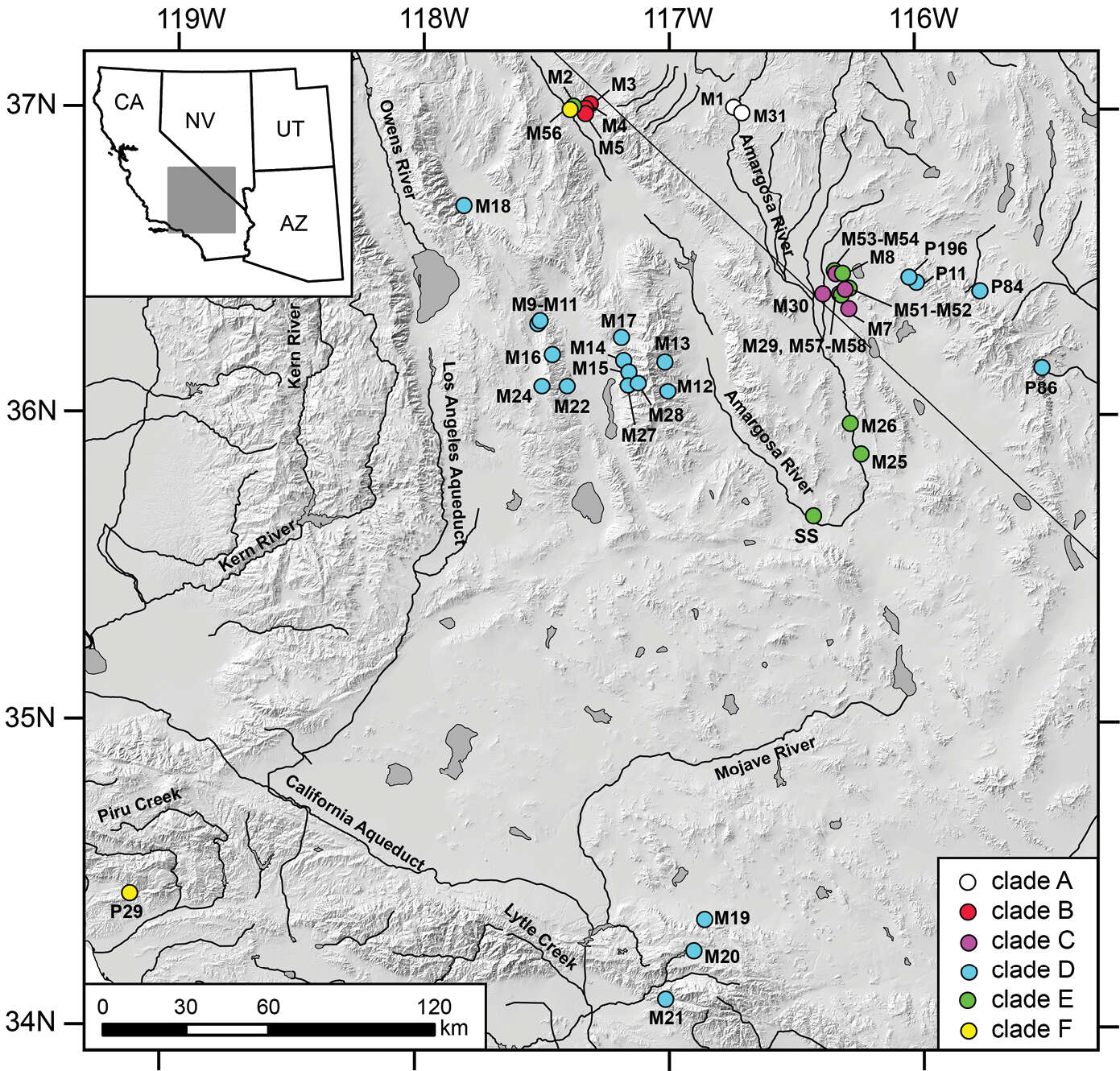
Figure 2.Map showing the distribution of mtDNA clades A–F with color codes matching those in Fig. 1.
-
Robert Hershler, Hsiu-Ping Liu, Corbin Bradford
Zookeys
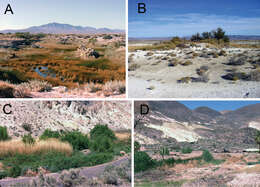
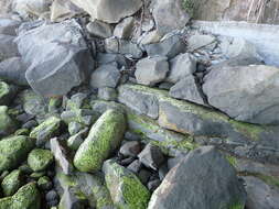
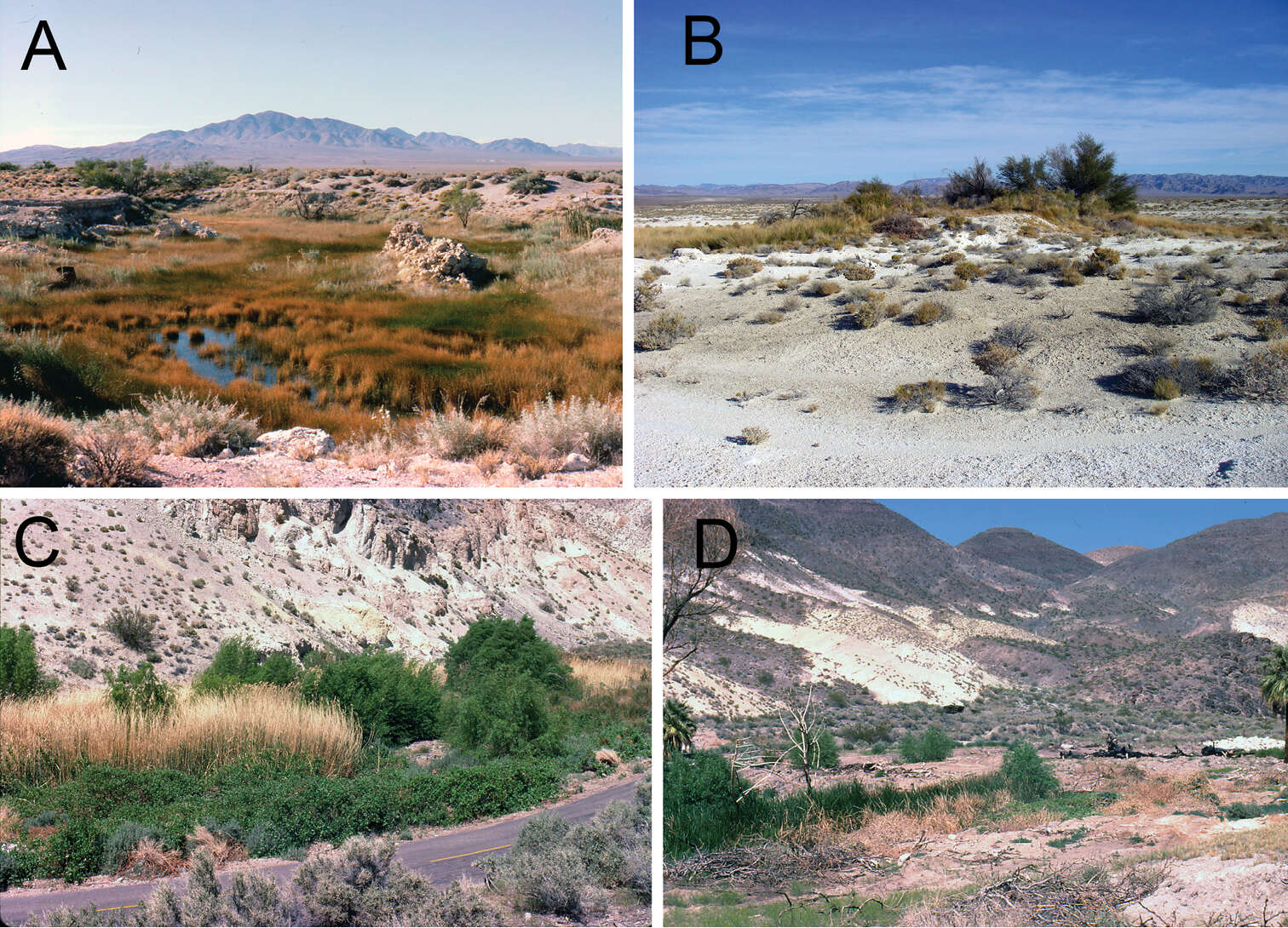
Figure 5.Photographs of habitats. A Spring south of Clay Pits, Ash Meadows, Nye County, Nevada, type locality of Pyrgulopsis licina sp. n. (photograph taken on 7/VII/1986) B Purgatory Spring, Ash Meadows, Nye County, Nevada, type locality of Pyrgulopsis sanchezi (15/XI/2011) C, D Uppermost spring east of Scotty’s Castle, Death Valley, Inyo County, California, type locality of Pyrgulopsis perforata sp. n. (18/IV/1980).
-
Robert Hershler, Hsiu-Ping Liu, Corbin Bradford
Zookeys
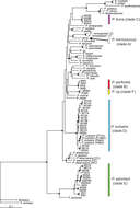
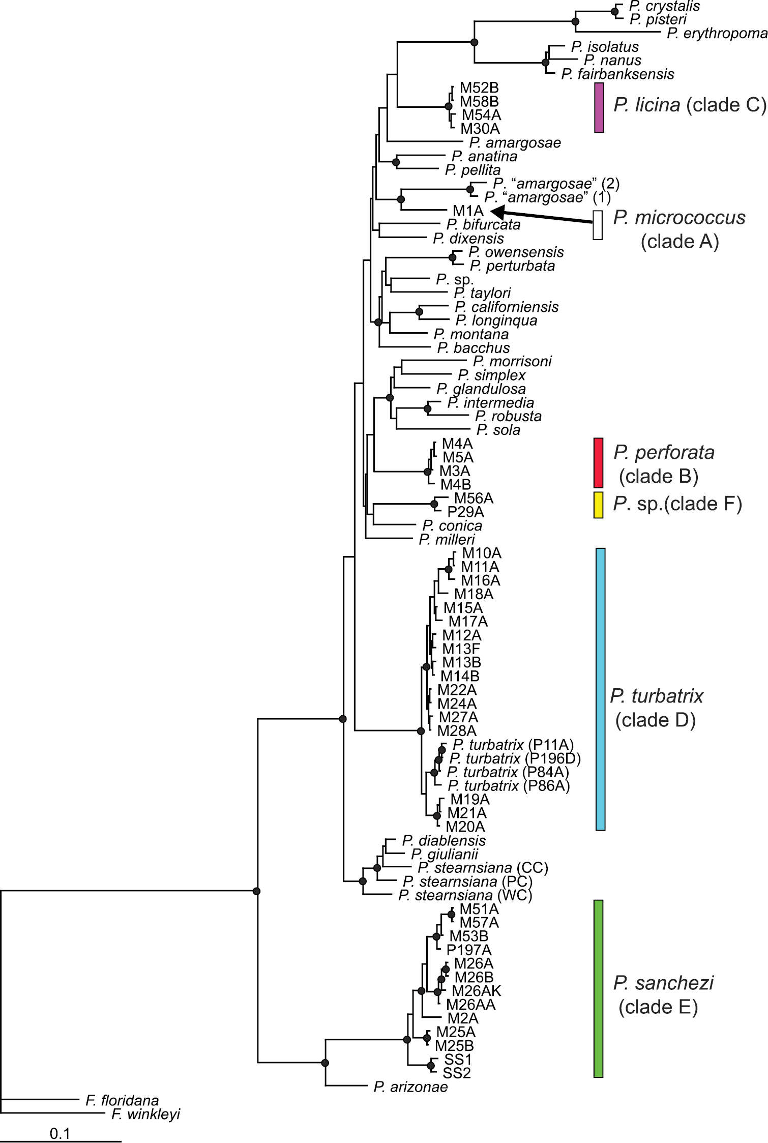
Figure 1.Bayesian tree based on the combined (COI, NDI) dataset. Nodes having posterior probabilities >95% are identified by filled circles. Specimen codes are from Appendix I.
-
Robert Hershler, Hsiu-Ping Liu, Corbin Bradford
Zookeys
Figure 4.Penes (dorsal, ventral surfaces). A, B Pyrgulopsis licina sp. n., USNM 850346 C, D Pyrgulopsis perforata sp. n., BellMNH 20891 E, F Pyrgulopsis sanchezi sp. n., USNM 883361 G, H Pyrgulopsis micrococcus, BellMNH 20663 I, J Pyrgulopsis turbatrix, USNM 860699 K, L Pyrgulopsis turbatrix, USNM 883373. Scale bars A–C 250 µm; D–L 500 µm. Pd penial duct Pf penial filament Pl penial lobe Tg terminal gland.
-
Robert Hershler, Hsiu-Ping Liu, Corbin Bradford
Zookeys
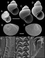
Figure 8.Shells, opercula and radula, Pyrgulopsis micrococcus. A Lectotype, ANSP 67279a B–D Shells, USNM 1004185, USNM 905091, USNM 1004183 E, F Opercula (outer, inner sides), USNM 847246 G Portion of radular ribbon, USNM 847246 H Central teeth, USNM 847246 I Lateral and inner marginal teeth, USNM 847246. Scale bars A–D 1.0 mm; E, F 250 µm; G–I 10 µm.
-
Robert Hershler, Hsiu-Ping Liu, Corbin Bradford
Zookeys
Figure 2.Map showing the distribution of mtDNA clades A–F with color codes matching those in Fig. 1.
-
Robert Hershler, Hsiu-Ping Liu, Corbin Bradford
Zookeys
Figure 1.Bayesian tree based on the combined (COI, NDI) dataset. Nodes having posterior probabilities >95% are identified by filled circles. Specimen codes are from Appendix I.
-
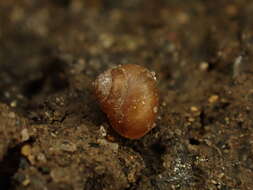
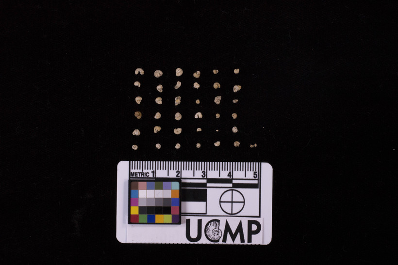
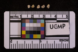
2017 University of California Museum of Paleontology
CalPhotos
-
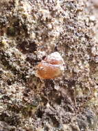
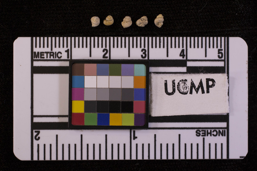
2017 University of California Museum of Paleontology
CalPhotos
-
-
-
-
-
-
-
-
-
-
-
-