-
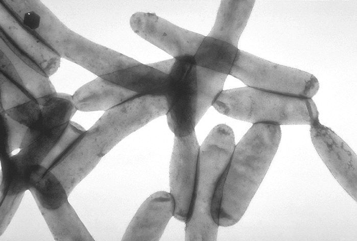
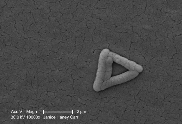
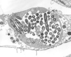
Transmission electron micrograph of Legionella pneumophila.Created:
-
Under a high magnification of 10000X, this colorized scanning electron micrograph (SEM) revealed the presence of four Gram-negative Salmonella typhimurium bacteria, that had been isolated from a pure culture. See PHIL 10976 for a black and white version of this image.How do people catch Salmonella??Salmonella live in the intestinal tracts of humans and other animals, including birds. Salmonella are usually transmitted to humans by eating foods contaminated with animal feces. Contaminated foods usually look and smell normal. Contaminated foods are often of animal origin, such as beef, poultry, milk, or eggs, but any food, including vegetables, may become contaminated. Thorough cooking kills Salmonella. Food may also become contaminated by the hands of an infected food handler who did not wash hands with soap after using the bathroom.Created: 2009
-
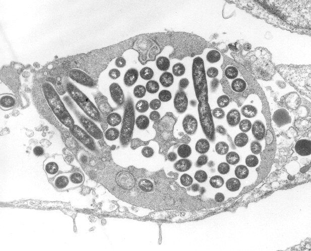
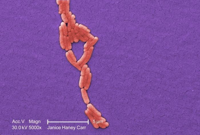
Legionella pneumophila multiplying inside a cultured human lung fibroblast.Created: 1979
-
Under a high magnification of 10000X, this scanning electron micrograph (SEM) revealed the presence of four Gram-negative Salmonella typhimurium bacteria, that had been isolated from a pure culture. See PHIL 10977 for a colorized version of this image.How do people catch Salmonella??Salmonella live in the intestinal tracts of humans and other animals, including birds. Salmonella are usually transmitted to humans by eating foods contaminated with animal feces. Contaminated foods usually look and smell normal. Contaminated foods are often of animal origin, such as beef, poultry, milk, or eggs, but any food, including vegetables, may become contaminated. Thorough cooking kills Salmonella. Food may also become contaminated by the hands of an infected food handler who did not wash hands with soap after using the bathroom.Created: 2009
-
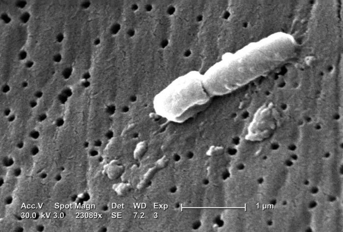
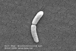
This scanning electron micrograph (SEM) revealed some of the ultrastructural morphologic features of a Klebsiella pneumoniae bacterium.Created: 2005
-
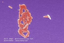
Under a moderate magnification of 5000X, this scanning electron micrograph (SEM) revealed the presence of numbers of clustered Gram-negative Salmonella typhimurium bacteria, that had been isolated from a pure culture. See PHIL 10975 for a colorized version of this image.What sort of germ is Salmonella?Salmonella is actually a group of bacteria that can cause diarrheal illness in humans. They are microscopic living creatures that pass from the feces of people or animals to other people or other animals. There are many different kinds of Salmonella bacteria. Salmonella serotype Typhimurium and Salmonella serotype Enteritidis are the most common in the United States. Salmonella germs have been known to cause illness for over 100 years. They were discovered by an American scientist named Salmon, for whom they are named.Created: 2009
-
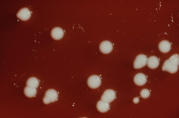
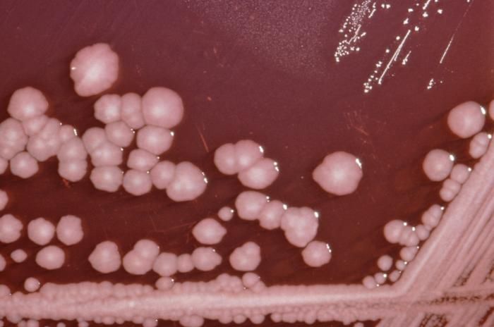
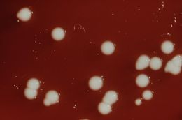
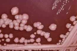
This blood agar plate (BAP) grew colonies of Gram-negative, small rod-shaped and facultatively anaerobic Klebsiella pneumoniae bacteria.Created: 1976
-
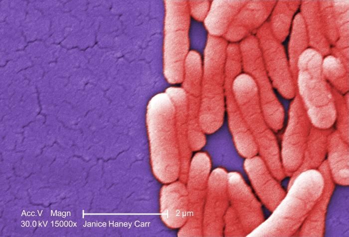
Under a very high magnification of 15000X, this colorized scanning electron micrograph (SEM) revealed the presence of numbers of clustered Gram-negative Salmonella typhimurium bacteria, which had been grown in a pure culture. See PHIL 10973 for a black and white version of this image.What sort of germ is Salmonella?Salmonella is actually a group of bacteria that can cause diarrheal illness in humans. They are microscopic living creatures that pass from the feces of people or animals to other people or other animals. There are many different kinds of Salmonella bacteria. Salmonella serotype Typhimurium and Salmonella serotype Enteritidis are the most common in the United States. Salmonella germs have been known to cause illness for over 100 years. They were discovered by an American scientist named Salmon, for whom they are named.Created: 2009
-
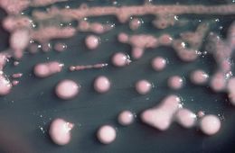
This inoculated MacConkey agar culture plate cultivated colonial growth of Gram-negative, small rod-shaped and facultatively anaerobic Klebsiella pneumoniae bacteria.Created: 1976
-
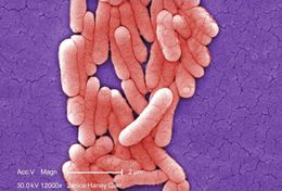
Under a high magnification of 12000X, this colorized scanning electron micrograph (SEM) revealed the presence of numbers of clustered Gram-negative Salmonella typhimurium bacteria, which had been grown in a pure culture. See PHIL 10970 for a black and white version of this image.What sort of germ is Salmonella?Salmonella is actually a group of bacteria that can cause diarrheal illness in humans. They are microscopic living creatures that pass from the feces of people or animals to other people or other animals. There are many different kinds of Salmonella bacteria. Salmonella serotype Typhimurium and Salmonella serotype Enteritidis are the most common in the United States. Salmonella germs have been known to cause illness for over 100 years. They were discovered by an American scientist named Salmon, for whom they are named.Created: 2009
-
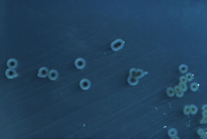
This photograph depicts the colonial morphology displayed by Shigella boydii bacteria cultivated on a Hektoen enteric (HE) agar surface; colonies of S. boydii bacteria grown on HE agar display a raised, green, and moist appearance.Created: 1976
-
Under a magnification of 5000X, this colorized scanning electron micrograph (SEM) revealed the presence of numbers of clustered Gram-negative Salmonella typhimurium bacteria that were isolated from what was a pure culture specimen. See PHIL 10968 for a black and white version of this image.What sort of germ is Salmonella?Salmonella is actually a group of bacteria that can cause diarrheal illness in humans. They are microscopic living creatures that pass from the feces of people or animals to other people or other animals. There are many different kinds of Salmonella bacteria. Salmonella serotype Typhimurium and Salmonella serotype Enteritidis are the most common in the United States. Salmonella germs have been known to cause illness for over 100 years. They were discovered by an American scientist named Salmon, for whom they are named.Created: 2009
-
This photograph depicts the colonial morphology displayed by Gram-negative Shigella boydii bacteria on a blood agar plate (BAP).Created: 1976
-
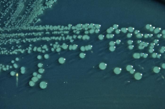
This photograph depicts the colonial growth pattern displayed by Salmonella typhimurium bacteria cultured on a Hektoen enteric (HE) agar medium; S. typhimurium colonies grown on HE agar are blue-green in color, for this organism is a lactose non-fermenter, but it does produce hydrogen sulfide, (H2S), therefore there can be black-colored deposits present.Created: 1976
-
This photograph depicts the colonial morphology displayed by Shigella boydii bacteria cultivated on a Hektoen enteric (HE) agar surface; colonies of S. boydii bacteria grown on HE agar display a raised, green, and moist appearance.Created: 1976
-
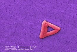
Under a very high magnification of 25000X, this scanning electron micrograph (SEM) revealed the presence of a single Gram-negative Salmonella typhimurium bacterium, which was imaged right at the point where it was undergoing the process of cell division, resulting in the formation of two separate organisms. This dividing bacterium had been isolated from a pure culture. See PHIL 10995 for a colorized version of this image.Created: 2008
-
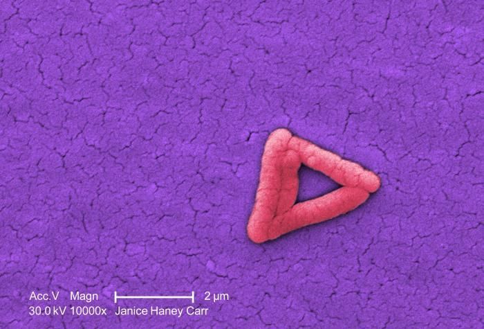
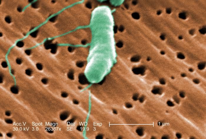
This colorized scanning electron micrograph (SEM) depicted a flagellated Vibrio vulnificus bacterium; Mag. 26367x.Created: 2005
-
Under a very high magnification of 15000X, this colorized scanning electron micrograph (SEM) revealed the presence of a single Gram-negative Salmonella typhimurium bacterium, which was imaged right at the point where it was undergoing the process of cell division, resulting in the formation of two separate organisms. This dividing bacterium had been isolated from a pure culture. See PHIL 10992 for a black and white version of this image.Created:
-
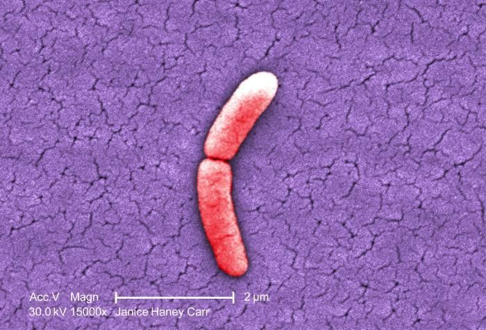
This photograph depicts the colonies of Proteus mirabilis bacteria grown on a Xylose Lysine Sodium Deoxycholate (XLD) agar plate.Created: 1976
-
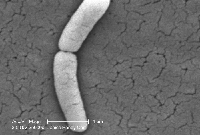
Under a very high magnification of 15000X, this scanning electron micrograph (SEM) revealed the presence of a single Gram-negative Salmonella typhimurium bacterium, which was imaged right at the point where it was undergoing the process of cell division, resulting in the formation of two separate organisms. This dividing bacterium had been isolated from a pure culture. See PHIL 10993 for a colorized version of this image.Created: 2009
-
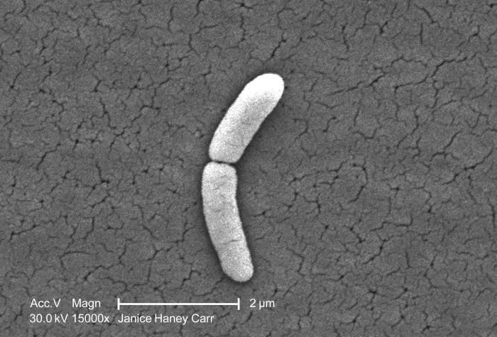
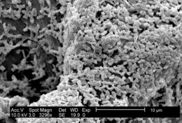
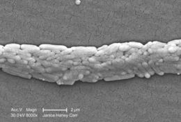
This SEM depicts a P. mirabilis (ATCC 29906) biofilm growing on PC (polycarbonate) coupons using a CDC biofilm reactor.Created: 2003
-
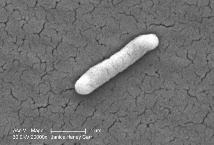
Under a very high magnification of 20000X, this scanning electron micrograph (SEM) revealed the presence of a single Gram-negative Salmonella typhimurium bacterium that had been isolated from a pure culture. See PHIL 10991 for a colorized version of this image.What sort of germ is Salmonella?Salmonella is actually a group of bacteria that can cause diarrheal illness in humans. They are microscopic living creatures that pass from the feces of people or animals to other people or other animals. There are many different kinds of Salmonella bacteria. Salmonella serotype Typhimurium and Salmonella serotype Enteritidis are the most common in the United States. Salmonella germs have been known to cause illness for over 100 years. They were discovered by an American scientist named Salmon, for whom they are named.Created: 2009
-
This SEM depicts a P. mirabilis (ATCC 29906) biofilm growing on PC (polycarbonate) coupons using a CDC biofilm reactor.Created: 2003
-
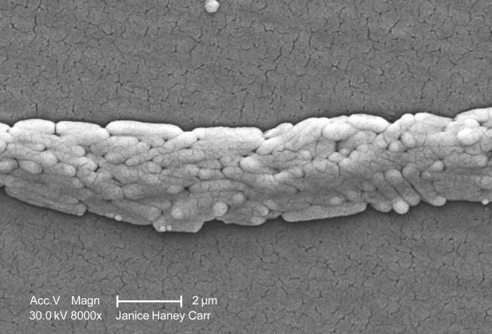
Under a moderately-high magnification of 8000X, this scanning electron micrograph (SEM) revealed the presence of a grouping of Gram-negative Salmonella typhimurium bacteria that had been isolated from a pure culture. See PHIL 10989 for a colorized version of this image.How do people catch Salmonella?Created: 2009