If a worm gets into an improper host such as humans the juveniles migrate through the body. The juveniles begin a typical tissue migration. They do not undergo development nor do they complete the normal migration, instead they will randomly wander through the body. Visceral larva migrans (VLM) is the resulting disease.
Toxocara canis infection is largely preventable. Worming pets often, with worming agents (such as antihelmintics: fenbendazole, piperazine, and Dichlorvos) from a veterinarian will reduce the possibility of human infection. This drugs also help in human treatment. Also careful and prompt disposal of dog feces will help. Humans should also wash their hands and the hands of children after handling dogs or dog feces, and especially before handling food. Lastly, parents and childcare givers need to watch out for toddlers eating soil and try to prevent it.
Nematodes within the Secernentea have phasmids, which are unicellular glands. Phasmids likely function as chemoreceptors. Females may produce pheromones to attract males.
Nematodes in general have papillae, setae and amphids as the main sense organs. Setae detect motion (mechanoreceptors), while amphids detect chemicals (chemoreceptors).
Communication Channels: tactile ; chemical
Other Communication Modes: pheromones
Perception Channels: tactile ; chemical
US Federal List: no special status
CITES: no special status
Toxocara canis is a canid parasite. Humans acquire the parasite as accidental hosts. In the tissues of all dogs, in many birds, and other mammals, the larval form of T. canis is found. The dog or canid host is the definitive host and only there will T. canis develop further than the larval stage. The name for the disease when in T. canis is in a host is Toxocariasis. Many animals such as mice, rabbits, and monkeys can serve as paratenic hosts.
Regardless of the path T. canis larvae take to get to the canid intestine once there the third stage larvae molt into adults. The adult worm remains in the intestine and produces an enormous number of eggs each day. Not until the fifth day post-infection do the eggs begin to appear in the canid feces.
Toxocara canis has a complex life cycle. Similar to other nematodes, T. canis is not infectious immediately when it leaves the definitive host. It needs to grow and develop into the stage that is infectious, ensheathed L3. Only this stage can infect other definitive hosts. There is strong evidence of two molts taking place inside the developing eggs, before the eggs even hatch. The molting process involves a separation of the cuticle from the epidermis. This causes a formation of the new cuticle, which is arising from the outermost surface of the epidermis. It also includes the shedding of the old cuticle.
Toxocara canis is widespread, causing disease in many mammals including humans. Many humans are infected with T. canis larvae. The larvae can cause serious damage to the human paratenic host. It can be found in wealthy and well-developed countries just as much as poor and under-developed places. In the United States about 98% of puppies and 20% of adult dogs are infected with T. canis. The means the risk of exposure to humans in the United States is very high. Most cases go unreported or are unrecognized. All small mammals can be paratenic hosts especially small children. The disease is most common in children between the ages one and three. Ingesting embryonated eggs from the feces of dogs and other canids spreads the diease. Often pet owners take their dog for a walk in the park. During the walk the dog may deposit egg-bearing feces in the park soil or sand. The next day an unsuspecting parent brings their small child to play in the park. The young child is at an age where everything is picked up and tasted, including the contaminated soil. The eggs of T. canis are most commonly ingested this way. The eggs once excreted from the definitive host can survive for 10-20 days in the external environment. This means even long after the dog has been in the park humans can still be infected. In Britain, one study showed that the climate conditions there allow for some T. canis eggs to survive in the soil for up to three years!
Many of the wandering larvae end up in the brain causing serious reactions, which can lead to death of the host. The most common place in the body of infection is the liver, but it can be found in every organ. The amount of damage is related to the number of juveniles in the body of the host. One of the more serious results of visceral larva migrans is blindness. The worms that infect the eye are called ocular larval migrans. Blindness occurs from the infection when a larva becomes trapped in the blood vessels at the back of the eye.
Negative Impacts: injures humans (causes disease in humans ); causes or carries domestic animal disease
Toxocara canis is a canid parasite. Humans acquire the parasite as accidental hosts. In the tissues of all dogs, in many birds, and other mammals, the larval form of T. canis is found. The dog or canid host is the definitive host and only there will T. canis develop further than the larval stage. The name for the disease when in T. canis is in a host is Toxocariasis. Many animals such as mice, rabbits, and monkeys can serve as paratenic hosts.
Ecosystem Impact: parasite
Species Used as Host:
The location of T. canis in hosts is in the small intestine. There they feed on intestinal contents. The adults have a specialized anaerobic metabolism. This specialized metabolism gives the adult worms an extra ATP. Adult T. canis worms are very host specific.
Pharyngeal glands and intestinal epithelium produce digestive enzymes to feed on the hosts’ body fluids. Extracellular digestion begins within the lumen and is finished intracellularly.
Animal Foods: body fluids
Primary Diet: carnivore (Eats body fluids)
Toxcocara canis has a worldwide distribution. It is prevalent in all locations that have domestic dogs, puppies, and other canids. Toxocara canis is also found in places that have other various mammals such as mice, pigs, birds, and foxes, but these hosts are only paratenic hosts. Hosts are terrestrial mammals and therefore T. canis is mainly found in terrestrial terrain.
Biogeographic Regions: nearctic ; palearctic ; oriental ; ethiopian ; neotropical ; australian
Other Geographic Terms: cosmopolitan
The eggs of T. canis are excreted in the feces of an infected canid host. The embryonated eggs can live in the feces for up to three weeks. The feces are often deposited in soil or sandy areas. A host must ingest the eggs for the life cycle to continue. If ingested, the new habitat becomes the internal organs of the host. The gut is the first area T. canis larvae reside. If the host has not been previously infected, hatched juveniles go throught the circulation to the lungs, then back to the gut. If in a canid host, they take up residence in the intestine and develop into adults. If hosts have been previously "immunized" junveniles go to the body tissues and become dormant as if they were in a paratenic host. Often the infectious larvae stay in the mammary glands until a pregnancy where they are passed on to a nursing pup. If in a human or other non-canid host the larvae will wonder throughout the organs. These wandering larvae are called visceral larva migrans. They may travel to the eyes, lungs, brain, heart, muscles, liver, and other organs. Here they do not develop further but can cause severe local reactions.
Habitat Regions: terrestrial
Terrestrial Biomes: desert or dune ; savanna or grassland ; chaparral ; forest ; rainforest ; scrub forest ; mountains
Other Habitat Features: urban ; suburban ; agricultural
Toxocara canis is smaller than most of the other species in the family Ascarididae. It has a complete gut in the form of a simple tube. It is a "round worm" implying the shape of the outer layer to be round (if seen in a cross section ). Depending on the host the worm gets into T. canis will have different number of larval stages. Most worms have three larval stages before becoming infective.
Toxocara canis is dioecious having morphology distinctly different for the male and female. Males, 4-6 cm long, are smaller than females. The male's posterior end is curved ventrally and the tail is bluntly pointed. The male has a single tubular testis. He also has simple spicules, which allows for direct sperm transfer. The female worms are generally around 6.5 cm but can be as long as 15 cm long. In the female the vulva is about one-third the body length from the anterior end. The ovaries are very large and extensive. The uteri contain up to 27 million eggs at a time.
Both males and females have three prominent lips. Each lip has a dentigerous ridge. The lateral hypodermal cords are visible with the naked eye. No gubernacullum is present. In both sexes there are prominent cervical alae. The eggs are brownish and almost spherical. The eggs measure 75-90 micrometers. The eggs are embryonated when laid and have surficial pits. These eggs are very resistant to various weather and chemical conditions.
Range length: 4 to 15 cm.
Other Physical Features: ectothermic ; heterothermic ; bilateral symmetry
Sexual Dimorphism: female larger; sexes shaped differently
These parasites are usually not preyed on directly, but are ingested from host to host.
Females may produce a phermomone to attract males. The male coils around a female with his curved area over the female genital pore. The gubernaculum, made of cuticle tissue, guides spicules which extend through the cloaca and anus. Males use spicules to hold the female during copulation. Nematode sperm are amoeboid-like and lack flagella. The adult worm remains in the intestine and produces an enormous number of eggs each day. Not until the fifth day post-infection do the eggs begin to appear in the canid feces. There is strong evidence of two moults taking place inside the developing eggs.
Key Reproductive Features: sexual ; fertilization (Internal ); oviparous
Parental Investment: pre-fertilization (Provisioning); pre-hatching/birth (Provisioning: Female)
Infection with larvae of the nematode (roundworm) Toxocara canis, which occurs worldwide, is the most common cause of toxocariasis in humans (less frequently, it is caused by T. cati, a parasite of cats). Both dogs and cats can acquire their respective nematode parasites at any age by ingesting eggs or paratenic hosts ("transport hosts"). The most widely recognized source of infection by Toxocara in humans is ingestion of contaminated soil, often by toddlers, but infection is also possible via the consumption of partial or whole paratenic hosts, such as raw livers of domestic animals (chickens, ducks, cows, and pigs), as well as earthworms. Uncooked vegetables have also been reported as a possible source of infection, especially those from farms that utilize animal or human excrement as fertilizer. One additional possible source of infection reported is contact with embryonated eggs on a dog’s hair coat. (Lee et al. 2010 and references therein)
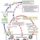
Toxocara canis completes its life cycle in dogs, with humans acquiring the infection as accidental hosts. Unembryonated eggs are shed in the feces of the definitive host. Eggs embryonate and become infective in the environment. Following ingestion by dogs, the infective eggs hatch and larvae penetrate the gut wall. In younger dogs, the larvae migrate through the lungs, bronchial tree, and esophagus; adult worms develop and oviposit in the small intestine. In older dogs, patent infections can also occur, but larval encystment in tissues is more common (an infection is "patent" when direct evidence of the organism can be detected, e.g., in the patient’s feces or blood, regardless of whether symptoms have appeared). Encysted stages are reactivated in female dogs during late pregnancy and infect by the transplacental and transmammary routes the puppies, in whose small intestine adult worms become established. Puppies are a major source of environmental egg contamination. Toxocara canis can also be transmitted through ingestion of paratenic hosts: eggs ingested by small mammals (e.g., rabbits) hatch and larvae penetrate the gut wall and migrate into various tissues where they encyst. The life cycle is completed when dogs eat these hosts and the larvae develop into egg-laying adult worms in the small intestine. Humans are accidental hosts who become infected by ingesting infective eggs in contaminated soil or infected paratenic hosts. After ingestion, the eggs hatch and larvae penetrate the intestinal wall and are carried by the circulation to a wide variety of tissues (liver, heart, lungs, brain, muscle, eyes). While the larvae do not undergo any further development in these sites, they can cause severe local reactions that are the basis of toxocariasis. The two main clinical presentations of toxocariasis are visceral larva migrans and ocular larva migrans. Diagnosis is usually made by serology or the finding of larvae in biopsy or autopsy specimens.
Toxocara canis (also known as dog roundworm) is a worldwide-distributed helminth parasite of dogs and other canids. The name is derived from the Greek word "toxon," meaning bow or quiver, and the Latin word "caro," meaning flesh.[1] They live in the small intestine of the definitive host. In adult dogs, the infection is usually asymptomatic but may be characterized by diarrhea. By contrast, massive infection with Toxocara canis can be fatal in puppies, causing diarrhea, vomiting, an enlarged abdomen, flatulence, and poor growth rate.[2][3]
As paratenic hosts, a number of vertebrates, including humans, and some invertebrates can become infected. Humans are infected, like other paratenic hosts, by ingestion of embryonated T. canis eggs.[4] The disease (toxocariasis) caused by migrating T. canis larvae results in two syndromes: visceral larva migrans and ocular larva migrans.[5] Owing to transmission of the infection from the mother to her puppies, preventive anthelmintic treatment of newborn puppies is strongly recommended. Several anthelmintic drugs are effective against adult worms, for example pyrantel, fenbendazole, and selamectin.[6]
T. canis is dioecious, having morphology distinctly different between the male and female. Male worms measure 4 to 6 cm (1.5" to 2.3"), typically smaller than female worms who measure at 6.5 to 15 cm (2.6" to 5.9"). The male's posterior end is curved ventrally and the tail is bluntly pointed. The male has a single tubular testis.[7] They also have simple spicules, which allow for direct sperm transfer. In the female, the vulva is about one-third the body length from the anterior end. The ovaries are very large and extensive. The uteri contain up to 27 million eggs at a time.[7]
Both males and females have three prominent lips. Each lip has a dentigerous ridge. The lateral hypodermal chords are visible with the naked eye. No gubernaculum is present. In both sexes there are prominent cervical alae. The adult T. canis has a round body with spiky cranial and caudal parts, covered by yellow cuticula. Toxocara canis is gonochoristic. The cranial part of the body contains two lateral alae (length 2 to 3.5 mm, width 0.1 mm). The eggs are brownish and almost spherical.T. canis eggs have oval or spherical shapes with granulated surfaces, are thick-walled, and measure from 72 to 85 μm.[2] The eggs are very resistant to various weather and chemical conditions typically found in soil.[8]
Eggs are deposited in feces of dogs, becoming infectious after 2–4 weeks.[9] Dogs ingest infectious eggs, allowing the eggs to hatch and the larval form of the parasite to penetrate through the gut wall. In dogs under 3 months of age, the larvae hatch in the small intestine, get into the bloodstream, migrate through the liver, and enter the lungs. Once in the lungs, the larvae crawl up the trachea. The larvae are then coughed up and swallowed, leading back down to the small intestine, where they mature to adulthood. This process is called tracheal migration. In dogs older than 3 months of age, the larvae hatch in the small intestine and enter the bloodstream, where they are carried to somatic sites throughout the body (muscles, kidney, mammary glands, etc.) where they become encysted second stage larvae. This process is called somatic migration. At the height of pregnancy, the encysted eggs in an infected female dog will migrate from the mother to the developing fetus, where they will reside in the liver. After parturition, the larvae migrate from the pup's liver to the lungs to undergo tracheal migration. Alternatively, the migrating larvae in the mother may encyst within the mammary glands, becoming active during lactation and passing directly to the nursing puppy via the milk. Larvae transmitted in this manner do not migrate once they are within the small intestine of the puppy; they will develop directly into the adult stage in the small intestine.[1] Once infected, a female dog will usually harbor sufficient larvae to subsequently infect all of her litters, even if she never again encounters an infection. A certain amount of the female dog's dormant larvae penetrate into the intestinal lumen, where molting into adulthood takes place again, thus leading to a new release of eggs containing L1 larvae.[9]
Another possible route of infection is the ingestion of paratenic hosts that contain encysted larvae from egg consumption, allowing the parasite to escape from the paratenic host and grow to adulthood within the small intestine of its definitive host, the dog.
Four modes of infection are associated with this species. These modes of infection include direct transmission, prenatal transmission, paratenic transmission, and transmammary transmission.[10][1]
Transmammary transmission occurs when the suckling pup becomes infected by the presence of L3 larvae in the milk during the first three weeks of lactation. There is no migration in the pup via this route.[10]
L2 larvae may also be ingested by a variety of animals like mice or rabbits, where they stay in a dormant stage inside the animals' tissue until the intermediate host has been eaten by a dog, where subsequent development is confined to the gastrointestinal tract.[11][12]
Consumption of eggs from feces-contaminated items is the most common method of infection for humans especially children and young adults under the age of 20 years.[13] Although rare, being in contact with soil that contains infectious eggs can also cause human infection, especially handling soil with an open wound or accidentally swallowing contaminated soil, as well as eating undercooked or raw meat of an intermediate host of the parasite such as lamb or rabbit.[11]
Humans can be infected by this roundworm, a condition called toxocariasis, just by stroking an infected dog's fur and accidentally ingesting infective eggs that may be present on the dog's fur. When humans ingest infective eggs, diseases like hepatomegaly, myocarditis, respiratory failure and vision problems can result depending on where the larvae are deposited in the body.[13] In humans, this parasite usually grows in the back of the eye, which can result in blindness, or in the liver or lungs.[14] However, a 2004 study showed, of 15 infected dogs, only seven had eggs in their coats, and no more than one egg was found on each dog. Furthermore, only 4% of those eggs were infectious.[15] Given the low concentration of fertile eggs on infected dogs' coats (less than 0.00186% per gram), it is plausible that such eggs were transferred to the dog's coat by contact with fecal deposits in the environment, making dog coats the passive transport host vehicle.[15] However, although the risk of being infected by petting a dog is extremely limited, a single infected puppy can produce more than 100,000 roundworm eggs per gram of feces.[16]
Humans suffering from visceral infection of T. canis, the drugs albendazole (preferred), mebendazole and thiabendazole are highly effective. For other treatments, refer to the disease pages: visceralis larva migrans and ocularis larva migrans.
Anthelminthic drugs are used to treat infections in dogs and puppies for adult worms. Treatment protocol will vary based on the dog's age, production level and activity level. There are different treatment paths for puppies, pregnant bitches, lactating bitches, dogs with increased risk of infection, professional dogs, and dogs sharing homes with young children or immunocompromised individuals.
Puppies: from the age of two weeks, then every 14 days up to two weeks after weaning with fenbendazole/febantel, flubendazole, pyrantel, or nitroscanate, followed by monthly treatments for up to six months of age.
Pregnant bitches: to prevent transmission to the puppies, pregnant females can be given macrocyclic lactones on the 40th and 55th day of pregnancy or genbendazole daily from the 40th day of pregnancy continuing until the 14th day postpartum.
Lactating bitches: should be treated concurrently with the first treatment of puppies.
Dogs with increased risk of infection: i.e. those used in sports, competitions, shows, or those kept in kennels can be given two treatments 4 weeks before and 2–4 weeks after the event.
Professional dogs: i.e. therapy, rescue, or police dogs: 12 times a year, if excretion of worm eggs is to be excluded.
Dogs sharing homes with young children or immunocompromised individuals: 12 times a year, if excretion of worm eggs is to be excluded. [17]
There are several ways to prevent a T. canis infection in both dogs and humans. Regular deworming by a veterinarian is important to stop canine re-infections, especially if the dog is frequently outdoors.[9] Removing dog feces from the yard using sealed disposable bags will help control the spread of T. canis. Good practices to prevent human infections include: washing hands before eating and after disposing of animal feces, teaching children not to eat soil, and cooking meat to a safe temperature in order to kill potentially infectious eggs.[11][13]
{{cite web}}: CS1 maint: bot: original URL status unknown (link) Toxocara canis (also known as dog roundworm) is a worldwide-distributed helminth parasite of dogs and other canids. The name is derived from the Greek word "toxon," meaning bow or quiver, and the Latin word "caro," meaning flesh. They live in the small intestine of the definitive host. In adult dogs, the infection is usually asymptomatic but may be characterized by diarrhea. By contrast, massive infection with Toxocara canis can be fatal in puppies, causing diarrhea, vomiting, an enlarged abdomen, flatulence, and poor growth rate.
As paratenic hosts, a number of vertebrates, including humans, and some invertebrates can become infected. Humans are infected, like other paratenic hosts, by ingestion of embryonated T. canis eggs. The disease (toxocariasis) caused by migrating T. canis larvae results in two syndromes: visceral larva migrans and ocular larva migrans. Owing to transmission of the infection from the mother to her puppies, preventive anthelmintic treatment of newborn puppies is strongly recommended. Several anthelmintic drugs are effective against adult worms, for example pyrantel, fenbendazole, and selamectin.


