-
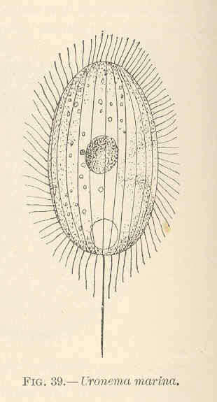
Uronema marina.
-
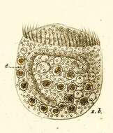
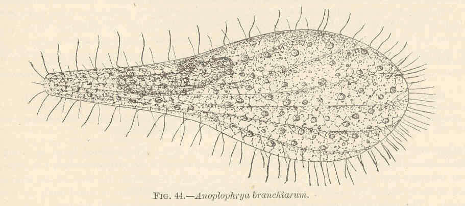
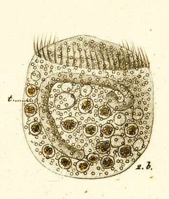
Anoplophrya.
-
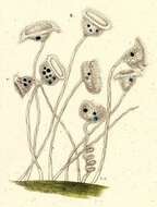
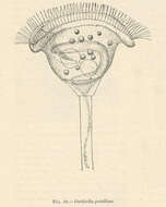
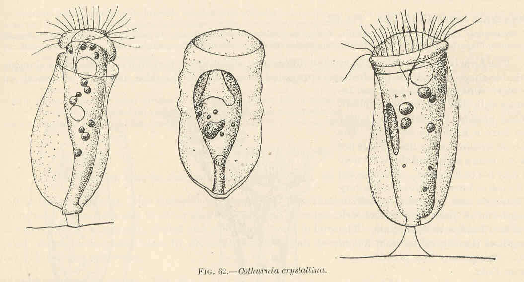
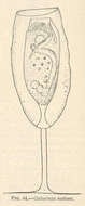
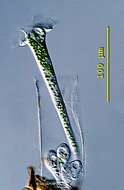
Cothurnia crystallini.
-
-
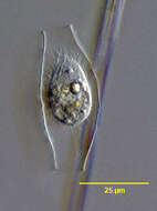
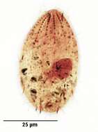
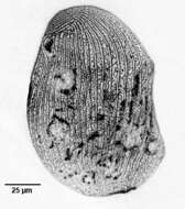
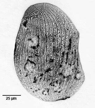
Portrait of the loricate pleuronematid ciliate, Calyptotricha pleuronemoides (Phillips, 1882). The transparent lorica of this species is open at both ends and dilated in the center where the cell resides. The cell is bluntly pointed anteriorly and broadly rounded posteriorly. The peristome is about 3/4 cell length. There is a prominent undulating membrane on the right margin of the peristome curving around its posterior end to form a shallow pouch (seen well here). There are three inconspicuous adoral membranelles. The longitudinal somatic kineties are uniformly distributed. There is a preoral and postoral suture. There is a single long caudal cilium. There is a single posterior contractile vacuole. The spherical macronucleus is centrally located. C. lanuginosa is similar in appearance of the cell except that it has two long anterior apical cilia and a cylindrical lorica with parallel sides. Collected from a freshwater dredge pond near Idaho City, Idaho June 2003. DIC.
-
Stained by the silver carbonate technique (see Foissner, W. Europ. J. Protistol., 27:313-330;1991).Brightfield.
-

Right lateral infraciliature of the hymenostome ciliate Colpidium kleini (Foissner, 1969). C. kleini is very similar in overall appearance to C. colpoda although usually more slender and with fewer somatic kineties. The cytostome is in the anterior 1/4 of the cell. There is a curved paraoral membrane along the convex right margin of the cytostome. The left margin is slightly concave. There are three adoral membranelles. There are 32 to 44 somatic kineties. The kineties to the right and left of the oral aperture meet at a curved preoral suture. The right somatic kineties bend leftward at the level of the cytostome. There is an anterior apical area bare of cilia. There are rows of inconspicuous mucocysts between the somatic kineties. The ellipsoid macronucleus and adjacent micronucleus are centrally located. The single contractile vacuole is located in the midbody with a single excretory pore on the right surface. The feature most clearly distinguishing Colpidium kleini from C. coploda is the silverline system (as demonstrated by silver nitrate staining).Stained by the silver carbonate technic (see Foissner, W.Europ. J. Protistol.27,313-330;1991). Collected from an organically enriched freshwater pond near Boise, Idaho.Brightfield.
-
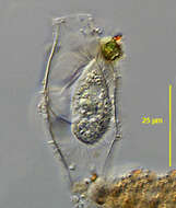
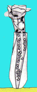
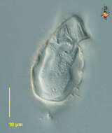
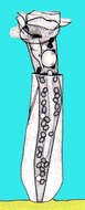
Thuricola (thurr-ick-owe-la) is a peritrich ciliate which lives within a lorica. Contractile and this cell has withdrawn into the lorica. A flap has closed over the contractile cell and this features distinguishes this genus. Differential interference contrast.
-
-
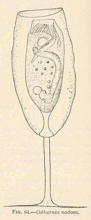
Cothurnia nodosa.
-
-
Portrait of the loricate pleuronematid ciliate, Calyptotricha pleuronemoides (Phillips, 1882). The transparent lorica of this species is open at both ends and dilated in the center where the cell resides. The cell is bluntly pointed anteriorly and broadly rounded posteriorly. The peristome is about 3/4 cell length. There is a prominent undulating membrane on the right margin of the peristome curving around its posterior end to form a shallow pouch. There are three inconspicuous adoral membranelles. The longitudinal somatic kineties are uniformly distributed. There is a preoral and postoral suture. There is a single long caudal cilium. There is a single posterior contractile vacuole. The spherical macronucleus is centrally located. C. lanuginosa is similar in appearance of the cell except that it has two long anterior apical cilia and a cylindrical lorica with parallel sides. Collected from a freshwater dredge pond near Idaho City, Idaho June 2003. DIC.
-
Stained by the silver carbonate technique (see Foissner, W. Europ. J. Protistol., 27:313-330;1991).Brightfield.
-
Right lateral view of the silverline system of the hymenostome ciliate Colpidium kleini (Foissner, 1969). C. kleini is very similar in overall appearance to C. colpoda although usually more slender and with fewer somatic kineties. The cytostome is in the anterior 1/4 of the cell. There is a curved paraoral membrane along the convex right margin of the cytostome. The left margin is slightly concave. There are three adoral membranelles. There are 32 to 44 somatic kineties. The kineties to the right and left of the oral aperture meet at a curved preoral suture.The right somatic kineties bend leftward at the level of the cytostome. There is an anterior apical area bare of cilia. There are rows of inconspicuous mucocysts between the somatic kineties. The ellipsoid macronucleus and adjacent micronucleus are centrally located. The single contractile vacuole is located in the midbody with a single excretory pore on the right surface. The feature most clearly distinguishing Colpidium kleini from C. coploda is the silverline system. In C. kleini there is only one secondary meridian (silverline) between two primary meridians (primary meridians correspond to somatic kineties). In some cases short segments of the secondary meridians may be duplicated. Short transverse L or T-shaped branches arise from both primary and secondary meridians at irregular intervals.Stained by the dry silver nitrate technic (see Foissner, W.Europ. J. Protistol.27,313-330;1991). Collected from an organically enriched freshwater pond near Boise, Idaho. Brightfield. Black and white.
-
-
-
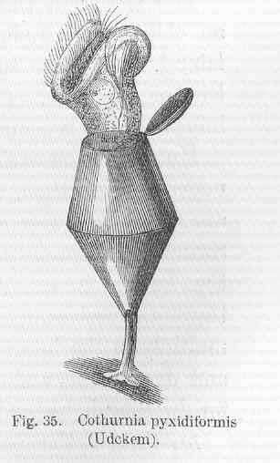
Cothurnia pyxidiformia (Udckem).
-
-
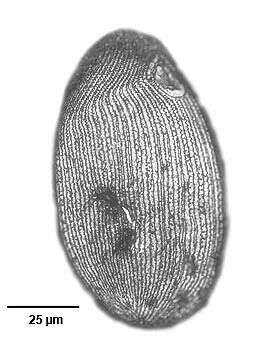
Dorsal infraciliature of Calyptotricha pleuronemoides (PHILLIPS,1882). Collected from organically enriched stagnant water at the edge of a freshwater stream near Boise, Idaho.Stained by the silver carbonate technique (Foissner,W. Europ. J. Protistol.27:313-330;1991).Brightfield.
-
Stained by the silver carbonate technique (see Foissner, W. Europ. J. Protistol., 27:313-330;1991).Brightfield.
-
Left lateral view of the silverline system of the hymenostome ciliate Colpidium kleini (Foissner, 1969). C. kleini is very similar in overall appearance to C. colpoda although usually more slender and with fewer somatic kineties. The cytostome is in the anterior 1/4 of the cell. There is a curved paraoral membrane along the convex right margin of the cytostome. The left margin is slightly concave. There are three adoral membranelles. There are 32 to 44 somatic kineties. The kineties to the right and left of the oral aperture meet at a curved preoral suture. There is an anterior apical area bare of cilia. There are rows of inconspicuous mucocysts between the somatic kineties. The ellipsoid macronucleus and adjacent micronucleus are centrally located. The single contractile vacuole is located in the midbody with a single excretory pore on the right surface. The feature most clearly distinguishing Colpidium kleini from C. coploda is the silverline system. In C. kleini there is only one secondary meridian (silverline) between two primary meridians (primary meridians correspond to somatic kineties seen here as the wavier lines). Short transverse L or T-shaped branches arise from both primary and secondary meridians at irregular intervals. In some cases short segments of the secondary meridians may be duplicated. Stained by the dry silver nitrate technic (see Foissner, W.Europ. J. Protistol.27,313-330;1991). Collected from an organically enriched freshwater pond near Boise, Idaho. Brightfield. Black and white.
-
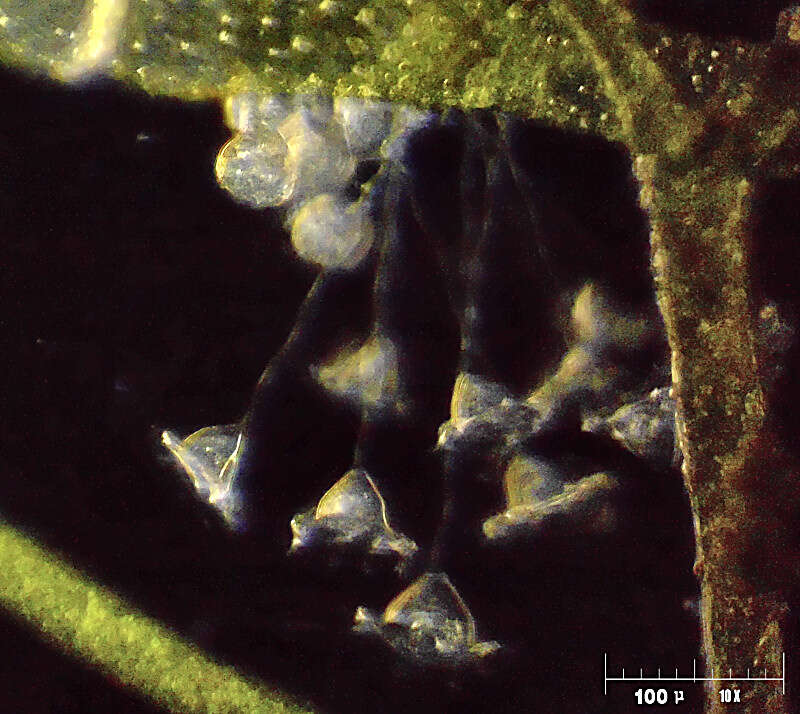
Thuricola (thurr-ick-cola) folliculata and 3 swarmers on the bottom of the lorica. The transparent lorica is equiped with a valve which closes the aperture as cell retracts. This specimen shows endosymbiotic algae. This specimen was collected in freshwater ponds near Konstanz, Germany. Differential interference contrast.
-
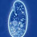
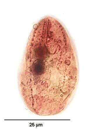
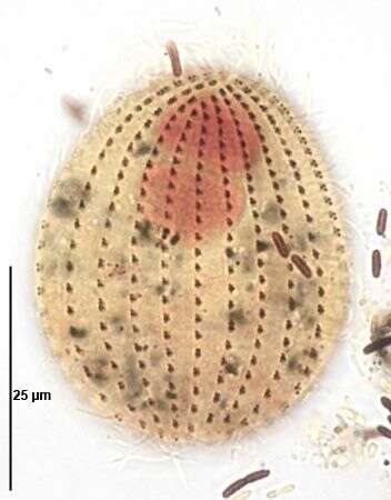
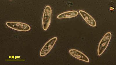
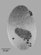
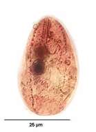
Paramecium (aurelia) (par-a-mee-see-um) is a very familiar genus of ciliates. They eat bacteria and have the mouth recessed in a buccal cavity, and the cell is often shaped with a scoop leading to the mouth. There are cilia all over the body with a caudal tuft of longer cilia at the back of the body. Usually with a layer of extrusomes (trichocysts) under the cell surface and a large oval macronucleus. Contractile vacuoles star-shaped. This species is P. aurelia, one of the smaller spindle-shaped (morpho)species. The (morpho) species is best distinguished by the presence of two small micronuclei pressed up against the macronucleus. Phase contrast.
-
Vorticella patellina.