-
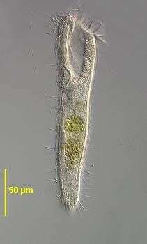
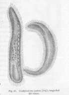
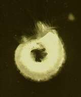
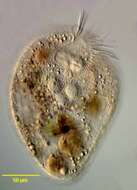
Condylostoma patens (Duj.), magnified....
-
Arboli, Catalonia, Spain
-
Ribadelago de Franco, Castille and Leon, Spain
-
Ribadelago de Franco, Castille and Leon, Spain
-
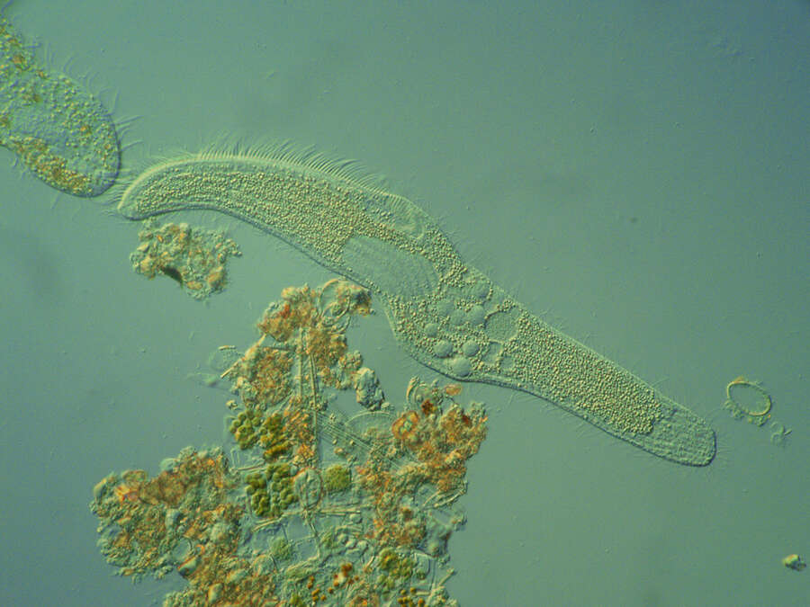
**En las aguas dulces cargadas de restos orgnicos bulle una intensa actividad en la que los protozoos ciliados adquieren un especial protagonismo por encontrar un medio ptimo para su desarrollo. Spirostomum intermedium, es uno de ellos y al igual que muchos ciliados, se alimenta de bacterias que son barridas hacia su citostoma con una fila de cilios fusionados especializados. Una de las caractersticas ms notables de Spirostomum es la forma en que puede contraerse. El organismo puede retraer su cuerpo a 1 / 4 de su longitud en un tiempo de 6-8 milisegundos, que es la ms rpida contraccin conocida en cualquier clula viva. La fotografa fue tomada con una cmara digital Olympus E-500 montada sobre un microscopio Leica DM 2000 utilizando un dispositivo de contraste de interferencia con un objetivo de x40. Para congelar el movimiento se utiliz el propio flash de la cmara.
-
Ribadelago de Franco, Castille and Leon, Spain
-
Urbanizacin Los Pinarejos, Comunidad de Madrid, Espaa
-
-
Sant Marti Sarroca, Catalonia, Spain
-
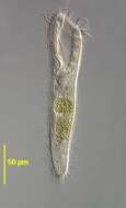
Portrait of the loricate marine heterotrich ciliate, Metafolliculina producta. Most folliculinid ciliates are found in marine habitats. Metafolliculina producta is greenish-blue in color. This large species resides in a lorica that has distinct annular ridges toward the fluted open end. The highly contractile cell withdraws instantly into the lorica when disturbed. The lorica is angulated but this feature is often not apparent due to compression by the coverslip. When fully protruded from the lorica the wing-like extensions of the peristomal area are evident. The cytostome lies at the central confluence of the peristomal wings. The somatic ciliation consists of uniform (holotrichous) longitudinal kineties. The genus Metafolliculina is distinguished from the similar Folliculina by its moniliform macronucleus (not well seen in this image). Collected from a commercial saltwater aquarium in Boise, Idaho February 2004. DIC optics.
-
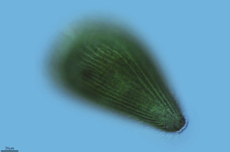
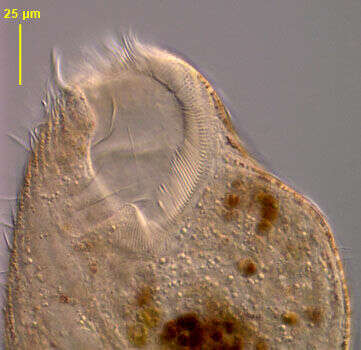
Portrait of the medium-size pigmented heterotrich ciliate, Blepharisma lateritium (Ehrenberg,1831; Stein, 1859). The cell is teardrop-shaped and pale pink in color. The peristome extends about 2/3 the cell length along the left side. The peristome is bordered on the left by serial polykinetids forming an adoral zone of membranelles and on the right by a narrow undulating membrane which is less conspicuous. The longitudinal somatic kineties bend to the left margin on the dorsal surface. The single ellipsoid to spherical macronucleus is located left of midline in the cell center. There are multiple very small micronuclei which are difficult to discern in vivo. These become swollen and densely stained in silver impregnated specimens. The contractile vacuole is at the posterior end. There are rows of pink cortical pigment granules between the somatic kineties. The pigment, blepharismin, is structurally similar to hypericin. When exposed to light, blepharismin causes a change in the cell's membrane potential and thus direction of ciliary beat causing light avoidance or photodispersal. Exposure to bright light for even short periods causes cell lysis. This is often observed in illuminated Blepharisma under the microscope. Blepharismin probably has a defensive function against predators such as the haptorid ciliate, Dileptus. Collected from an artificial freshwater dredge pond near Boise, Idaho October 2004. DIC optics.
-
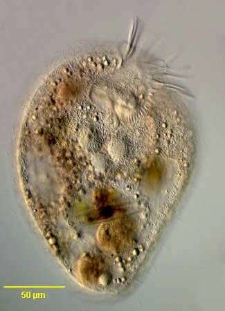
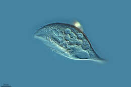
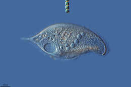
Ventral view of the marine spirotrich ciliate, Condylostoma psammophilum (Bock,1952).Collected from a commercial saltwater aquarium in Boise, Idaho.March 2006.DIC.
-
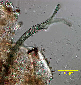
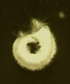
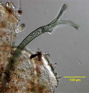
Eufolliculina swarmer has found a suitable place to build his chitinous lorica. Now the cell is flattening and starts to segregate material to build up the bottom of the lorica and to connect it to the substratum. Collected from Bodden, the brackish waters lying between the isles of Hiddensee and Ruegen (German Baltic Sea). This image was taken using Zeiss Universal with Olympus C7070 CCD camera.
-
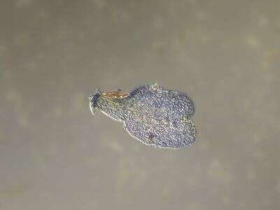
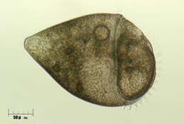
Image of a Spirorbis shell - a marine polychaete. Folliculinids often occupy the depressed centre of the cell where they presumably are more sheltered from currents. The type of folliculinid is not known, but the tests with a basal ampulla, a narrower neck are typical of this group of ciliates. Many species have this dark green colour.
-
Canencia, Madrid, Spain
-
-
Ribadelago, Castille and Leon, Spain
-
Mahide, Castille and Leon, Spain
-
Canencia, Comunidad de Madrid, Espaa
-
Melgar de Tera, Castille and Leon, Spain
-
Ribadelago de Franco, Castille and Leon, Spain
-
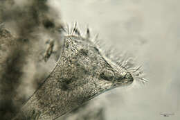
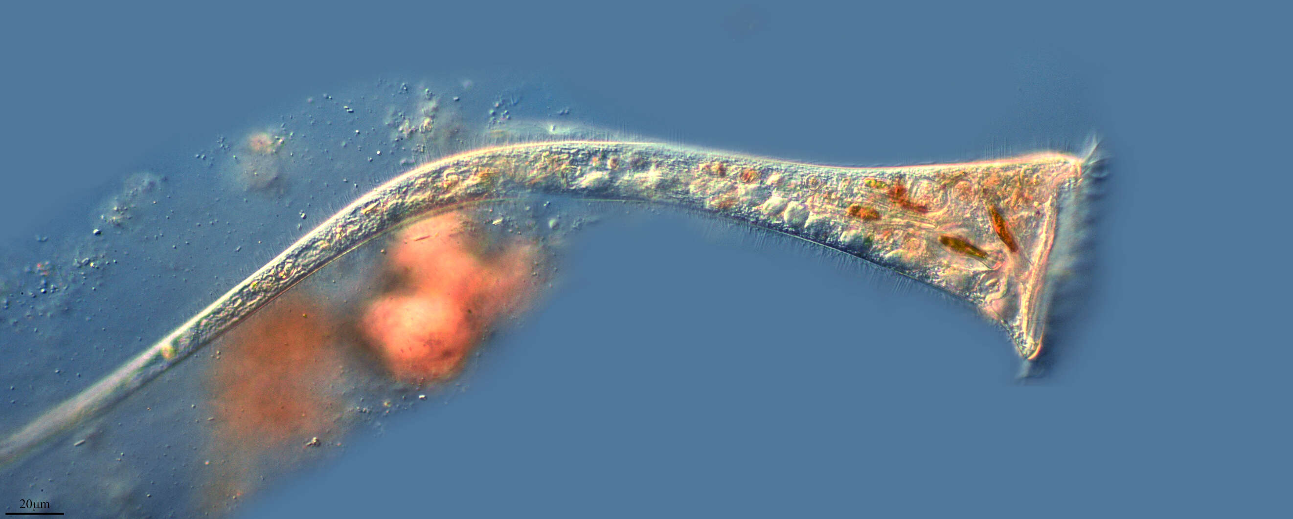
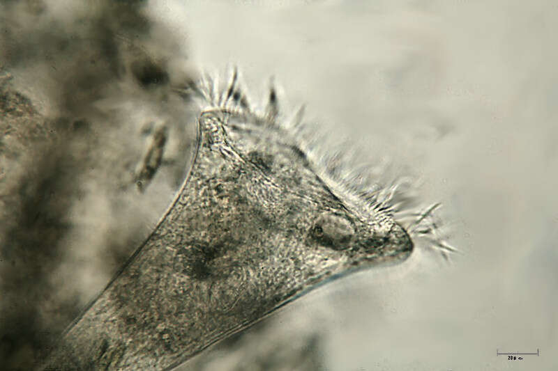
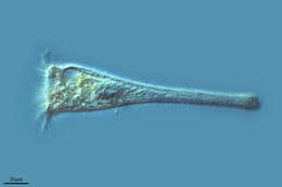
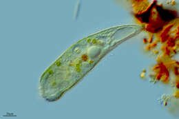
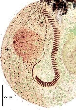
Anterior detail of the marine heterotrich ciliate, Condylostoma arenarium (Spiegel, 1926). The dorsoventrally flattened elongate cell body is very contractile (this cell is partly contracted). Contraction is probably mediated by calcium dependent subcortical myonemes and cell extension by interaction of cortical postciliary microtubular ribbons. The broad anterior V-shaped peristome is bordered on the right by a large undulating membrane. An adoral zone of membranelles (AZM) winds from right anterior clockwise around the left margin of the peristome. Several cirri are located at the right-most end of the AZM (seen well here). Uniform longitudinal somatic kineties are separated by strips of yellowish cortical granules (pigmentocysts). Pigmentocysts are extrusomes containing toxic substances. They play a role in cell defense against predators and may also function in photoreception. Pigmentocysts are also found in other heterotrichs (e.g. Blepharisma and Stentor species). The long moniliform macronucleus extends along the right cell margin (faintly visible here). No contractile vacuole. Brownish food vacuoles throughout the cytoplasm in this individual contain ingested dinoflagellates (Amphidinium). Collected from a seawater aquarium in Boise, Idaho January 2004. DIC optics.
-
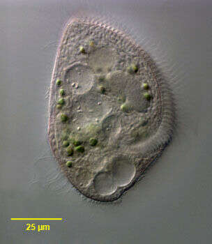
Infraciliature (ventral side) of the medium-size pigmented heterotrich ciliate, Blepharisma lateritium (Ehrenberg,1831; Stein, 1859). The cell is teardrop-shaped and pale pink in color.Green endosymbiotic zoochlorellae are visible in the cytoplasm.A zoochlorellae-bearing Blepharisma species otherwise indistinguishable from B. lateritium was reported from freshwater in Iowa (Johnson,L.P.A symbiotic Blepharisma.Proc. Iowa Acad. Sci. 55:391-393,1948). The peristome extends about 2/3 the cell length along the left side. The peristome is bordered on the left by serial polykinetids forming an adoral zone of membranelles and on the right by a narrow undulating membrane. The longitudinal somatic kineties bend to the left margin on the dorsal surface. The single ellipsoid to spherical macronucleus is located left of midline in the cell center. There are multiple very small micronuclei which are difficult to discern in vivo. These become swollen and densely stained in silver impregnated specimens (seen here). Stained by silver carbonate techniique (see Foissner, W.Europ. J. Protistol.27,313-330;1991). Collected from a freshwater pond near Boise, Idaho October 2004. Brightfield optics.
-
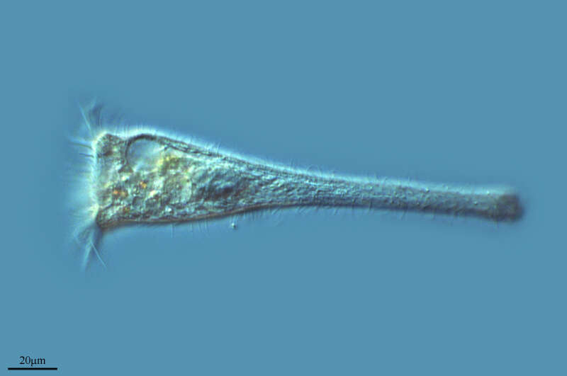
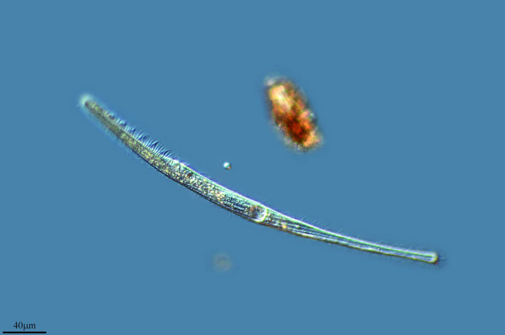
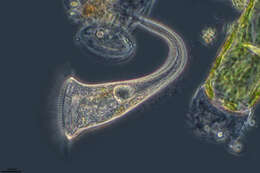
Ventral view of Condylostomides tardus (Penard,1922) Foissner,2002.DIC.