-
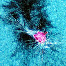
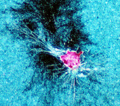
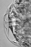
False-color image of a foraminiferan (pink) rending and consuming a bacterial biofilm (blue). The dark area is the region cleared by the foram in approximately 12 hours. Species not identified. Image courtesy of Joan Bernhard, WHOI. A version of this image appeared in Bernhard, J., and Bowser, S.S. (1992) Mar. Ecol. Prog. Ser. 83:263-272.
-
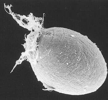
Notice the network of reticulopodia emerging from the aperture of the organic-walled test. Image courtesy of Susan T. Goldstein, University of Georgia. This image first appeared in J. Foram Res. 32:375-383 and is used with permission.
-
-
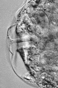
A brightfield image of a portion of a reticulopod and several rod-shaped bacteria. The oral zone of the foraminiferan is at upper left. Image courtesy of Samuel S. Bowser, Wadsworth Center.
-
Notice the distinctive "herringbone" texture of the organic test (the dark portion of the image). Part of the cell itself fills the upper right hand corner of the photo. Image courtesy of Susan T. Goldstein, University of Georgia. This image first appeared in J. Foram Res. 32:375-383 and is used with permission.
-
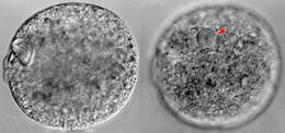
Bathyallogromia was found in deep water (1000 to 6300 m) in the Weddell Sea, off the coast of Antarctica. Both images are of the same foram. The left image has the focal plane through the aperture (the twin bumps on the left side); the right image has the focal plane through the nucleus (indicated by the red arrow). The cell is about 200 um across, and appears whitish in reflected light. Image courtesy of Andrew J. Gooday, Southampton Oceanography Centre.
-
-
The aperture is a passage through the foram's transparent test, and is the route that the reticulopodia take to extend into the environment. The aperture of this species is surrounded by a "collar" which protrudes from the rest of the test. Image courtesy of Andrew J. Gooday, Southampton Oceanography Centre.
-
Lieberkuehnia from the Severn Estuary as promised.
-
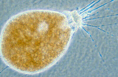
This image clearly shows the prominent "collar" around the oral zone for which the species is named. Image courtesy of Samuel S. Bowser, Wadsworth Center.
-
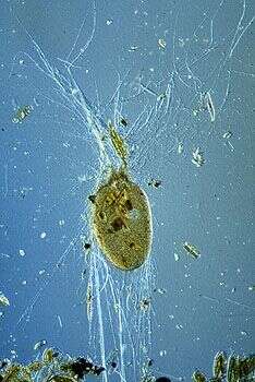
This species has an organic-walled test, which is thin, flexible and somewhat transparent. It is visible as the hazy "halo" around the cell body. Image courtesy of Samuel S. Bowser, Wadsworth Center.
-
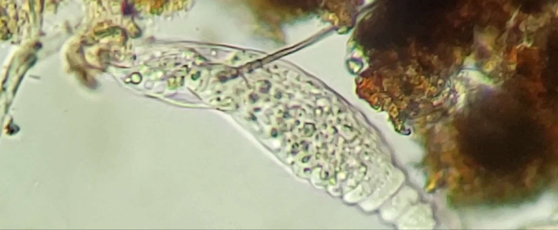
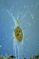
This individual is surrounded by the empty frustules of diatoms it has consumed. Image courtesy of Jeffrey L. Travis, University at Albany.
-
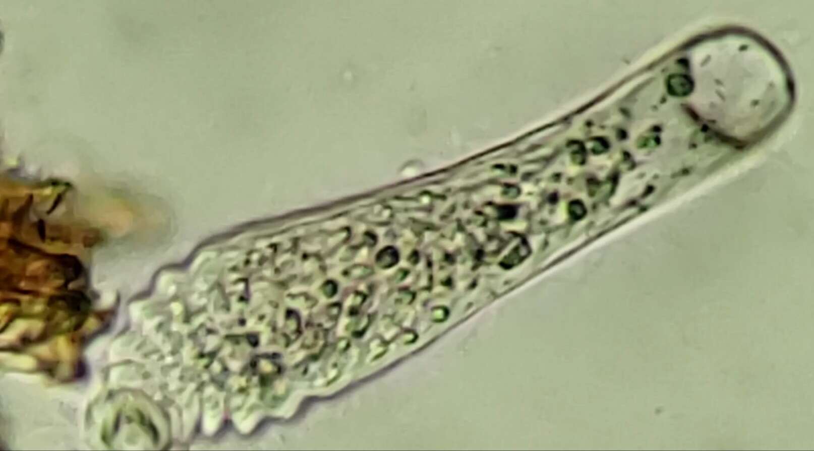
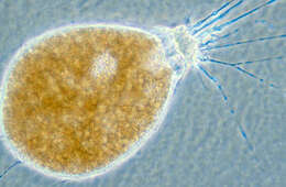
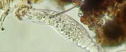
Marine thecate foraminiferan with reticulopods extended. Isolated from culture provided by Jeff L. Travis. Microscopy by L.W.Parfrey
-
-
-
-
-
-
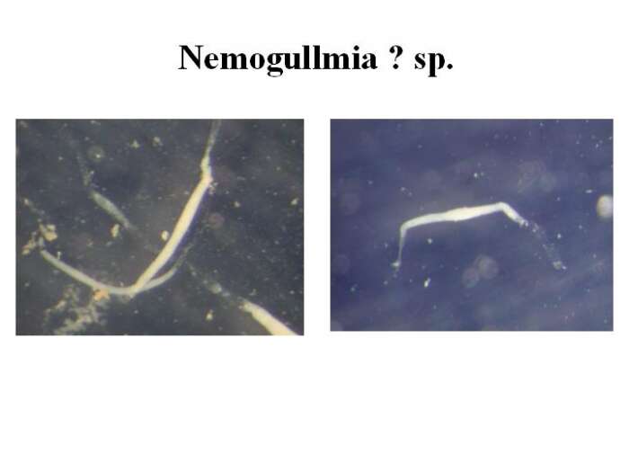
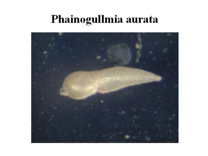
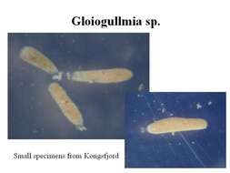
Specimens were isolated from surface sediments samples collected in Kongsfjorden, Isfjorden and Adventfjorden during the cruise of r/v « Oceania » between 22 July and 2 August 2004. The sediment samples were sieved at 500 um and 125 um sized meshes, and living specimens were picked under dissecting microscope on board. The specimens were photographed, measured and fixed for further DNA extraction. Source: http://www.iopan.gda.pl/projects/biodaff/EMBS-06.html
-
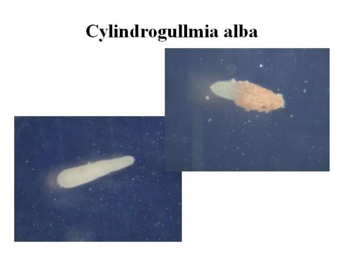
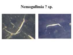
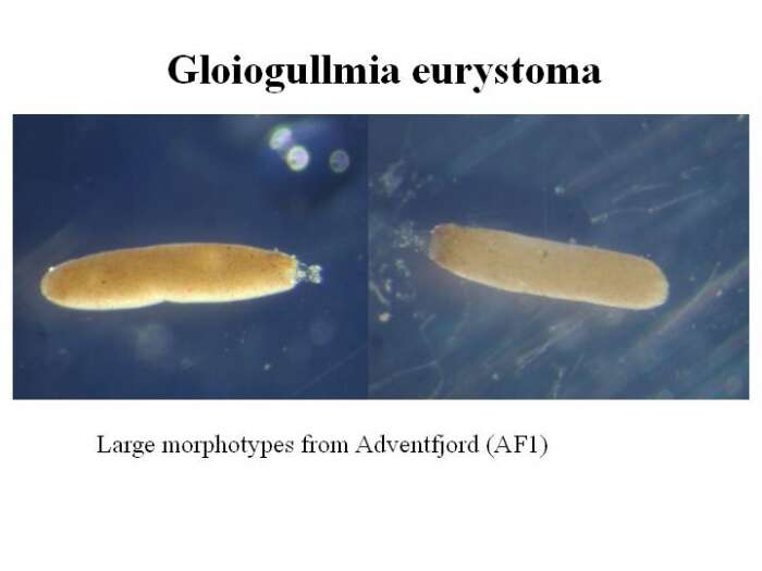
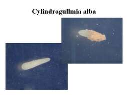
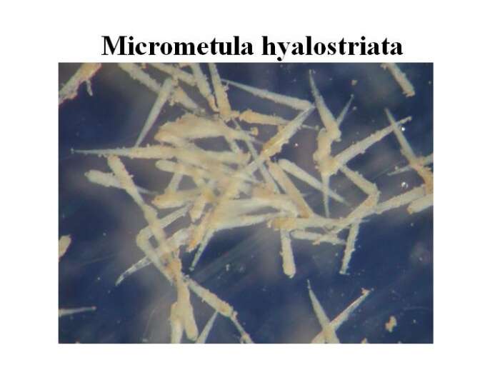
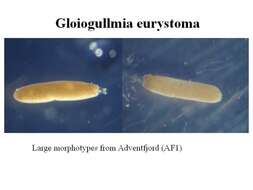
Specimens were isolated from surface sediments samples collected in Kongsfjorden, Isfjorden and Adventfjorden during the cruise of r/v « Oceania » between 22 July and 2 August 2004. The sediment samples were sieved at 500 um and 125 um sized meshes, and living specimens were picked under dissecting microscope on board. The specimens were photographed, measured and fixed for further DNA extraction. Source: http://www.iopan.gda.pl/projects/biodaff/EMBS-06.html