-
Ronald Vonk, Bert W. Hoeksema, Damia Jaume
Zookeys
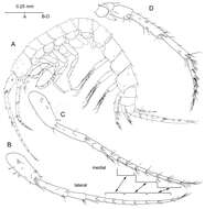
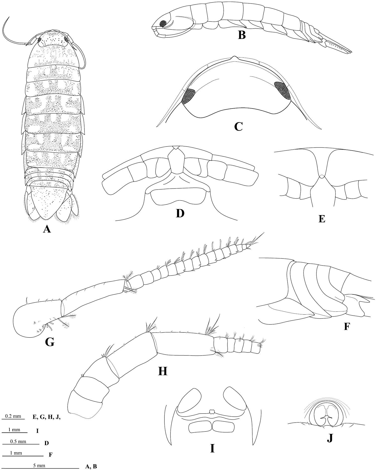
Figure 2.Psammogammarus wallacei sp. n., male paratype 2.53 mm, P5-P7 wanting. A body, lateral B left antennule, lateral C same showing medial armature D right antenna, lateral. Arrows pointing at serially-homologous pairs of articles of main flagellum of antennule.
-
Kristine N. White, James Davis Reimer
Zookeys
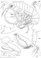
Figure 3.Leucothoe amamiensis sp. n., holotype male, 5.9 mm, RUMF-ZC-1654.
-
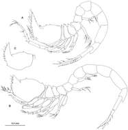
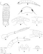
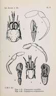
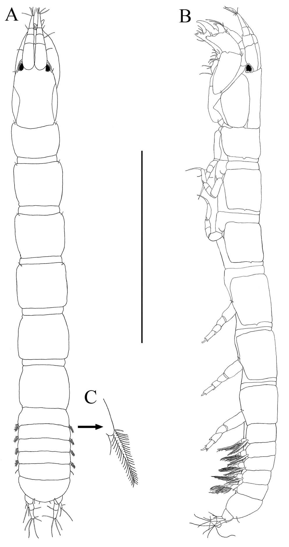
Figure 2.Ithyleucon sorbei gen. et sp. n. A preadult female holotype (ICMU12101901), whole animal in lateral view B preadult male paratype (ICMU12101904) C carapace of immature male paratype (ICMU12101906).
-
B.A.R. Azman, B.H.R. Othman
Zookeys
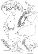
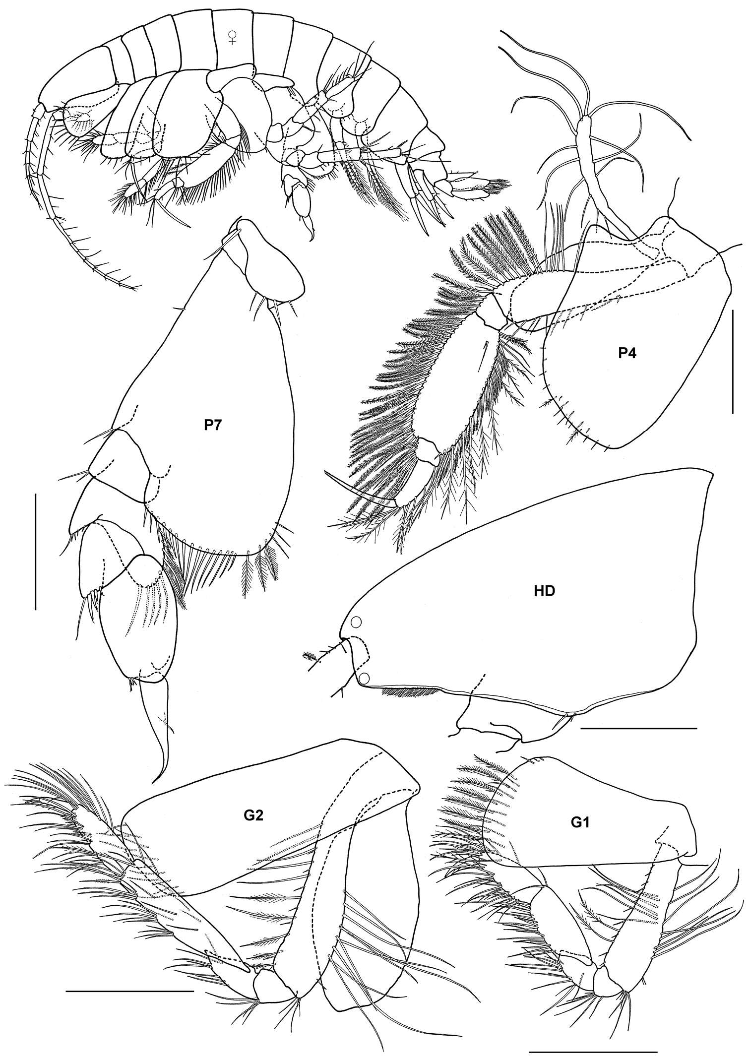
Figure 2.Ampelisca brevicornis (Costa), female (UKMMZ-1454), 4.8 mm. Renggis, Pulau Tioman. Scales for G1, G2, P4, P7 represent 0.5 mm; HD scale = 0.2 mm.
-
Young-Hyo Kim, Ed A. Hendrycks
Zookeys
Figure 1.Socarnes tongyeongensis sp. n., female, 8.8 mm, Gyeongpo, Pungwha-ri, Sanyang-eup, Tongyeong-si, Korea.
-
Eknarin Rodcharoen, Niel L. Bruce, Pornsilp Pholpunthin
Zookeys
Figure 2.Cirolana songkhla sp. n., male holotype (PSUZC-CR0281-01) (13.7 mm) (A–F), male paratype (PSUZC-CR0281-2) (11.2 mm) (G–H), male paratype (PSUZC-CR0281-2) (13.8 mm) (I–J). A dorsal view B lateral view C head, dorsal view D frons E detail of frontal lamina F pleon G antennule H antennal peduncle I antero-ventral view of penial opening J ventral view of penial opening.
-
Shigenori Karasawa, Kenshi Goto
Zookeys
Figure 1.Burmoniscus kitadaitoensis, male, holotype, TOYA-Cr 14899. A, B Pleopod 1 endopodite C pleopod 1 exopodite D pleopod 2 endopodite E pleopod 2 exopodite F genital papilla. Scale bars: A, C–E 200 μm, B 50 μm.
-
Andrés G. Morales-Núñez, Richard W. Heard
Zookeys
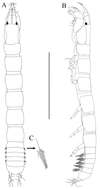
Figure 2.Paratanais rosadi sp. n., holotype female: A dorsal view B lateral view C enlargement of articulated setulate seta on pleonite-1. Scale bar A–B 1.0 mm.
-
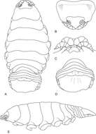
Kerry A. Hadfield, Niel L. Bruce, Nico J. Smit
Zookeys
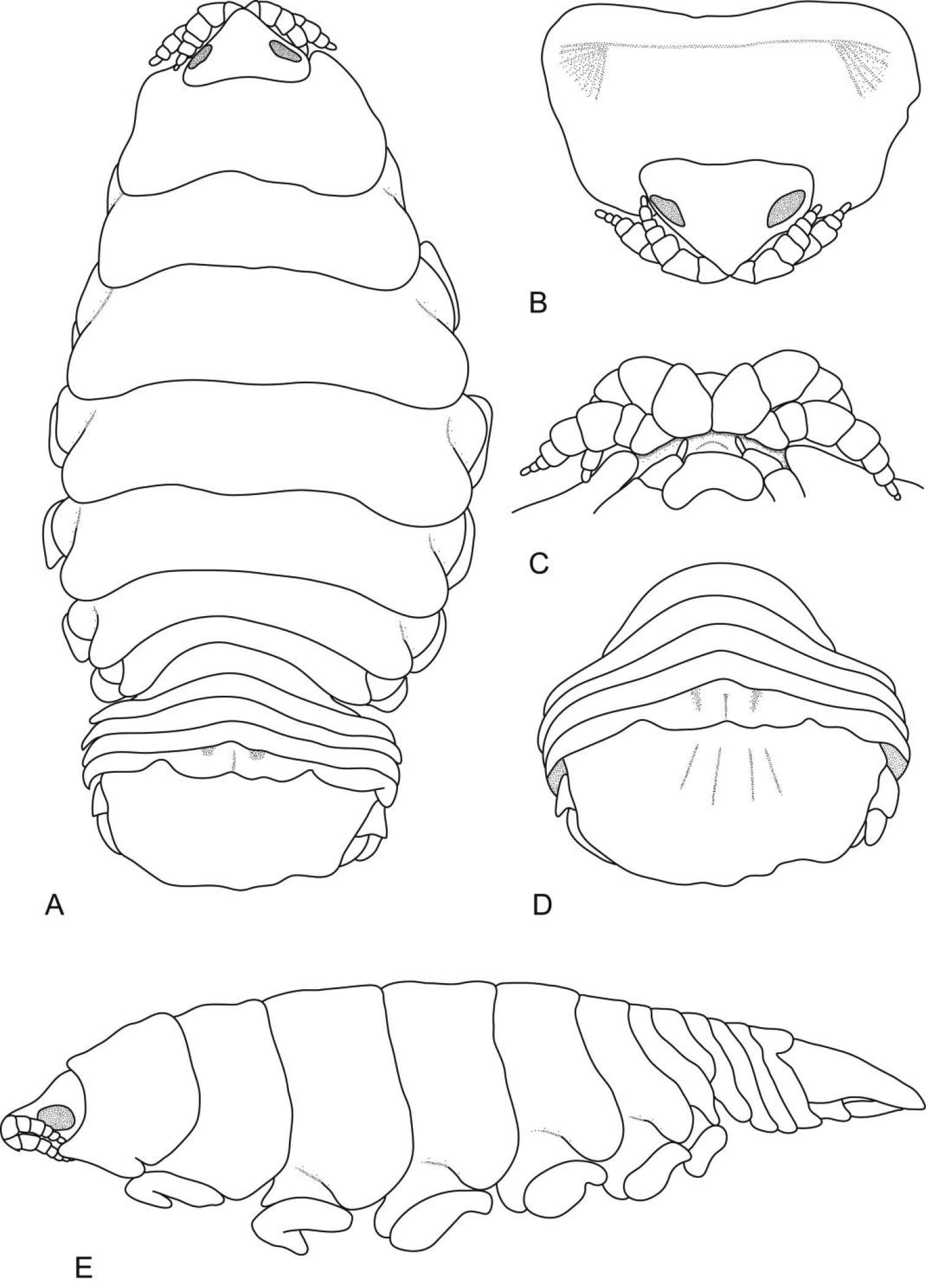
Figure 1.Ceratothoa africanae sp. n. female holotype (29 mm) (SAM A45937): A dorsal view B antero-dorsal view of pereonite 1 and cephalon C ventral view of cephalon D dorsal view of pleotelson E lateral view.
-
Patricia Cabezas, Enrique Macpherson
Zookeys
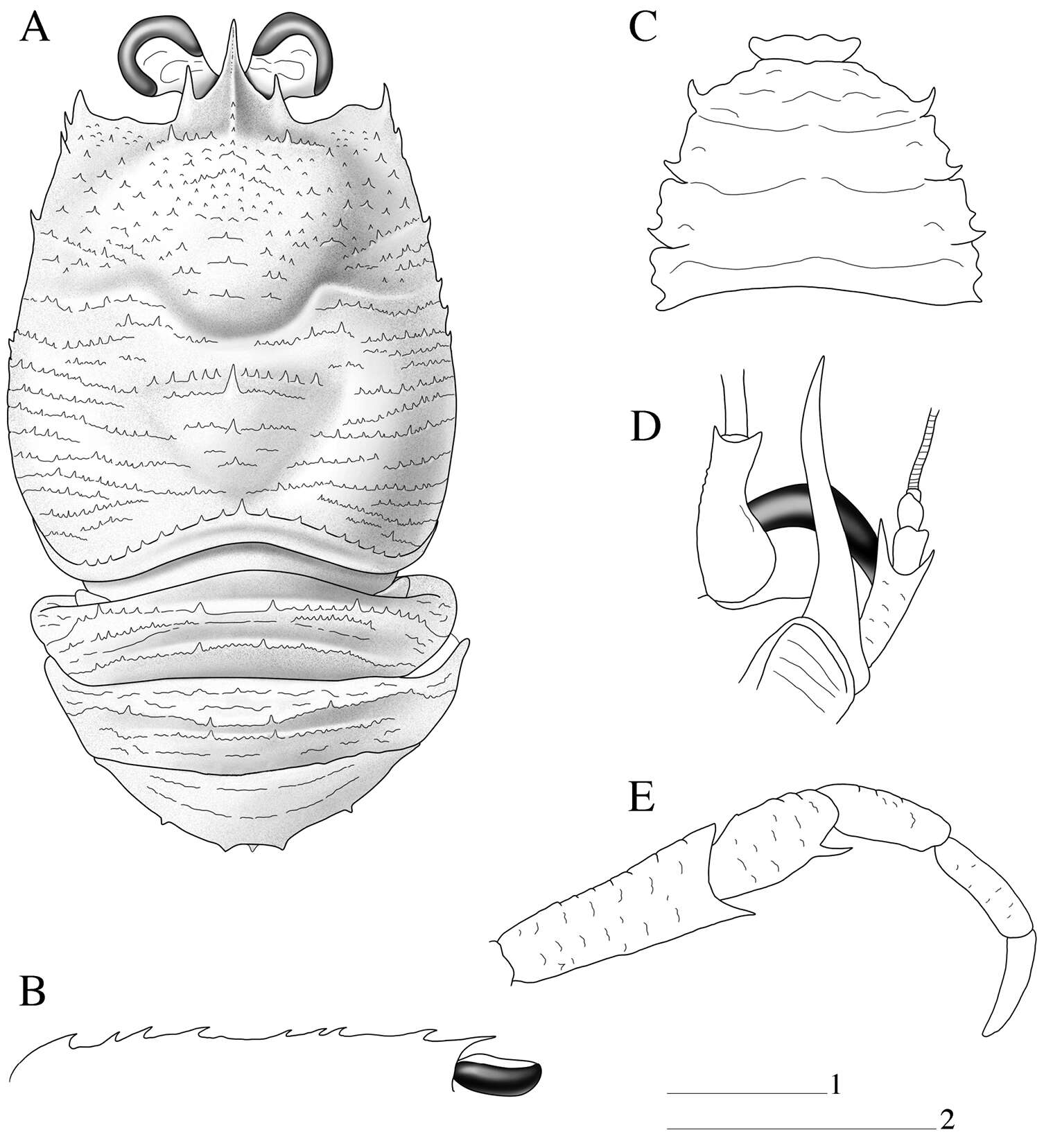
Figure 1.Paramunida haigae sp. n. male holotype, 16.6 mm (LACM–CR1973-3312). Christmas (Kiritimati) Island. A carapace and abdomen, dorsal view B carapace, lateral profile C sternum D left antennule and antenna, ventral view E right maxilliped 3, lateral view. Scale: 5 mm (scale 1 for A–C, E; scale 2 for D).
-
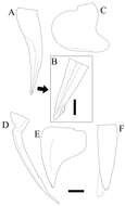
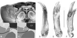
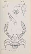
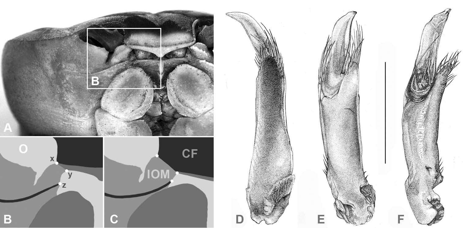
Figure 2.(CF) Carapace front; (O) orbit; (IOM) mesial lobe of infraorbital margin; (x) widest width of CF; (z) mesial end of suborbital crista; (x–y) width of IOM at point of contact with CF; (y–z) shortest distance between CF and mesial end of suborbital crista; A, B Atlantic Gecarcinus lateralis (Freminville 1835), male, carapace width (CW) 31 mm, Costa Rica, Puerto Viejo C Gecarcinus nobilii sp. n., holotype, male, CW 31 mm, Ecuador, Punta Galera (LACM CR 1968-477). First male gonopod: Gecarcinus nobilii sp. n., holotype: D mesial view E lateral view F Pacific Gecarcinus lateralis (sensu Türkay 1973), CW 31 mm, Costa Rica, Hermosa Beach, lateral view; Scale bar = 5 mm.
-
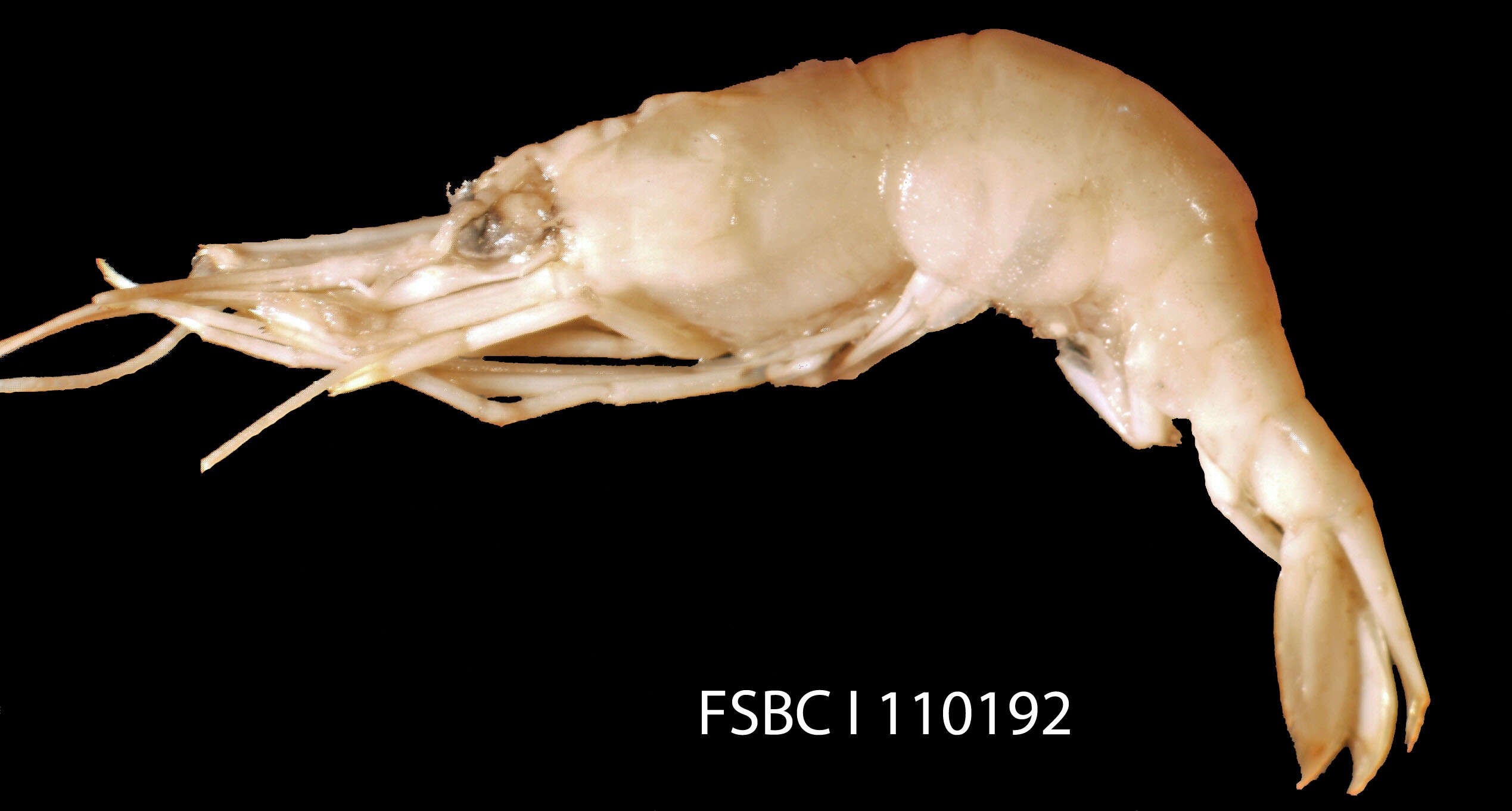
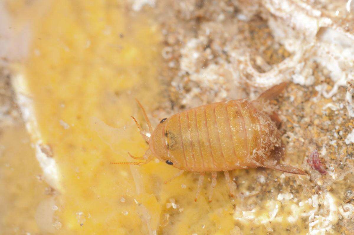
Scientific name: Processa profunda
-
Cape Paterson, Victoria, Australia
-
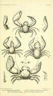
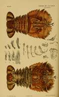
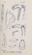
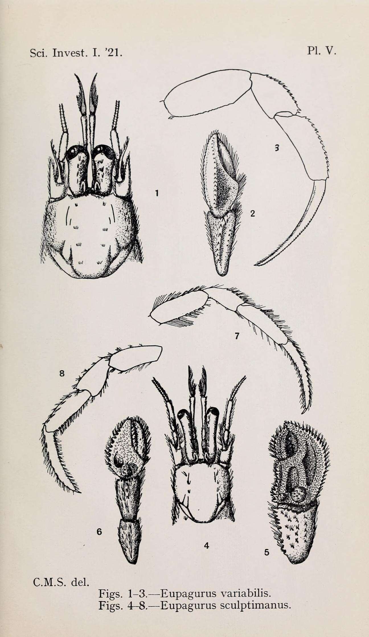
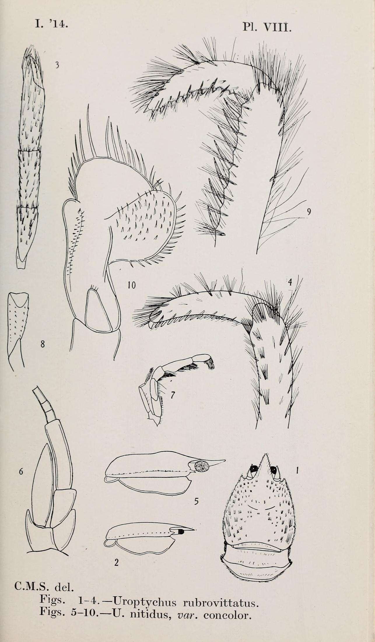
Report on the zoological collections made in the Indo-Pacific Ocean during the voyage of H.M.S. 'Alert' 1881-2.London :Printed by order of the Trustees,1884.
biodiversitylibrary.org/page/12067742
-
Cape Paterson, Victoria, Australia
-
-
-
-
-
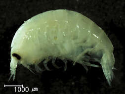
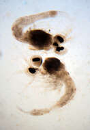
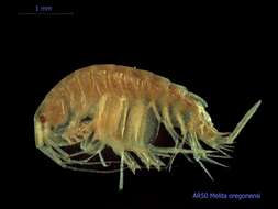
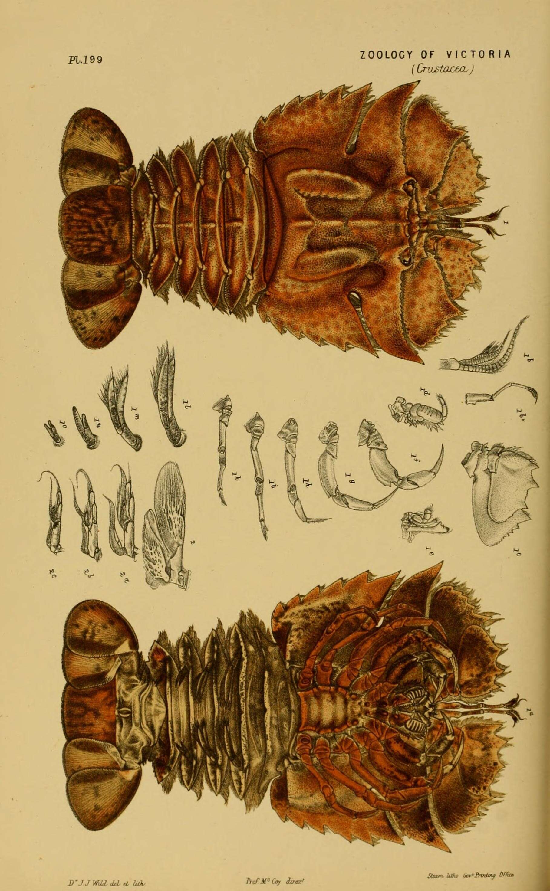
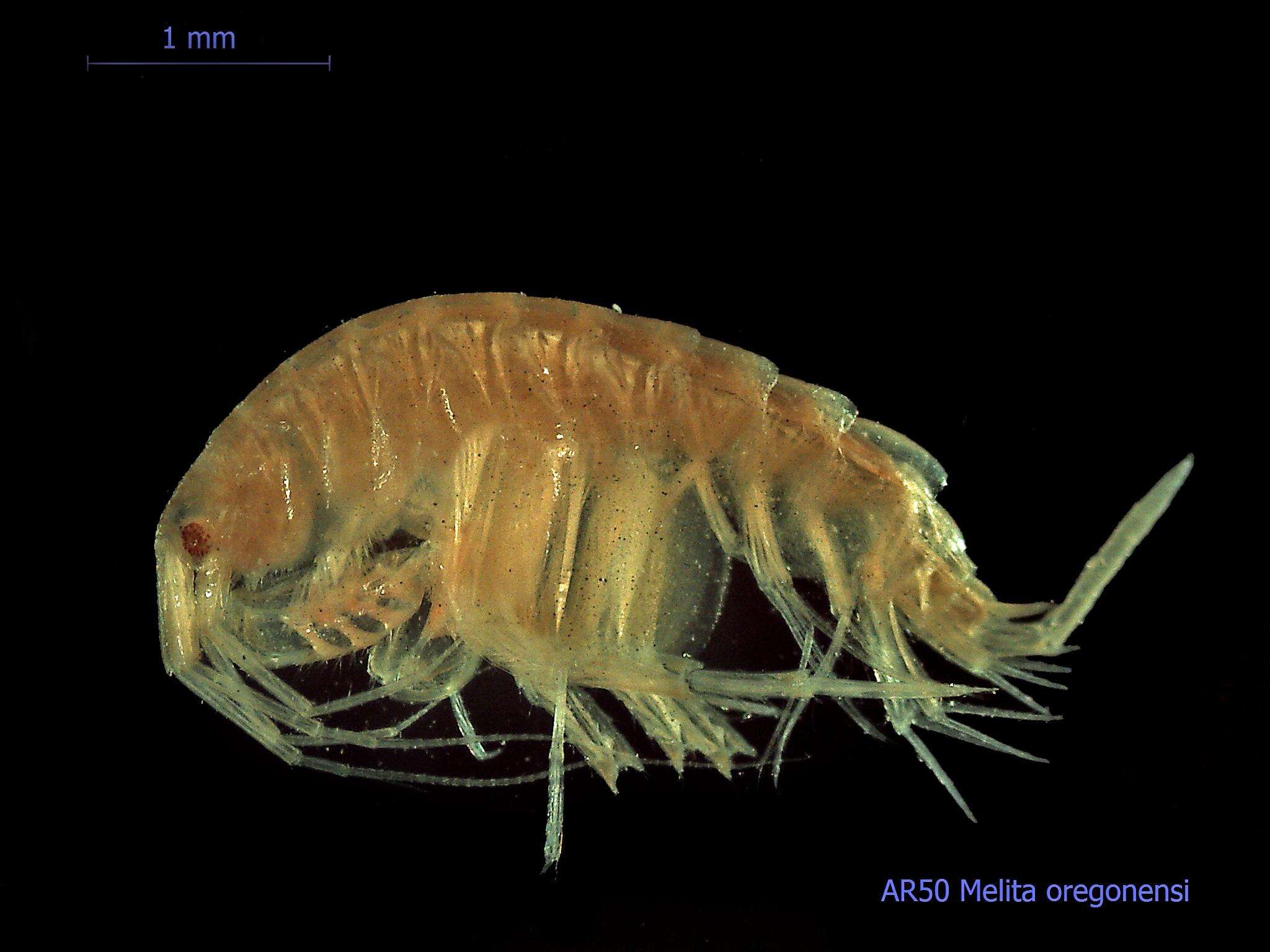
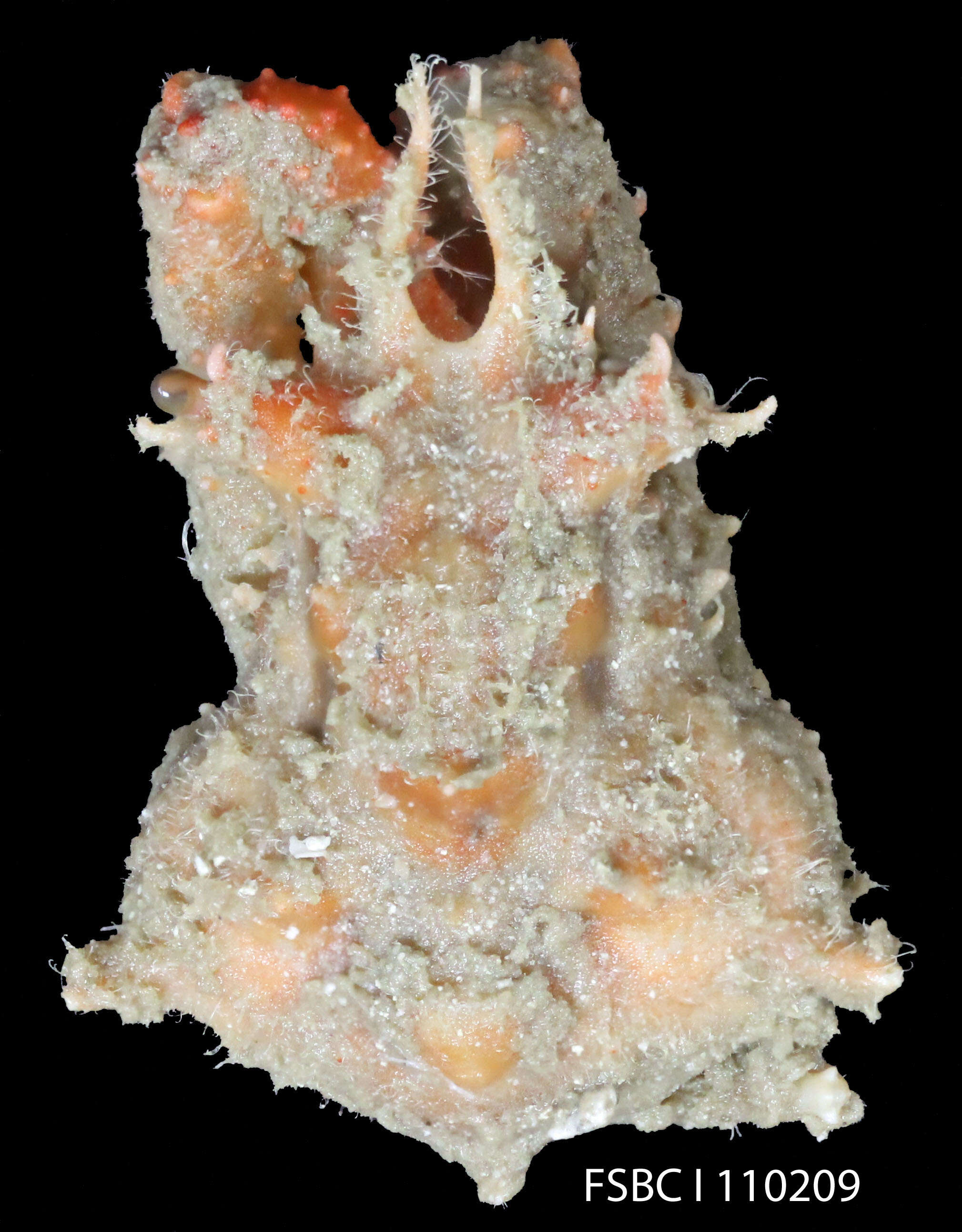
Collected from Puget Sound sediments and photographed by the Washington State Department of Ecologys Marine Sediment Monitoring Team. For more information about this teams work visit:
www.ecy.wa.gov/programs/eap/psamp/index.htm.
-
-
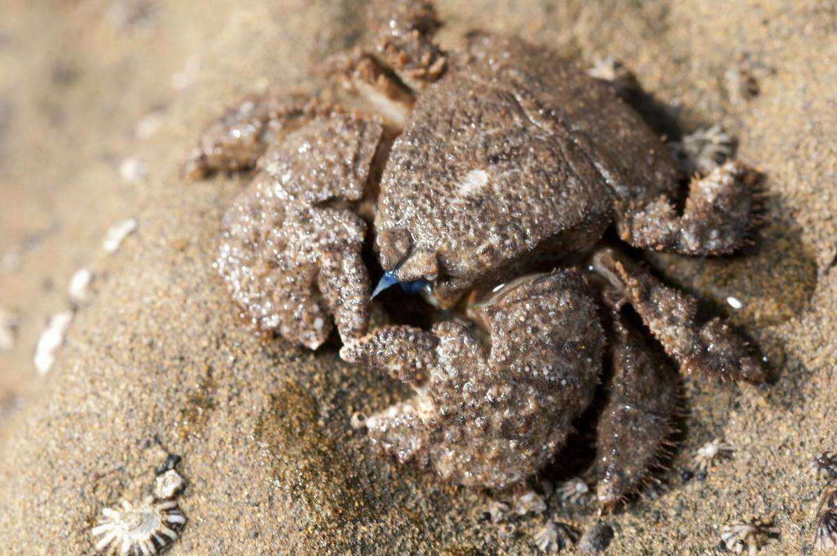
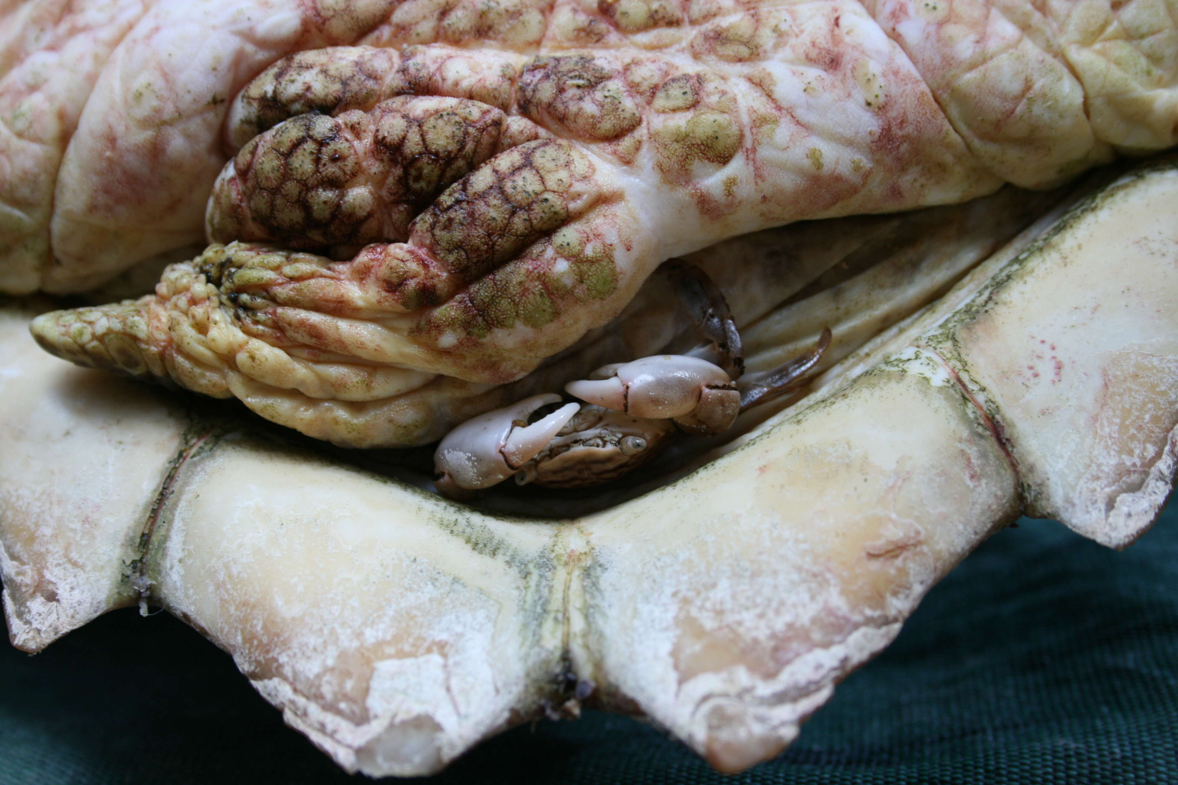
Scientific name: Macrocoeloma eutheca
-
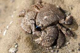
Attribution: Maristella D'Addario (University of Rome).A small crab, Planes minutus (Columbus crab), living on an individual of Caretta caretta (Loggerhead Sea Turtle). This crab is known to prey upon other sea turtles epibionts.Highly Commended in the BMC Ecology Image Competition 2012:BMC Ecology 2013, 13:6 doi:10.1186/1472-6785-13-6
www.biomedcentral.com/1472-6785/13/6
-
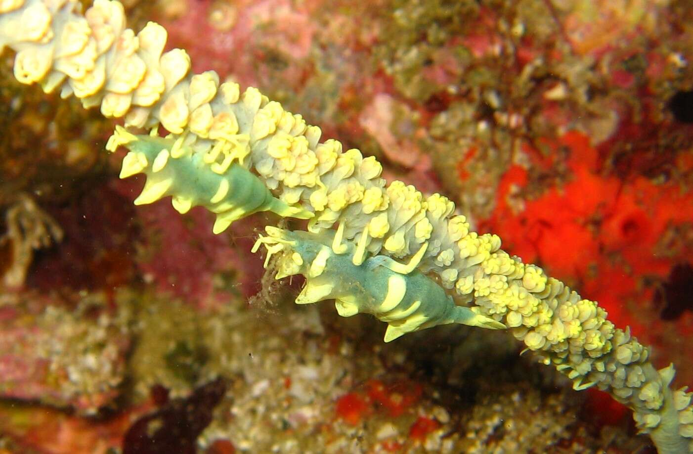
Otsuki-cho, Kochi Prefecture, Japan