-
Sergey G. Ermilov, Andrei V. Tolstikov, Nathalie Mary, Heinrich Schatz
Zookeys
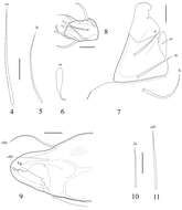
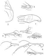
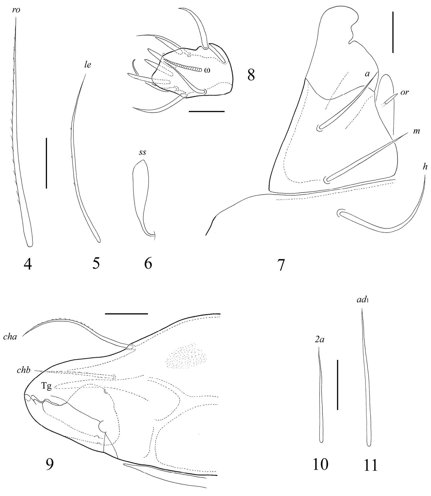
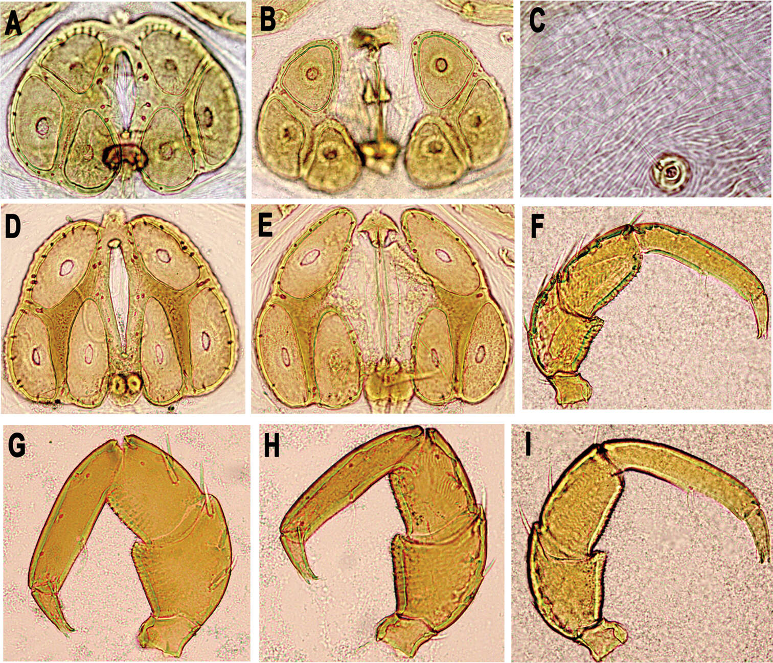
Figures 4–11. Fortuynia smiti sp. n., adult: 4 rostral seta 5 lamellar seta 6 sensillus 7 subcapitulum, right half of anterior part, ventro-lateral view 8 palptarsus 9 chelicera, anterior part 10 epimeral seta 2a 11 adanal setae ad1. Scale bar (4–7, 9–11) 20 μm, (8) 10 μm.
-
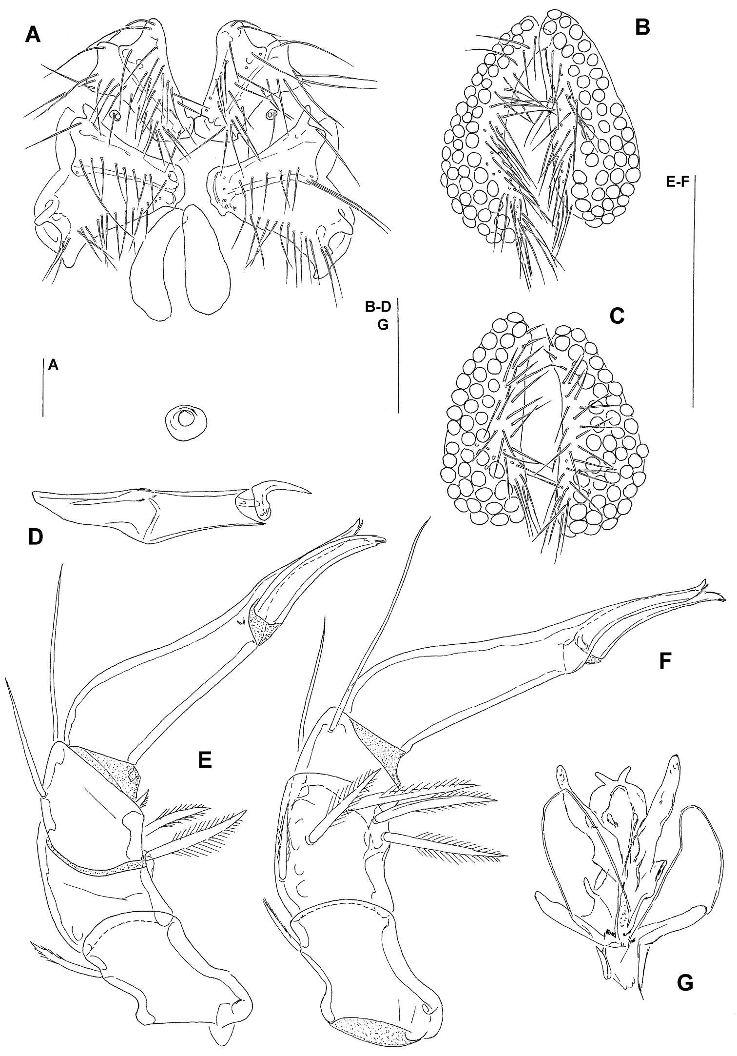
Sergey G. Ermilov, Jochen Martens, Andrei V. Tolstikov
Zookeys
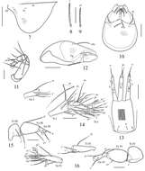
Figures 7–16.Lepidozetes acutirostrum sp. n., adult: 7 pteromorph, lateral view 8 notogastral seta c 9 notogastral seta p1 10 subcapitulum, ventral view 11 palp 12 chelicera 13 ovipositor 14 genu (Ge), tibia (Ti) and tarsus (Ta) of leg I, right, antiaxial view 15 trochanter (Tr), femur (Fe) and genu of leg III, left, antiaxial view 16 leg IV, right, antiaxial view. Scale bar (7) 100 μm, (8–10, 12, 14–16) 40 μm, (11) 20 μm, (13) 50 μm.
-
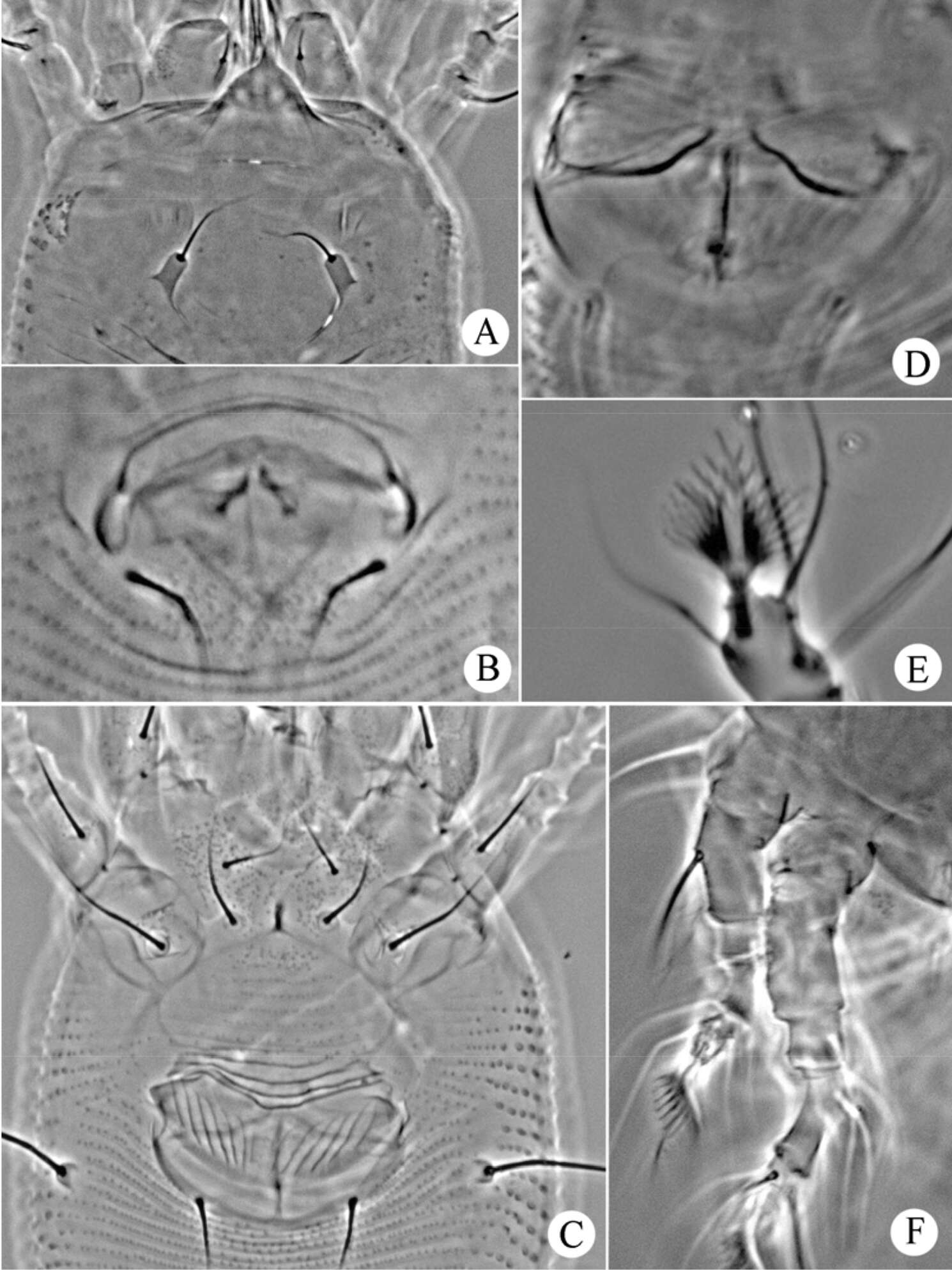
Sergey G. Ermilov, Badamdorj Bayartogtokh, Dorothee Sandmann, Franca Marian, Mark Maraun
Zookeys
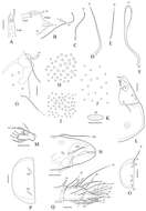
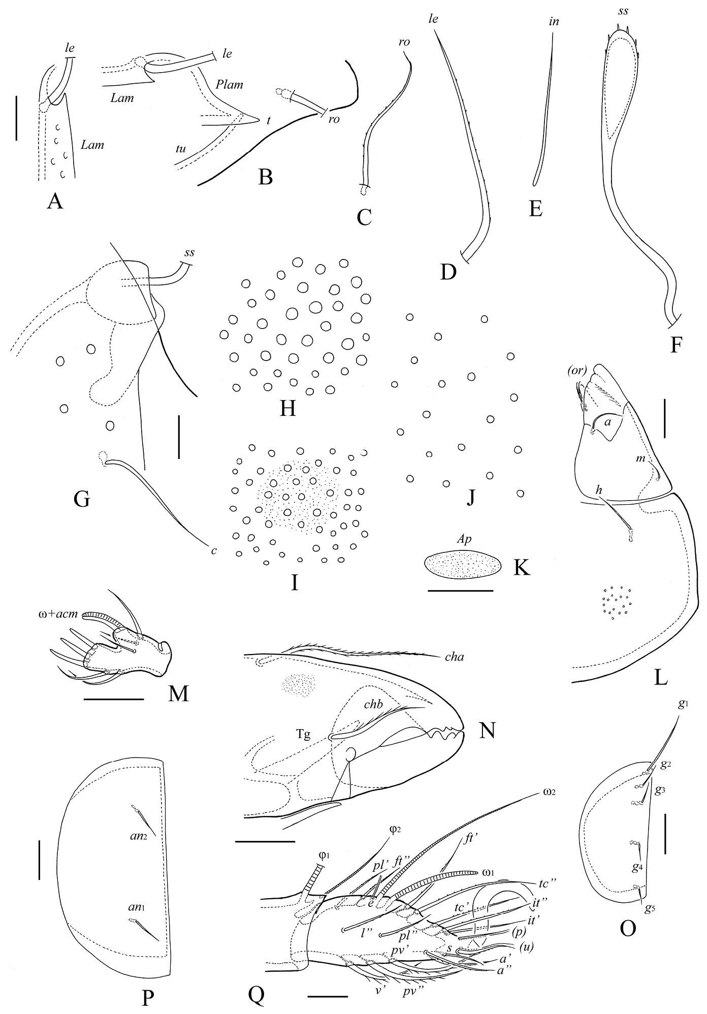
Figure 6.Trachyoribates (Rostrozetes) glaber (Beck, 1965), adult: A anterior part of lamella (medio-distal part of lamellar seta not illustrated) B anterior part of lamella and tutoria, and prolamellar line dorso-laterally (medio-distal part of rostral and lamellar seta not illustrated) C rostral seta D lamellar seta E interlamellar seta F sensillus G bothridium and notogastral seta c H foveolae on rostrum I foveolae in central part of prodorsum J foveolae on notogaster K postanal porose area L left half of subcapitulum M palptarsus N anterior part of chelicera O right genital plate P right anal plate Q tarsus and anterior part of tibia of leg I, right, antiaxial view. Scale bar 10 μm.
-
Yunus Esen, Vladimir Pešić, Orhan Erman, Yücel Kaya
Zookeys
Figure 2.A–C, F, I photographs of Hygrobates anatolicus sp. n. (A, F male B–C, I female), Göksu stream, Turkey D–E, G–H photographs of Hygrobates nigromaculatus Lebert, 1879 (D, G male E, H female), Ohrid Lake, Macedonia: A–B, D–E genital field C detail of dorsal integument F–I palp.
-
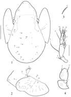
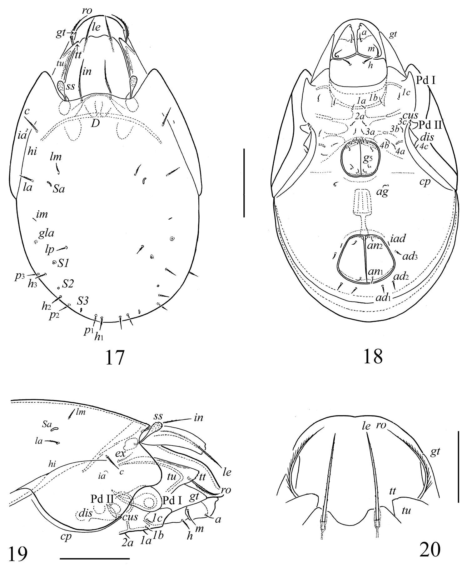
Sergey G. Ermilov, Alexander E. Anichkin, Andrei V. Tolstikov
Zookeys
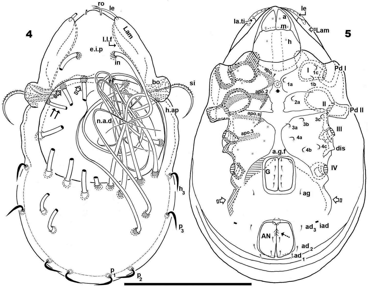
Figures 3–5.Papillocepheus primus sp. n., adult: 3 lateral view of prodorsum and anterior part of notogaster (legs not illustrated) 4 lateral view of posterior part of notogaster 5 tarsus and anterior part of tibia of leg I, right, antiaxial view. Scale bar (3, 4) 100 μm, (5) 20 μm.
-
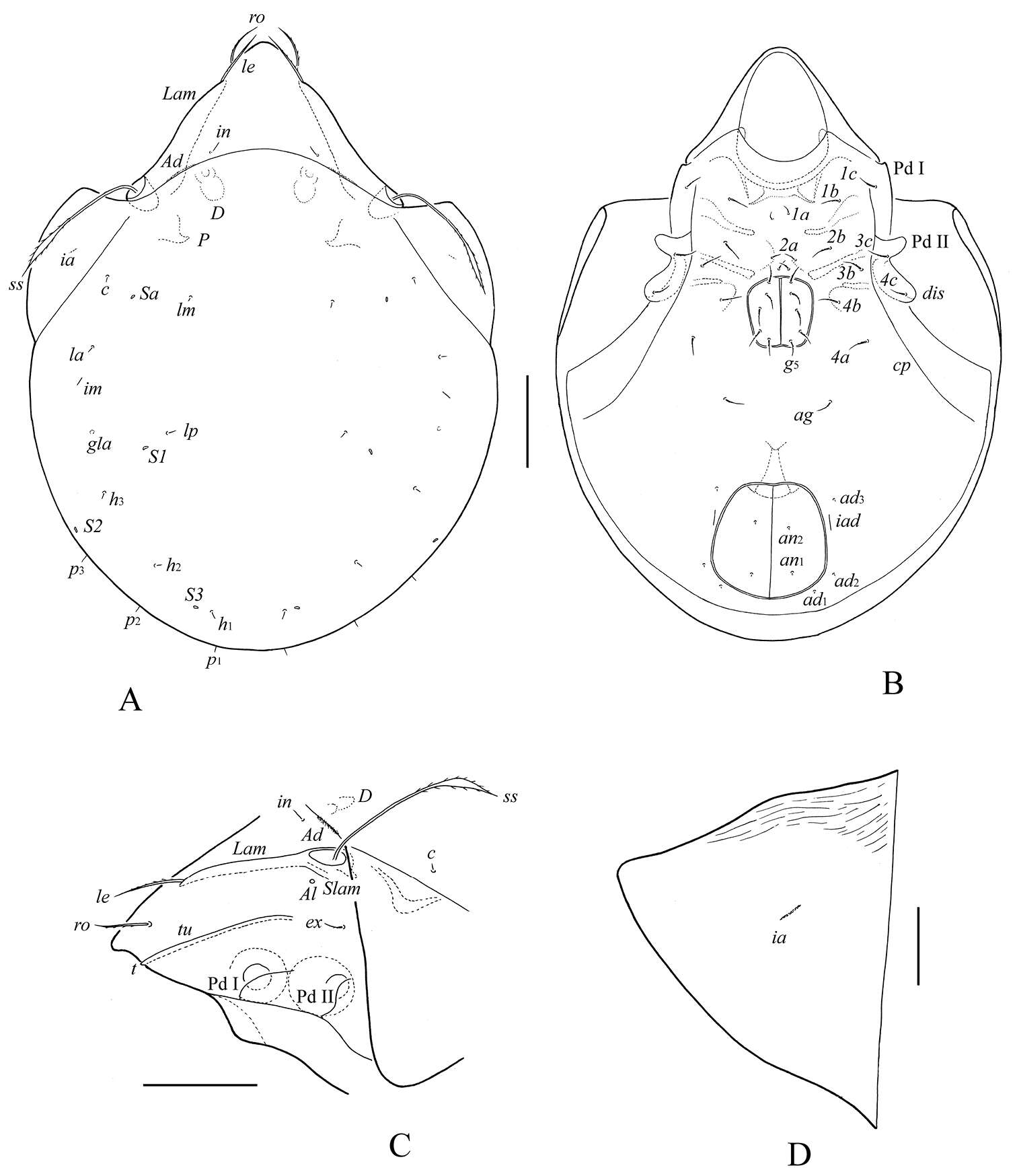
Sergey G. Ermilov, Umukusum Ya. Shtanchaeva, Luis S. Subías, Alexander E. Anichkin
Zookeys
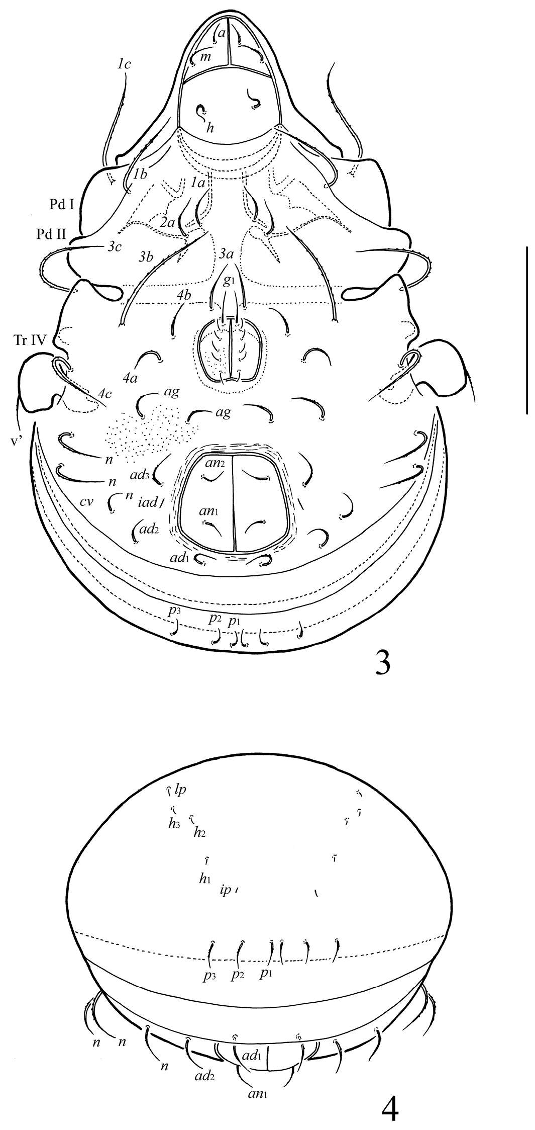
Figures 3–4.Ctenobelba (Berndamerus) bugiamapensis sp. n.: 3 ventral view (legs except trochanter IV not shown) 4 posterior view. Scale bar 100 μm.
-
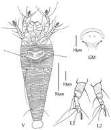
Qiong Wang, Xiao Han, Xiao-Feng Xue, Xiao-Yue Hong
Zookeys
Figure 6.Phyllocoptes setalsolenidion sp. n.: V ventral view of female GM male genital region L1 leg I L2 leg II.
-
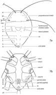
Michael J. Skvarla, J. Ray Fisher, Ashley P. G. Dowling
Zookeys
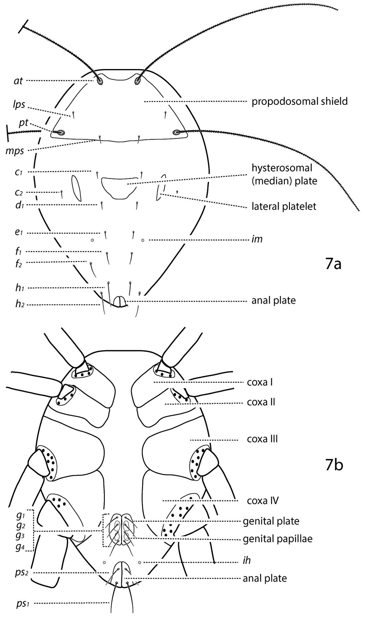
Figure 7.Generalized schematic of cunaxid idiosomal morphology. 7a Dorsal. 7b Ventral.
-
Sergey G. Ermilov, Umukusum Ya. Shtanchaeva, Luis S. Subías, Jochen Martens
Zookeys
Figure 2.Lasiobelba (Lasiobelba) daamsae sp. n.: ventral view (legs not illustrated). Scale bar 400 μm.
-
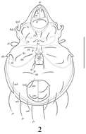
Nestor Fernandez, Pieter Theron, Christine Rollard, Elio Rodrigo Castillo
Zookeys
Figures 4–5.Malgasodes curvisetus Mahunka, 2000, adult. Optic observations. 4 dorsal view 5 ventral view. Notes: Abbreviations: see “Material and methods”. Scale bar 4, 5 = 250 μm.
-
Glenstrup Sø, Jylland, Danmark
-
Mushroom Observer Image 103176: Aculops
-
Figure 1. A–G Hydrodroma meridionalis sp.n. (A–B, D, G = male holotype, C, E, F = female paratype) A = coxal and genital field B–C = genital field D = chelicera E = palp, lateral view F = palp, medial view G = ejaculatory complex. Scale Bars = 100 μm.
-
Lixia Xie, Yi Yan, Rong Huang, Maofa Yang
Zookeys
Figure 3. Damaeus (Paradamaeus) yushuensis sp. n. – lateral view (100μm)
-
Hao-Sen Li, Xiao-Feng Xue, Xiao-Yue Hong
Zookeys
Figure 29.Diptacus berberinus sp. n.: A dorsal view of female B ventral view of female C lateral microtubercles D empodium E dorsal view of female posterior part F ventral view of female posterior part G leg I and leg II.
-
Vladimir Pešić, Ksenia A. Semenchenko, Wonchoel Lee
Zookeys
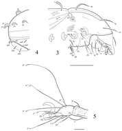
Figure 3.Torrenticola kimichungi sp. n., male holotype: A dorsal shield B ventral shield C ejaculatory complex D palp, lateral view E palp, medial view F gnathosoma. Scale bars = 100 μm.
-
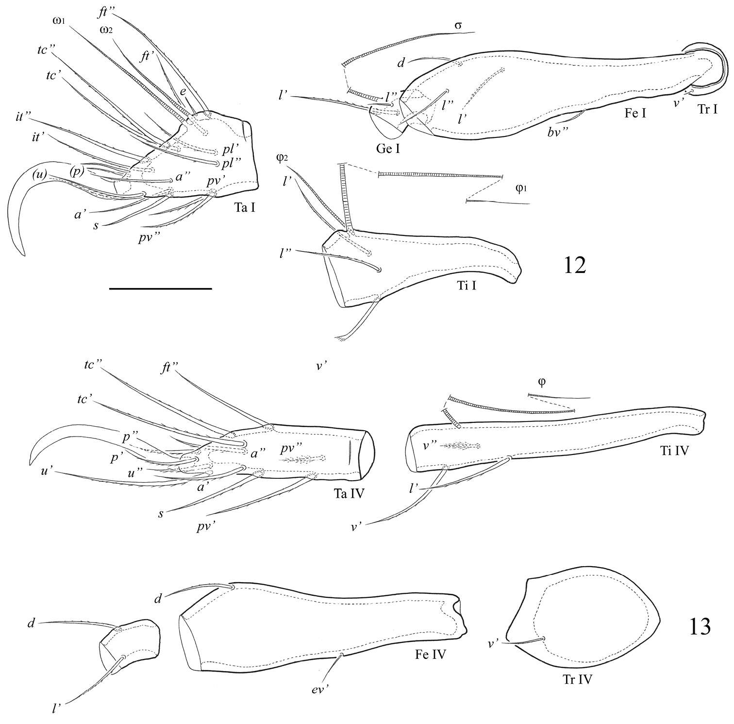
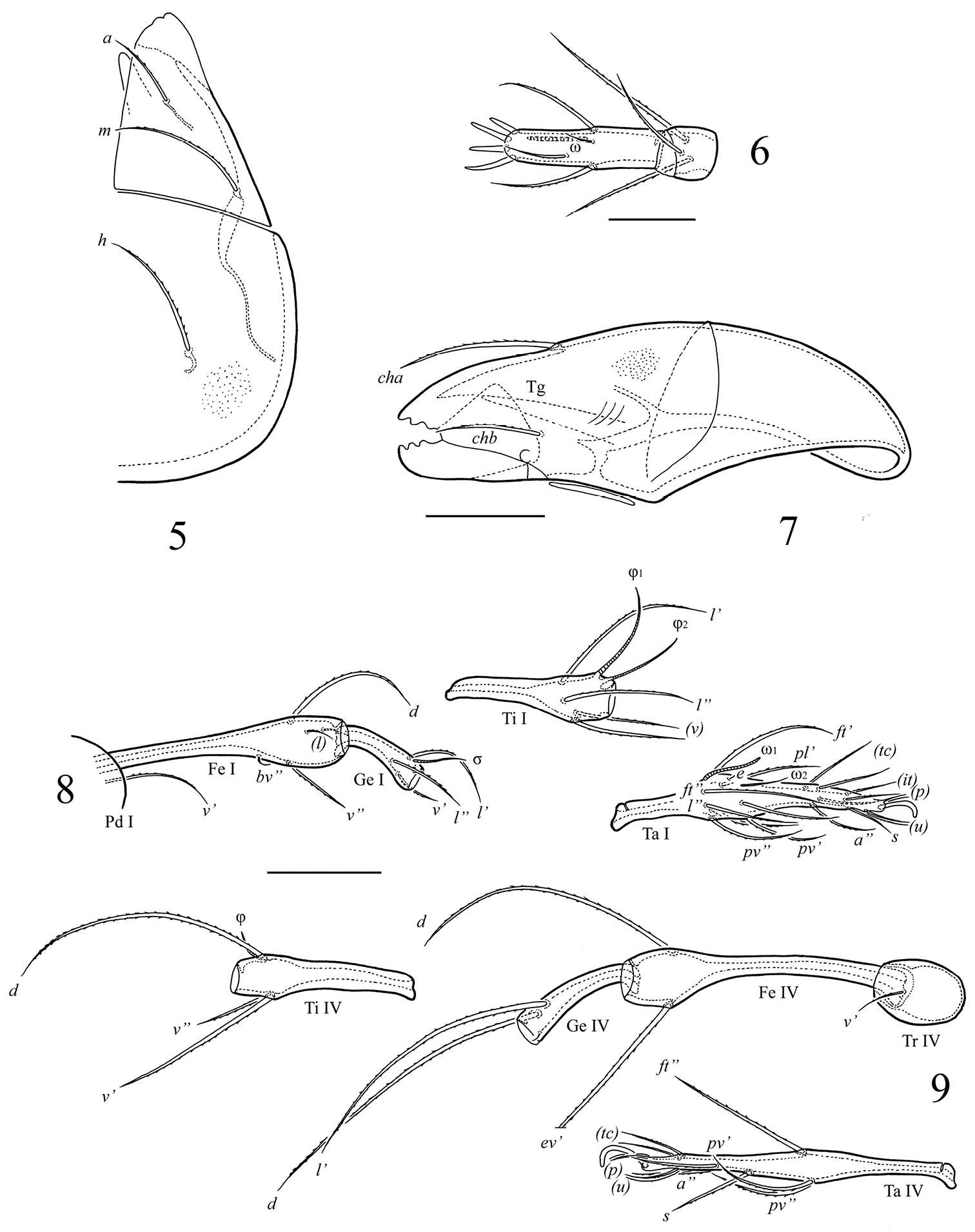
Sergey G. Ermilov, Andrei V. Tolstikov, Nathalie Mary, Heinrich Schatz
Zookeys
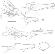
Figures 12–13. Fortuynia smiti sp. n., adult: 12 segments of leg I, left, antiaxial view 13 segments of leg IV, right, antiaxial view. Scale bar 50 μm.
-
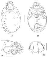
Sergey G. Ermilov, Jochen Martens, Andrei V. Tolstikov
Zookeys
Figures 17–20.Scutozetes clavatosensillus sp. n., adult: 17 dorsal view 18 ventral view (legs not illustrated) 19 anterior part of body, lateral view (legs not illustrated)20 rostrum, anterior margin of lamellae and tutoria, rostral and lamellar setae, dorso-anterior view. Scale bar (17–19) 100 μm, (20) 40 μm.
-
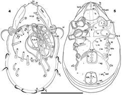
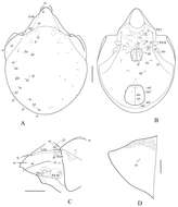
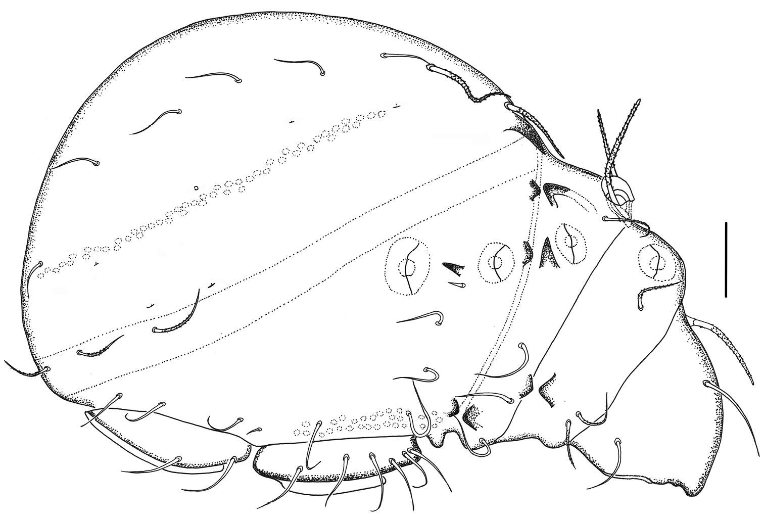
Sergey G. Ermilov, Badamdorj Bayartogtokh, Dorothee Sandmann, Franca Marian, Mark Maraun
Zookeys
Figure 1.Haplozetes paraminimicoma sp. n., adult: A body dorsally B body ventrally (gnathosoma and legs not illustrated) C prodorsum and anterior part of notogaster laterally D left pteromorph. Scale bar (A–C) 50 μm, scale bar (D) 20 μm.
-
Yunus Esen, Vladimir Pešić, Orhan Erman, Yücel Kaya
Zookeys
Figure 3.A–H Hygrobates (Rivobates) diversiporus Sokolow, 1927 (A–D male E–H female): A Palp, medial view B, F Coxal and genital field C–D, G–H Genital field E Palp, lateral view (Scale bars = 100 µm).
-
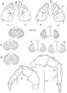
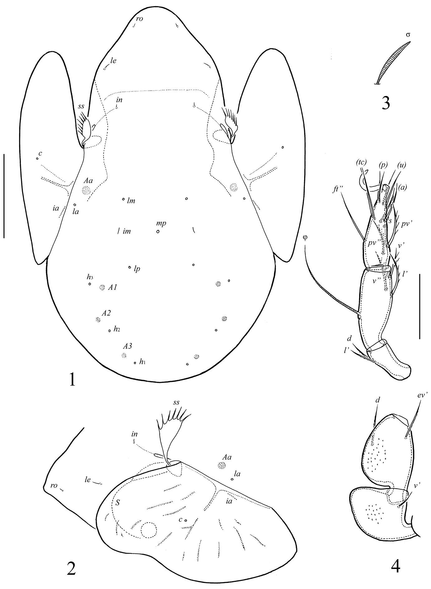
Sergey G. Ermilov, Alexander E. Anichkin
Zookeys
Figures 1–4.Allogalumna monodactyla sp. n., adult: 1 dorsal view 2 dorso-lateral view of prodorsum, pteromorph and anterior part of notogaster 3 solenidion of leg genu III 4 leg IV, left, antiaxial view. Scale bar (1, 2) 50 μm, (3, 4) 20 μm.
-
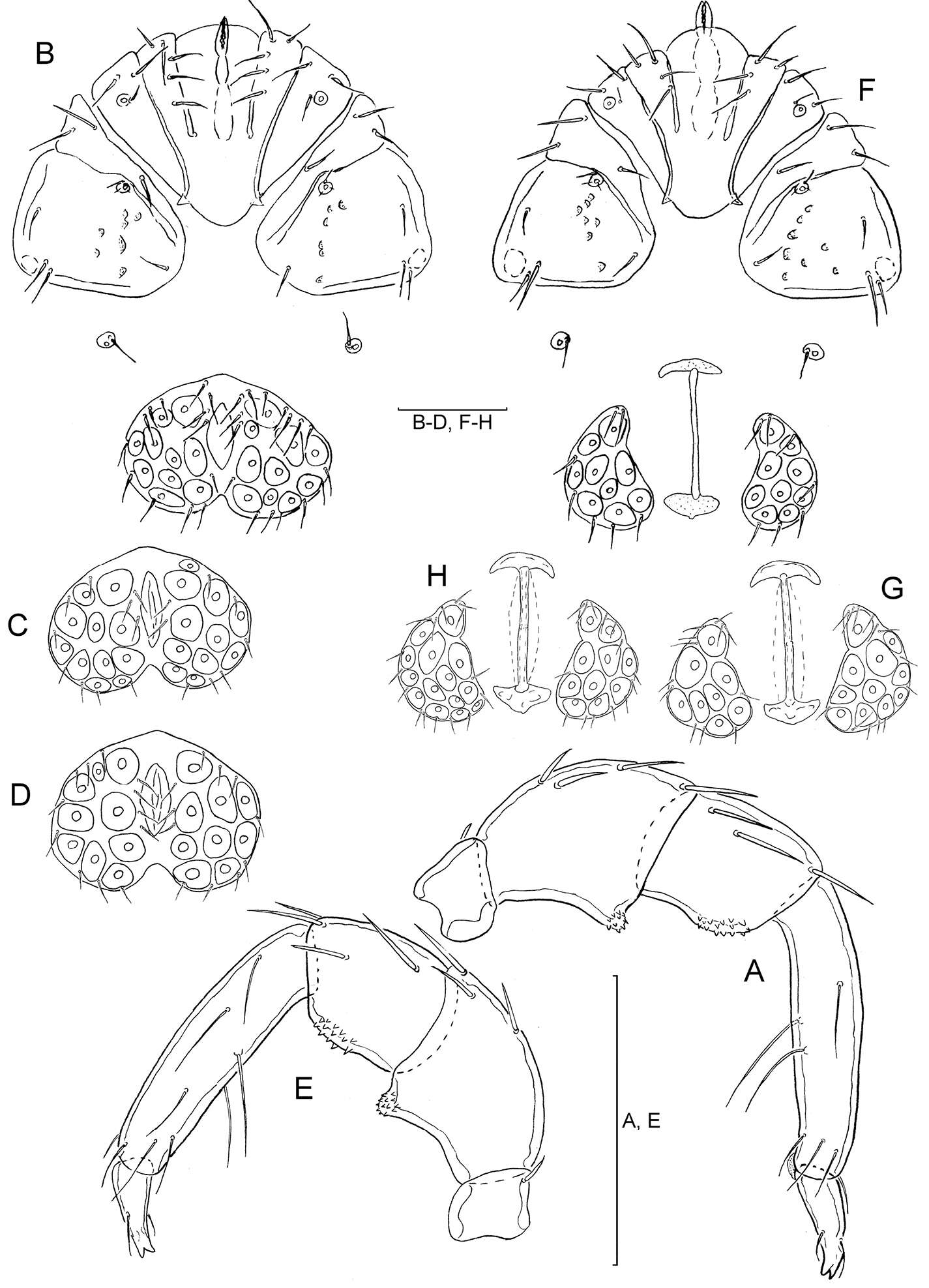
Sergey G. Ermilov, Umukusum Ya. Shtanchaeva, Luis S. Subías, Alexander E. Anichkin
Zookeys
Figures 5–9.Ctenobelba (Berndamerus) bugiamapensis sp. n., adult: 5 subcapitulum, left half, ventral view 6 tibia and tarsus of palp 7 chelicera 8 leg I (trochanter and basal part of femur not shown), right, antiaxial view 9 leg IV, right, antiaxial view. Scale bar 20 μm (5, 7); 10 μm (6); 50 μm (8–9).
-
Qiong Wang, Xiao Han, Xiao-Feng Xue, Xiao-Yue Hong
Zookeys
Figure 7.Phyllocoptes setalsolenidion sp. n.: A prodorsal shield B male genitalia C coxae and female genitalia D female internal genitalia E empodium F leg I and leg II.
-
Michael J. Skvarla, J. Ray Fisher, Ashley P. G. Dowling
Zookeys
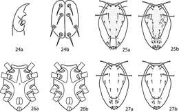
Figures 24–27.Lupaeus illustrations. 24a Pedipalp tibiotarsus 24b Genital setae not in a row, g3 out of line 25–27 Lupaeus key illustrations. Setae and cupules removed from figures 25a, b to increase clairity 25a Lupaeus longisetus, dorsal 25b Lupaeus polilloensis, dorsal 26a Ventral, small platelet present 26b Ventral, small platelet absent 27a Setae f1, f2 born on small platelets 27b Setae f1, f2 born on integument.