-
Sergey G. Ermilov, Olman Alvarado-Rodríguez, Axel P. Retana-Salazar
Zookeys
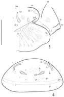
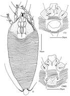
Figure 2.Pergalumna elongatiporosa sp. n.: ventral view (legs not illustrated). Scale bar 100 μm.
-
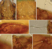
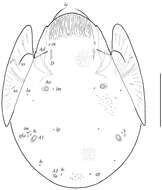
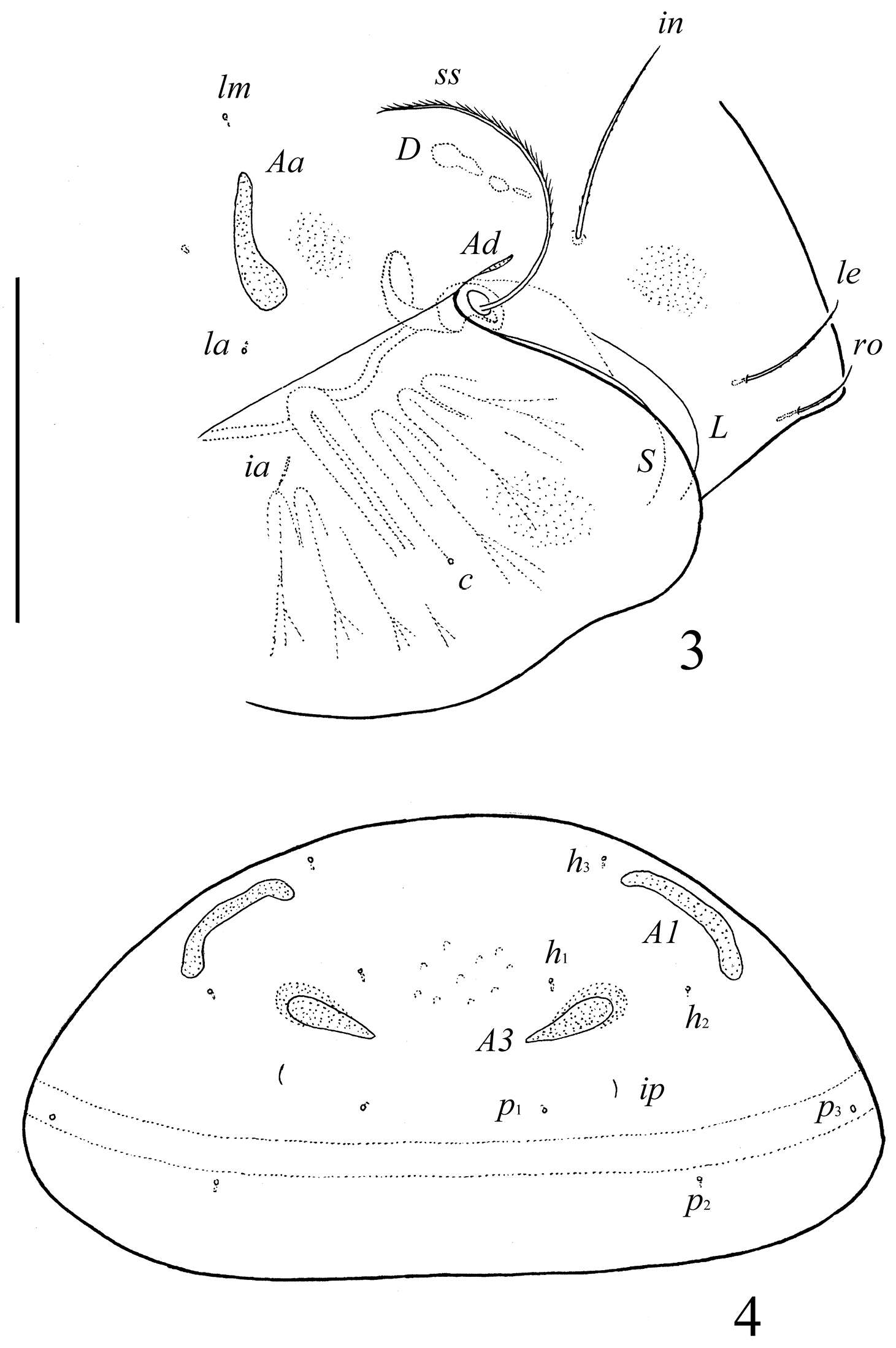
Figure 3.Conchogneta glabrisensillata sp. n. A Prodorsum, showing enantiophysis E, costula and bothridium B Central part of prodorsum, showing alveolus of interlamellar seta and interbothridial tubercle (indicated by arrow) C Part of laterial view of prodorsum, showing sensillus and granular tubercles on humeral region D Lateral view of prodorsal costula E Sensillus, lateral view F Slight variation of sensillus, lateral view G Granular tubercles on lateral part of prodorsum H Humeral region, showing tubercles Ha and Hp.
-
Hao-Sen Li, Xiao-Feng Xue, Xiao-Yue Hong
Zookeys
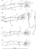
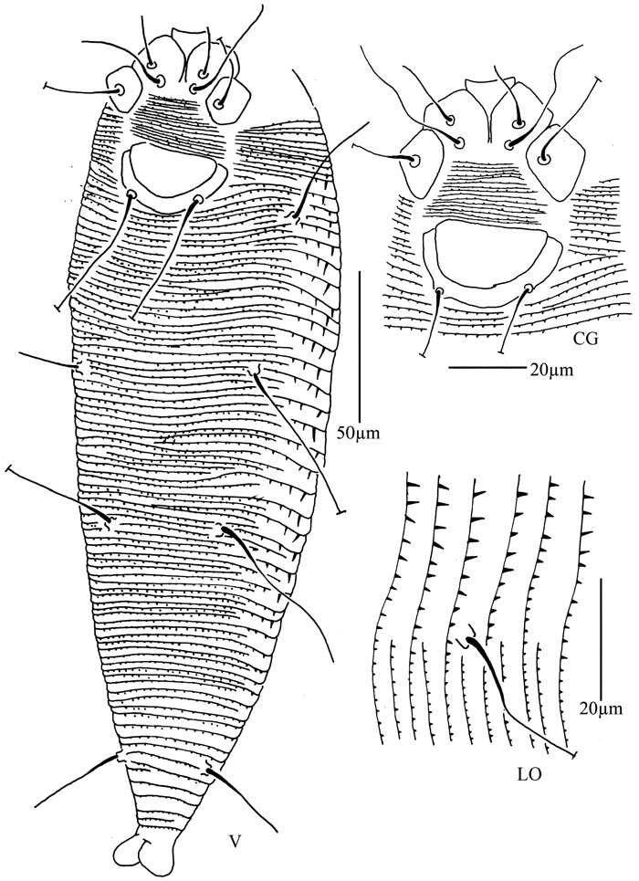
Figure 35.Rhyncaphytoptus spinus sp. n.: V ventral view of female CG coxae and female genitalia LO lateral microtubercles.
-
Vladimir Pešić, Ksenia A. Semenchenko, Wonchoel Lee
Zookeys
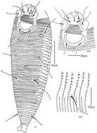
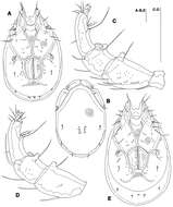
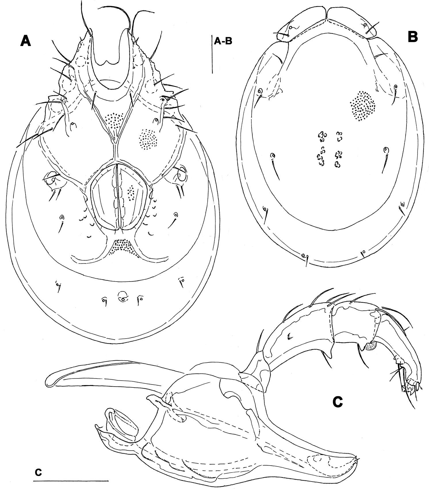
Figure 9.Torrenticola ussuriensis (Sokolow, 1940), female, Inje River, Korea: A ventral shield B dorsal shield C gnathosoma and palp, medial view. Scale bars = 100 μm.
-
Michael J. Skvarla, J. Ray Fisher, Ashley P. G. Dowling
Zookeys
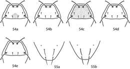
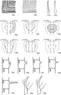
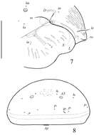
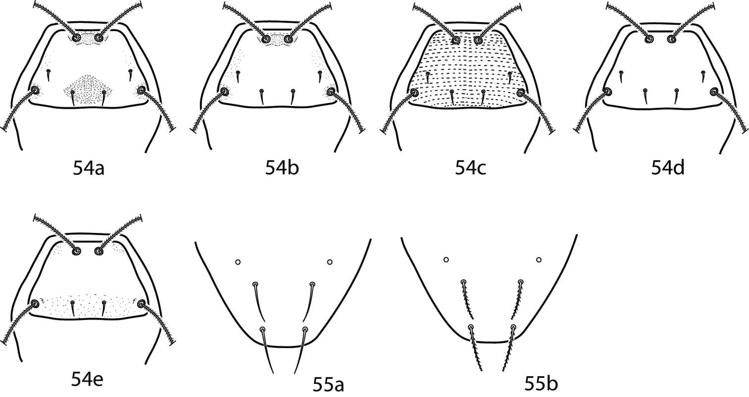
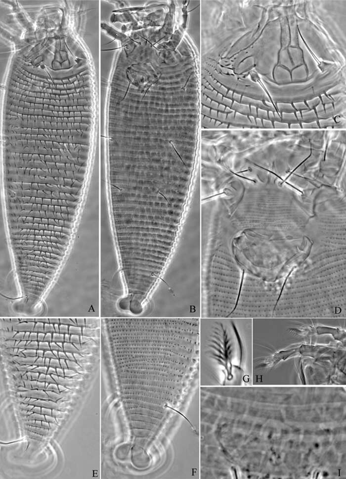
Figures 54, 55.Cunaxa key illustrations. 54a–e Proterosomal shield, dorsal 54a Proterosomal shield with oval area formed by broken striae around pt present, mps present 54b Proterosomal shield with oval area formed by broken striae around pt absent, mps present 54c Proterosomal shield striated, mps present 54d Proterosomal shield smooth, mps present 54e Proterosomal shield with lps absent 55a Smooth f1, h1 55b Spiculate f1, h1.
-
Ioana Cristina Constantinescu, Gabriel Chişamera, D. Khlur B. Mukhim, Costică Adam
Zookeys
Figure 3.Pedanodectes angustilobus sp. n., details: A–D legs I–IV of male, respectively, dorsal view.
-
Sergey G. Ermilov, Olman Alvarado-Rodríguez, Axel P. Retana-Salazar
Zookeys
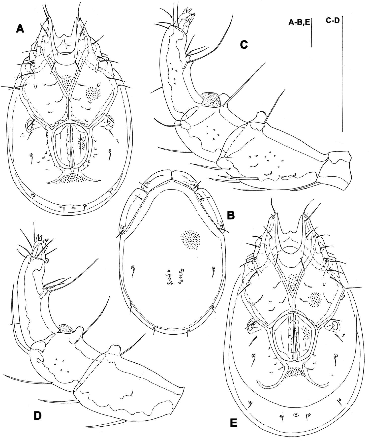
Figures 3–4.Pergalumna elongatiporosa sp. n.: 3 dorso-lateral view of prodorsum and anterior part of notogaster and pteromorph (gnathosoma and legs not illustrated) 4 posterior view of notogaster. Scale bars 100 μm.
-
Hao-Sen Li, Xiao-Feng Xue, Xiao-Yue Hong
Zookeys
Figure 36.Rhyncaphytoptus spinus sp. n.: A dorsal view of female B ventral view of female C prodorsal shield D coxae and female genitalia E dorsal view of female posterior part F ventral view of female posterior part G empodium H leg I and leg II I female internal genitalia.
-
Vladimir Pešić, Ksenia A. Semenchenko, Wonchoel Lee
Zookeys
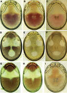
Figure 11.Photographs of dorsal shield: A–C Torrenticola recentis Tuzovskij, 2003 (A, B specimens from Dobong stream, Korea C specimen from River Kedrovaya, Russia): A male C–B female D–F Torrenticola ussuriensis (Sokolow, 1940), female (D specimen photographed immediately after dissection E–F specimens mounted in Hoyer’s medium): D–E specimen from Korea F specimen from Russia G–H Torrenticola turkestanica (Sokolow, 1926), specimens from River Inje, Korea: G male H female I Monatarctides abei sp. n., male holotype. Photos. V. Pešić (Figs A–B, D–E, G–I), K. Semenchenko (Figs C, F).
-
Michael J. Skvarla, J. Ray Fisher, Ashley P. G. Dowling
Zookeys
Figures 56–60.Cunaxa key illustrations. 56a, b Integumental striations 57a Chelicera with longitudinal striations present 57a Chelicera with longitudinal striations absent 58a–f Examples of variation in the hysterosomal median plate 59a Pedipalp telofemoral apophysis uncinated 59b Pedipalp telofemoral apophysis truncated 59c Pedipalp telofemoral apophysis short and cone-like 59d Pedipalp telofemoral apophysis short and finger-like 59e Pedipalp telofemoral femoral apophysis long 60a Pedipalp tibiotarsus with small teeth present 60b Pedipalp tibiotarsus with small teeth absent.
-
Ioana Cristina Constantinescu, Gabriel Chişamera, D. Khlur B. Mukhim, Costică Adam
Zookeys
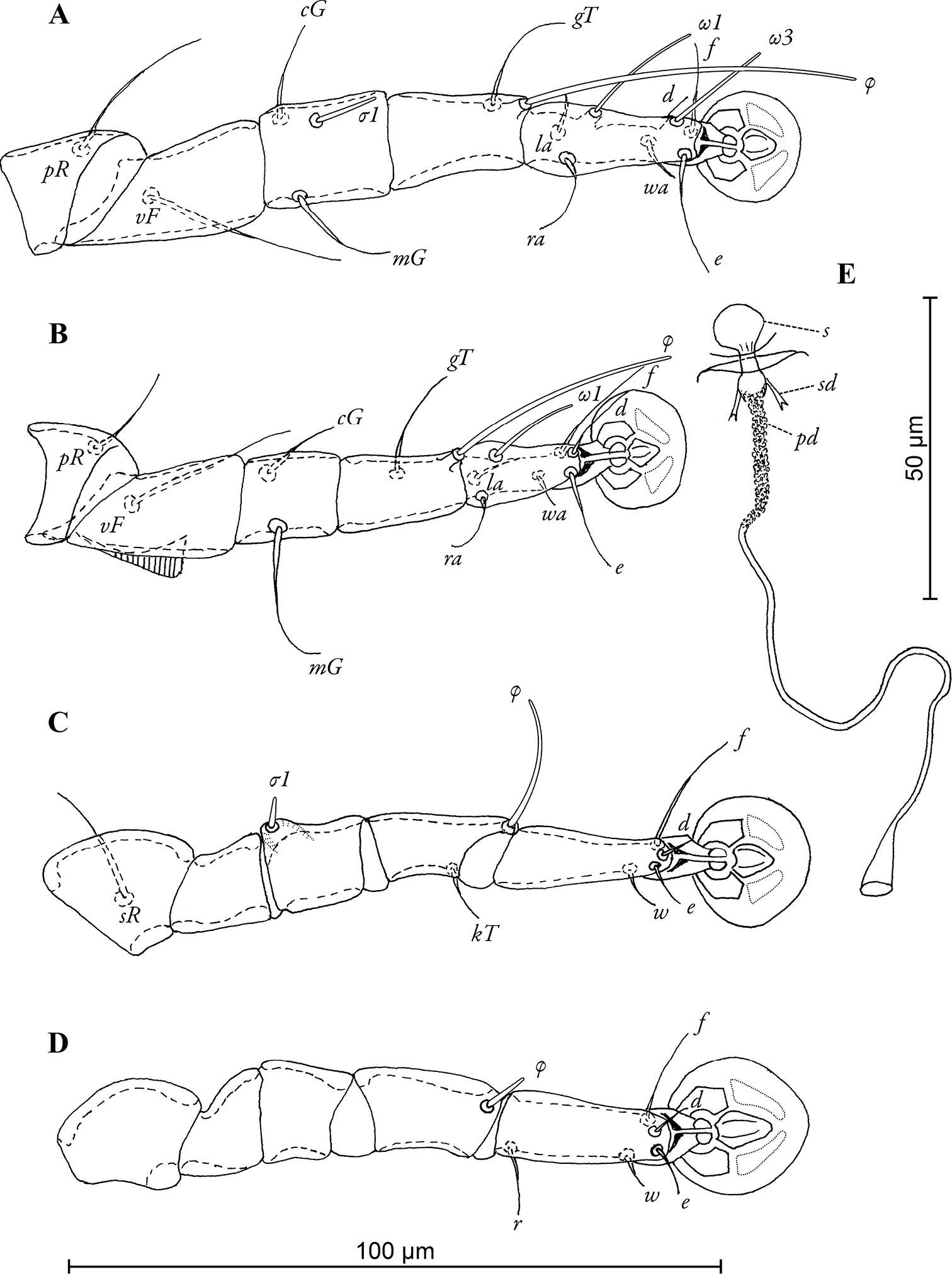
Figure 4.Pedanodectes angustilobus sp. n., details: A–D legs I–IV of female, respectively E spermatheca and spermaducts, dorsal view. Abbreviations: pd - primary spermaduct; s - spermatheca; sd - secondary spermaduct.
-
Sergey G. Ermilov, Olman Alvarado-Rodríguez, Axel P. Retana-Salazar
Zookeys
Figure 5.Pergalumna striatiprodorsum sp. n.: dorsal view. Scale bar 200 μm.
-
Hao-Sen Li, Xiao-Feng Xue, Xiao-Yue Hong
Zookeys
Figure 1.Acaphyllisa tuberculumae sp. n.: D dorsal view of female CG coxae and female genitalia CMG coxae and male genitalia.
-
Vladimir Pešić, Ksenia A. Semenchenko, Wonchoel Lee
Zookeys
Figure 10.Torrenticola turkestanica (Sokolow, 1926) (A–D male E female), Inje River, Korea: A, E ventral shield B dorsal shield C palp, lateral view D palp, medial view (P-1 missing). Scale bars = 100 μm.
-
Michael J. Skvarla, J. Ray Fisher, Ashley P. G. Dowling
Zookeys
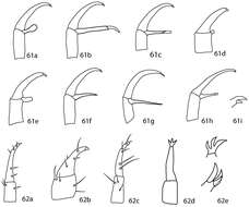
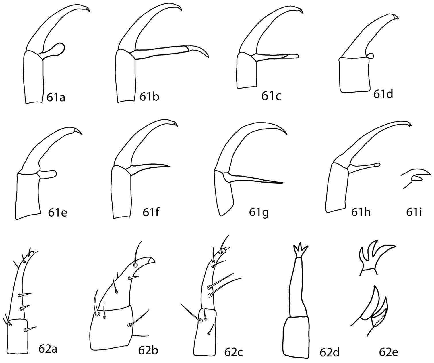
Figures 61–62.Dactyloscirus key illustrations. 61a–h Pedipalp genu and tibiotarsus with adjoining apophysis present 61i Close up of bifid claw 62a–d Pedipalp genu and tibiotarsus with adjoining apophysis absent 62e Close up of trifid claw.
-
Sergey G. Ermilov, Olman Alvarado-Rodríguez, Axel P. Retana-Salazar
Zookeys
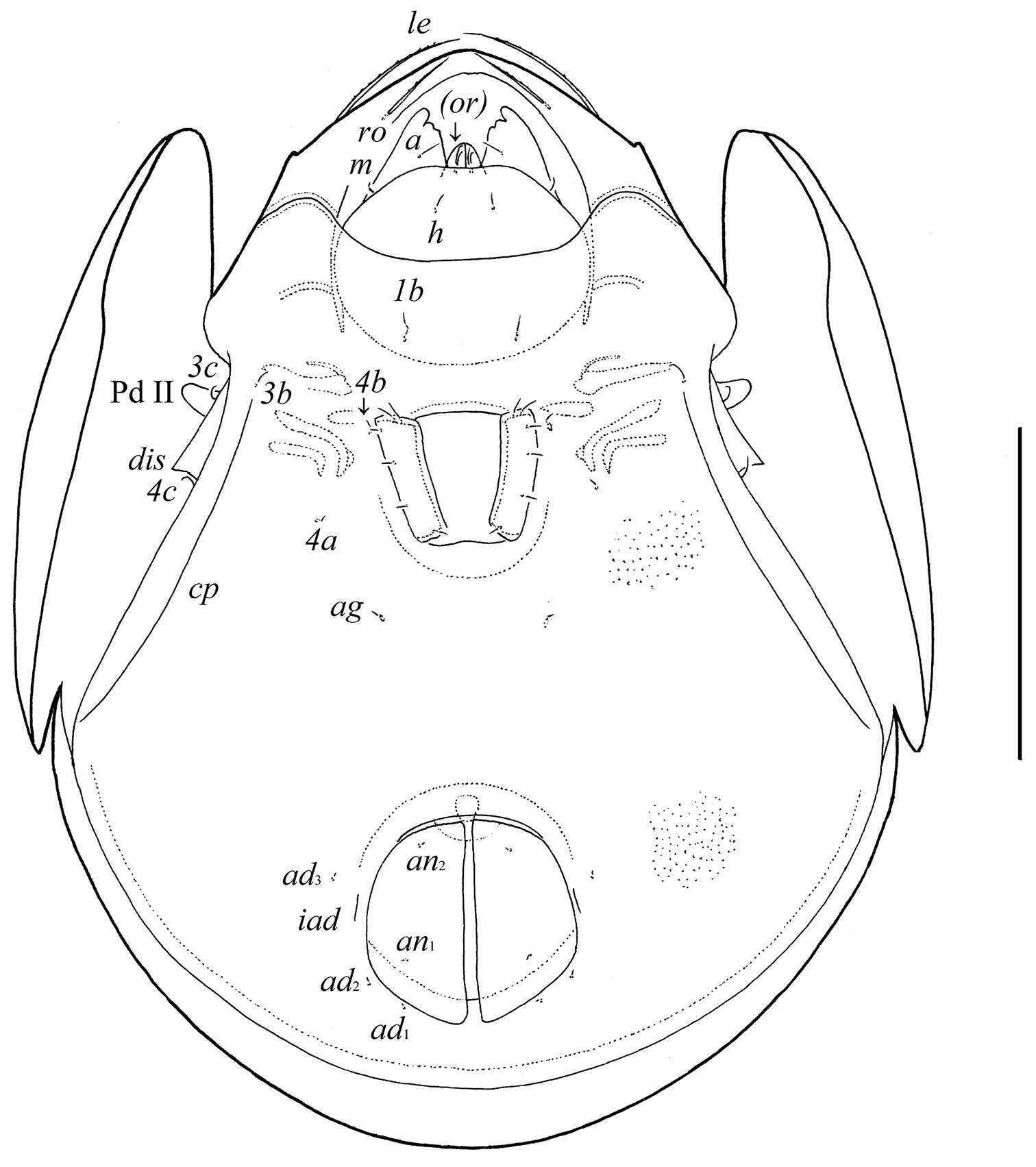
Figure 6.Pergalumna striatiprodorsum sp. n.: ventral view (legs not illustrated). Scale bar 200 μm.
-
Hao-Sen Li, Xiao-Feng Xue, Xiao-Yue Hong
Zookeys
Figure 2.Acaphyllisa tuberculumae sp. n.: V ventral view of female em empodium IG female internal genitalia L1 leg I L2 leg II.
-
Vladimir Pešić, Ksenia A. Semenchenko, Wonchoel Lee
Zookeys
Figure 11.Photographs of dorsal shield: A–C Torrenticola recentis Tuzovskij, 2003 (A, B specimens from Dobong stream, Korea C specimen from River Kedrovaya, Russia): A male C–B female D–F Torrenticola ussuriensis (Sokolow, 1940), female (D specimen photographed immediately after dissection E–F specimens mounted in Hoyer’s medium): D–E specimen from Korea F specimen from Russia G–H Torrenticola turkestanica (Sokolow, 1926), specimens from River Inje, Korea: G male H female I Monatarctides abei sp. n., male holotype. Photos. V. Pešić (Figs A–B, D–E, G–I), K. Semenchenko (Figs C, F).
-
Michael J. Skvarla, J. Ray Fisher, Ashley P. G. Dowling
Zookeys
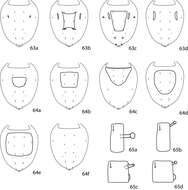
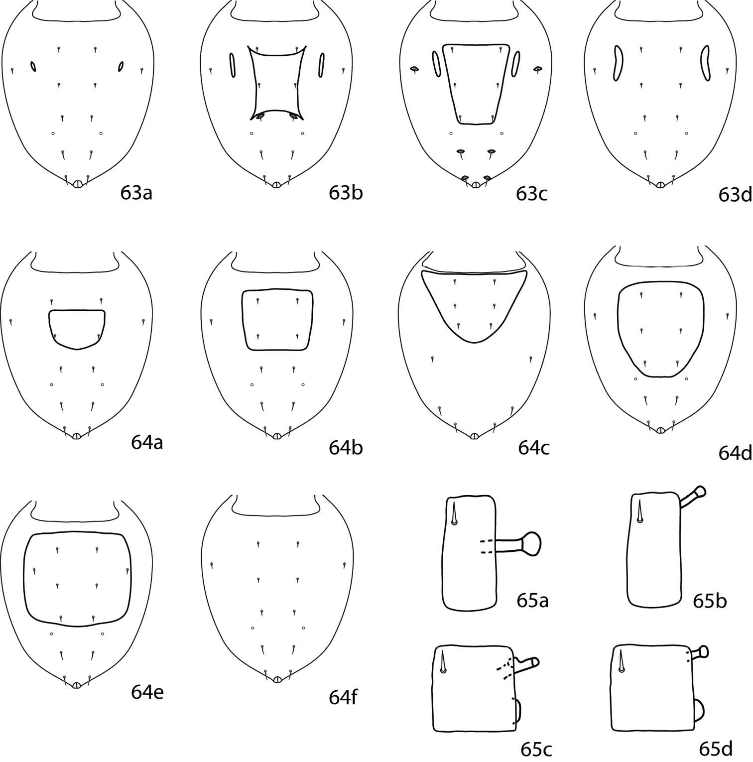
Figures 63–65.Dactyloscirus key illustrations. 63a–d Dorsal idiosoma, lateral hysterosomal platelets present 64a–f Dorsal idiosoma, lateral hysterosomal platelet absent 65a Pedipalp telofemur with one apophysis, which is about as long as the width of the telofemur 65b Pedipalp telofemur with one apophysis, which is shorter than the width of the telofemur 65c, d Pedipalp telofemur with two apophyses, one apical and one basal which is flattened and disc-shaped.
-
Sergey G. Ermilov, Olman Alvarado-Rodríguez, Axel P. Retana-Salazar
Zookeys
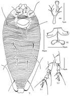
Figures 7–8.Pergalumna striatiprodorsum sp. n.: 7 dorso-lateral view of prodorsum and anterior part of notogaster and pteromorph (gnathosoma and legs not illustrated) 8 posterior view of notogaster. Scale bars 200 μm.
-
Hao-Sen Li, Xiao-Feng Xue, Xiao-Yue Hong
Zookeys
Figure 3.Acaphyllisa tuberculumae sp. n.: A dorsal view of female B ventral view of female C dorsal view of female posterior part D ventral view of female posterior part E prodorsal shield F coxae and female genitalia.
-
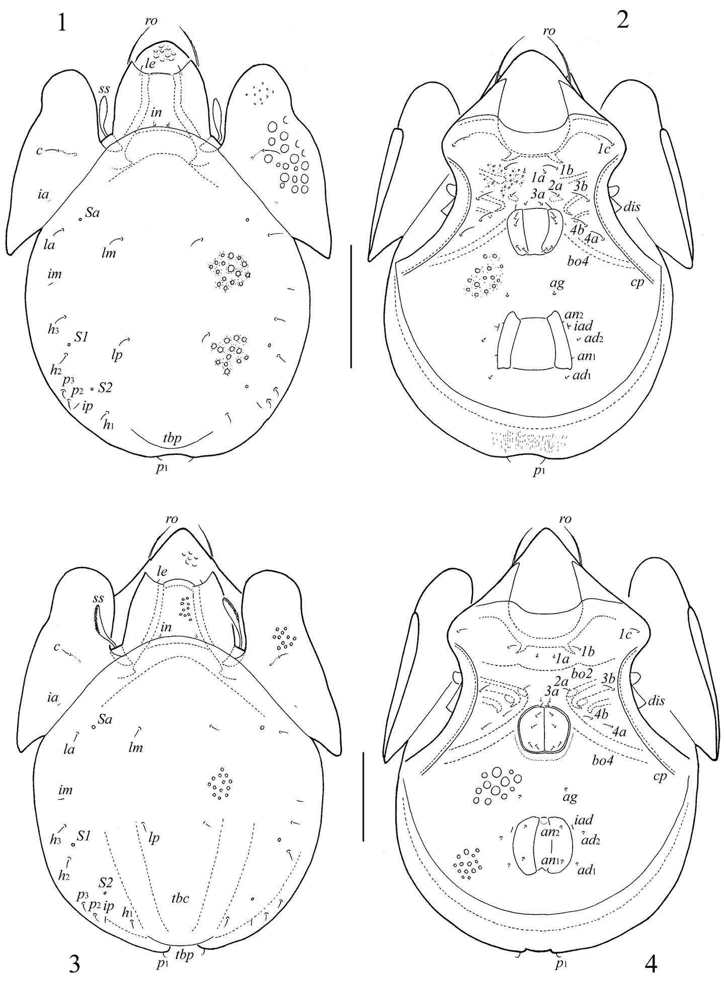
Sergey G. Ermilov, Dorothee Sandmann, Franca Marian, Mark Maraun
Zookeys
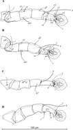
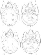
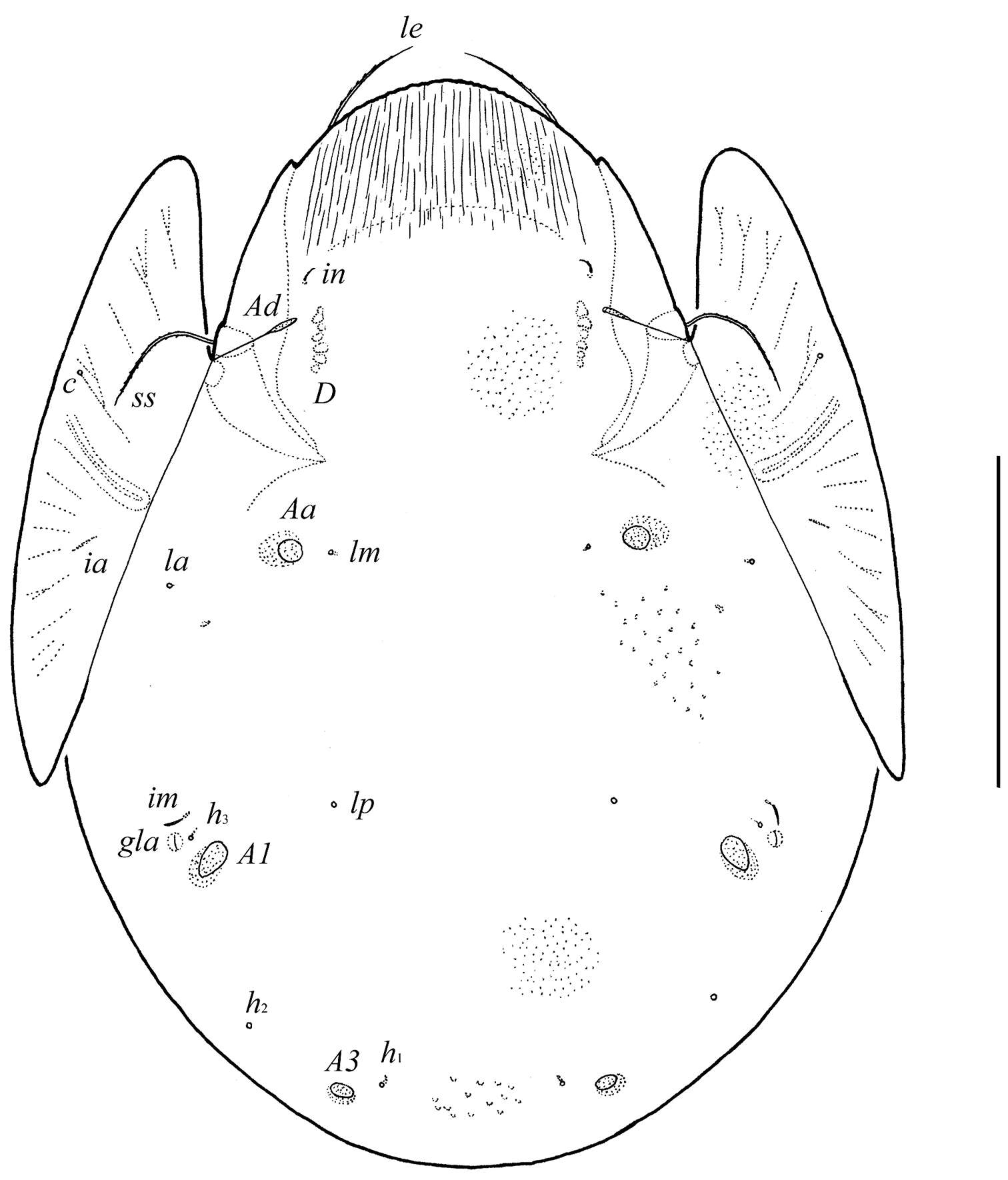
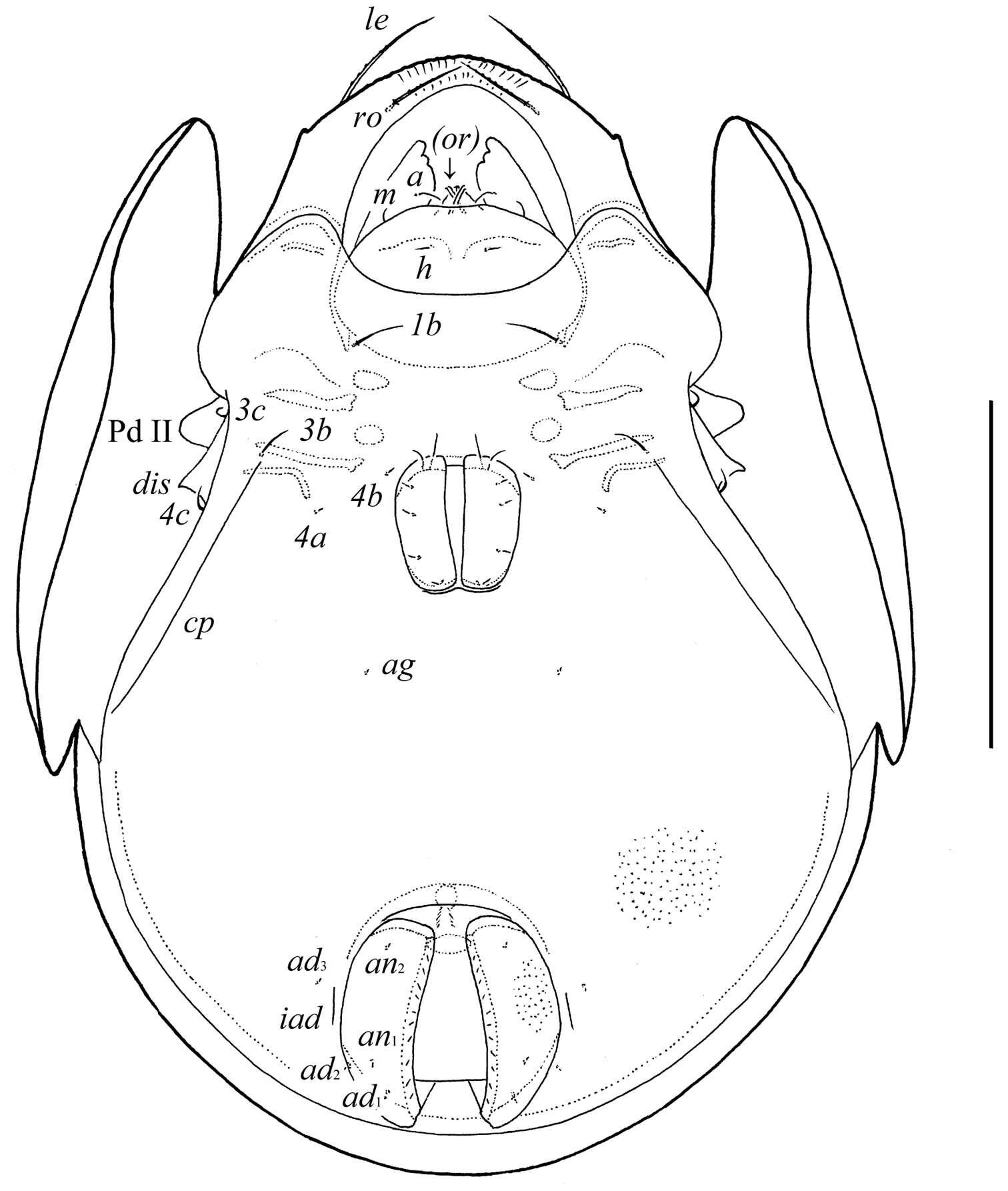
Figures 1–4.Truncozetes ecuadoriensis sp. n. (1, 2) and Truncozetes monodactylus sp. n. (3, 4), adults. 1, 3 body dorsally 2, 4 body ventrally (gnathosoma and legs not illustrated). Scale bars: (1, 2) 100 μm, (3, 4) 50 μm. Abbreviations in text.
-
Michael J. Skvarla, J. Ray Fisher, Ashley P. G. Dowling
Zookeys
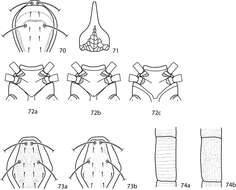
Figures 70–74.Coleoscirus key illustrations. 70 Dorsal idiosomal shield with horizontal reticulations present 71 Gnathosoma with extensive reticulations present 72a Sternal plate rounded posteriomedially, indentation absent 72b Sternal plate rounded posteriomedially, indentation present 72c Sternal plate truncated posteriomedially 73a Dorsal idiosomal shield even sclerotized, light reticulation present 73b Dorsal idiosomal shield unevenly sclerotized, light reticulation absent 74a Integumental dots on legs forming rows 74b Integumental dots on legs random.
-
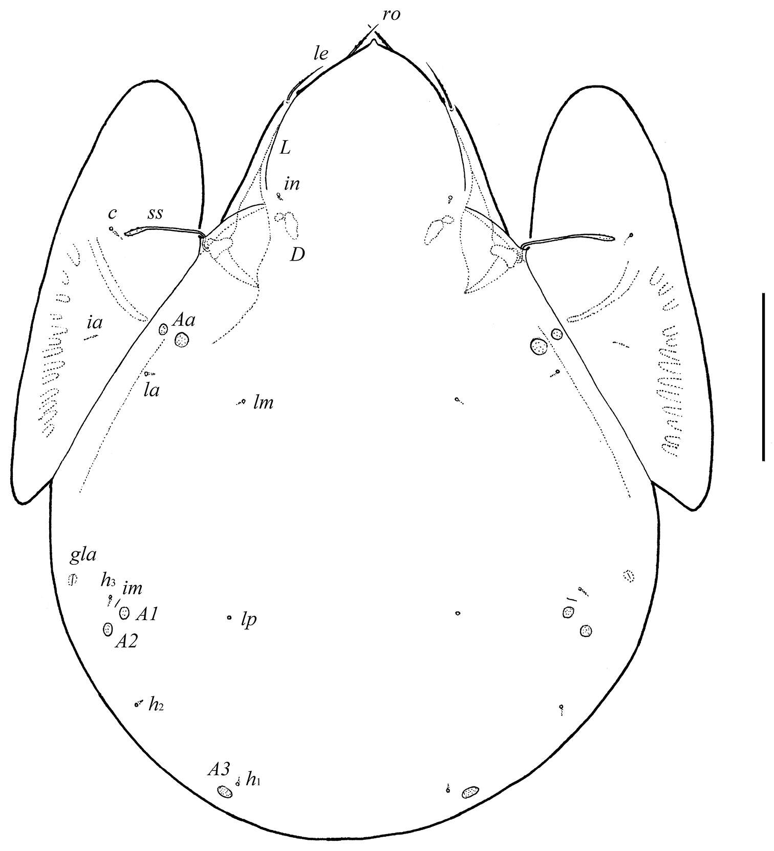
Sergey G. Ermilov, Jochen Martens, Andrei V. Tolstikov
Zookeys
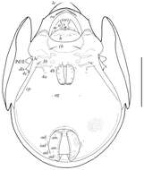
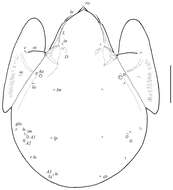
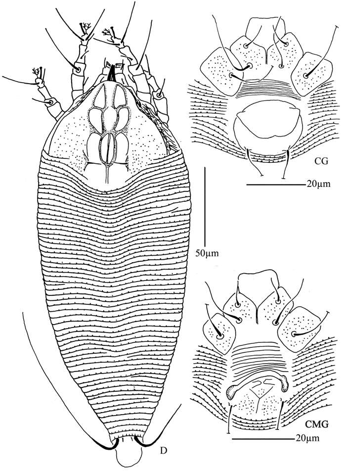
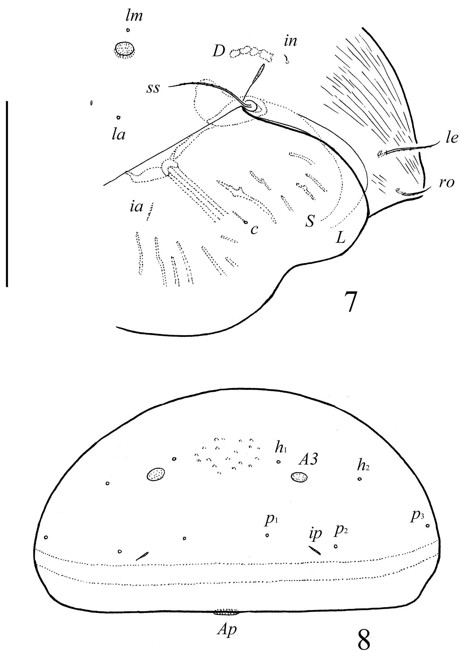
Figure 1.Galumna tetraporosa sp. n., adult: dorsal view. Scale bar 200 μm.