-
Nestor Fernandez, Pieter Theron, Christine Rollard, Elio Rodrigo Castillo
Zookeys
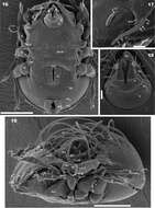
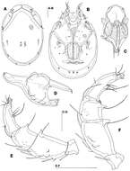
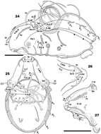
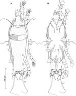
Figures 16–19.Malgasodes curvisetus Mahunka, 2000, adult. SEM observations. 16 ventral view 17 lamellae and lamellar tip 18 subcapitulum 19 lateral view, rotated. Abbreviations: see “Material and methods”. Scale bar: 16, 19 = 100 μm; 17, 18 = 10 μm.
-
Glenstrup Sø, Jylland, Danmark
-
Xiao-Feng Xue, Hussein Sadeghi, Xiao-Yue Hong, Samira Sinaie
Zookeys
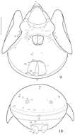
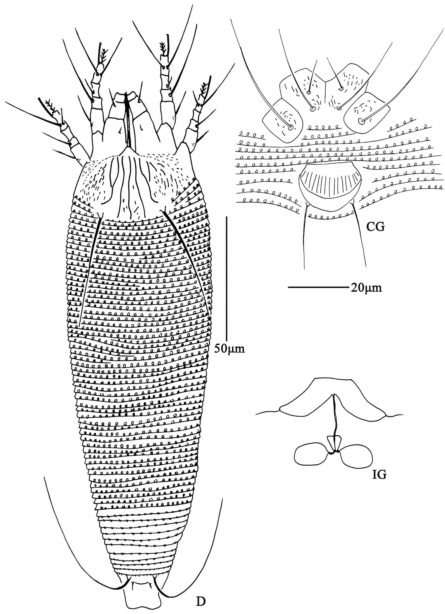
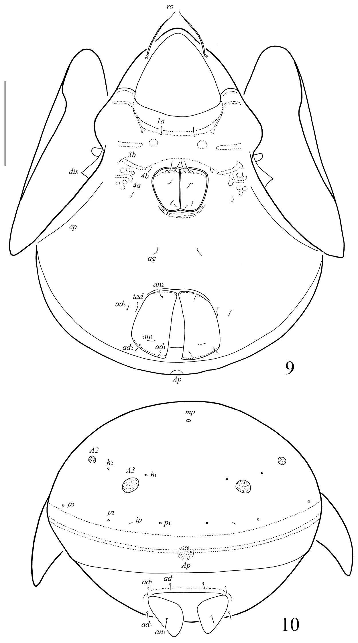
Figure 6. Aceria pulicaris sp. n. D dorsal view of female CG coxae and female genitalia IG female internal genitalia.
-
Lixia Xie, Yi Yan, Rong Huang, Maofa Yang
Zookeys
Figure 6. Damaeus (Paradamaeus) yushuensis sp. n. A trochanter, femur IV (100μm) B genu IV (100μm) C tibia, tarsus IV (100μm).
-
Hao-Sen Li, Xiao-Feng Xue, Xiao-Yue Hong
Zookeys
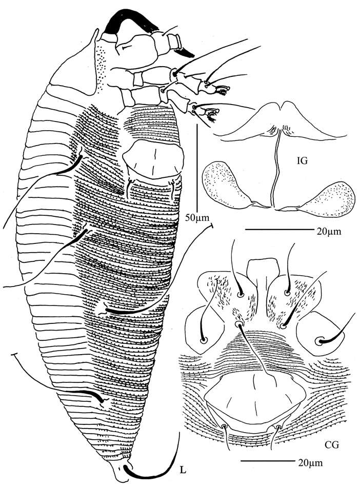
Figure 32.Diptacus mengdaensis sp. n.: L lateral view of female IG female internal genitalia CG coxae and female genitalia.
-
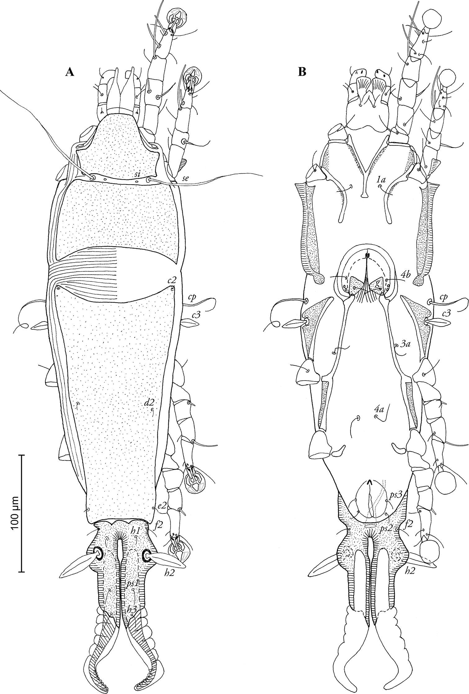
Vladimir Pešić, Ksenia A. Semenchenko, Wonchoel Lee
Zookeys
Figure 5.Torrenticola nipponica (Enami, 1940), male, Dobong stream, Korea: A dorsal shield B ventral shield C ejaculatory complex D gnathosoma E–F palp, medial view (E-smaller specimen, F-larger specimen). Scale bars = 100 μm.
-
Yunus Esen, Vladimir Pešić, Orhan Erman, Yücel Kaya
Zookeys
Figure 6.A–D Atractides (s. str.) nikooae Pešić, 2004, female: A Coxal and genital field B Palp, medial veiw C Vgl-1–2 D I–L-5–6 (Scale bars = 100 µm).
-
Sergey G. Ermilov, Alexander E. Anichkin
Zookeys
Figures 9–10.Galumna (Galumna) paracalcicola sp. n., adult: 9 ventral view (gnathosoma and legs not illustrated) 10 posterior view. Scale bar 100 μm.
-
Michael J. Skvarla, J. Ray Fisher, Ashley P. G. Dowling
Zookeys
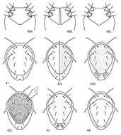
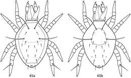
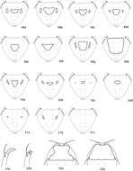
Figures 40–44.Scutopalus key illustrations. 40a Coxae I–II faintly divided 40b Coxae I–II totally divided 41 Coxae I–II fused medially 42 Dorsal shield with thick, rod-like setae present 43 Dorsal shield smooth or punctate 44a Dorsal shield rugose 44b Dorsal shield reticulate 44c Dorsal shield sparsely granulate 45a Setae mps, c1, c2, d1, e1, f1 clavate 45b Setae mps, c1, c2, d1, e1, f1 setiform 46 Setae lps, mps, c1, c2, d1, e1, f1 set on tubercles.
-
Sergey G. Ermilov, Umukusum Ya. Shtanchaeva, Luis S. Subías, Jochen Martens
Zookeys
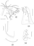
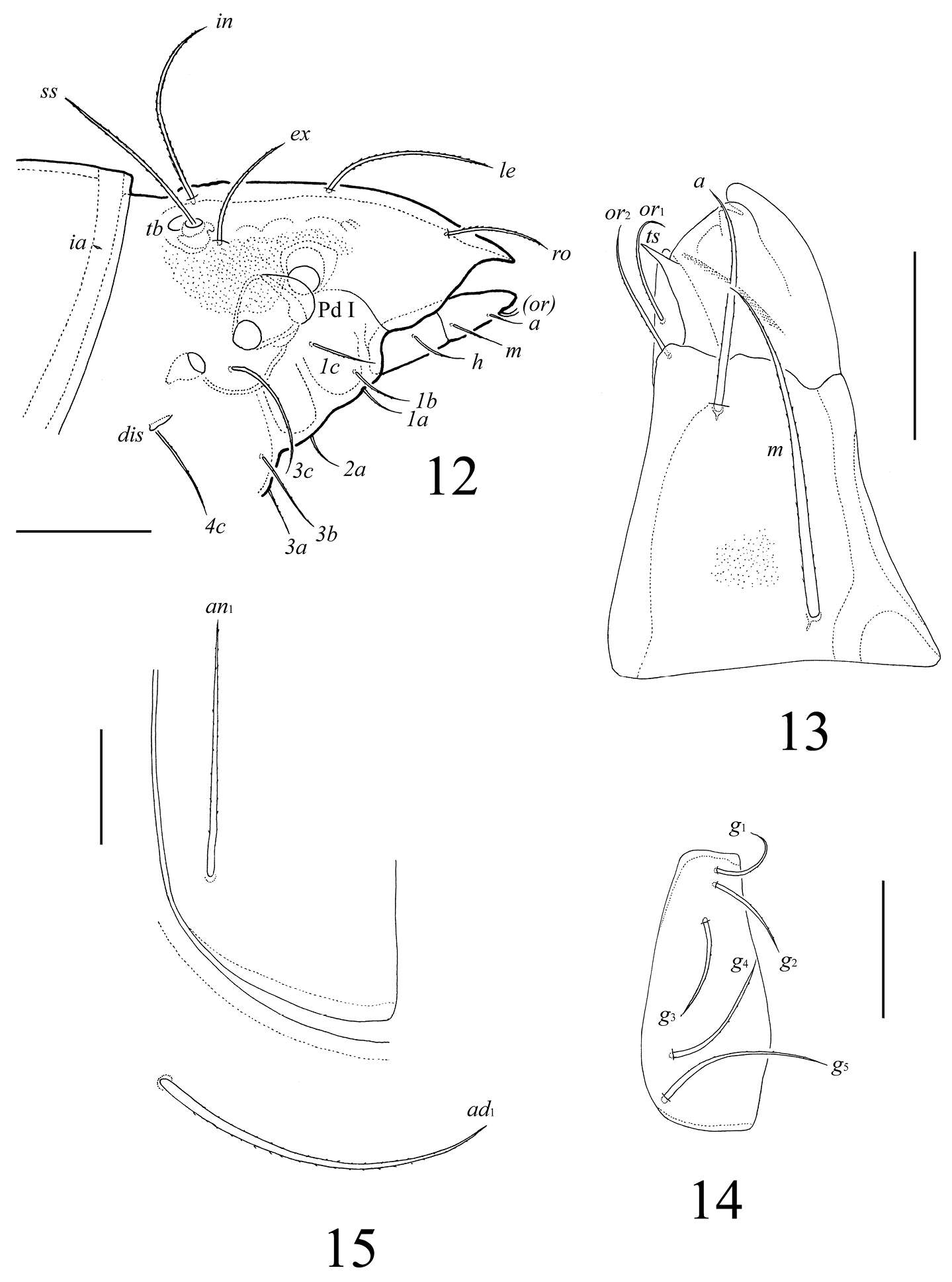
Figures 12–15.Lasiobelba (Antennoppia) nepalica sp. n.: 12 lateral view of prodorsum (legs not illustrated) and anterior part of notogaster 13 left rutellum and gena of subcapitulum, ventral view 14 genital plate, right 15 posterior part of anal plate with seta an1 and adanal seta ad1. Scale bar 50 μm.
-
Nestor Fernandez, Pieter Theron, Christine Rollard, Elio Rodrigo Castillo
Zookeys
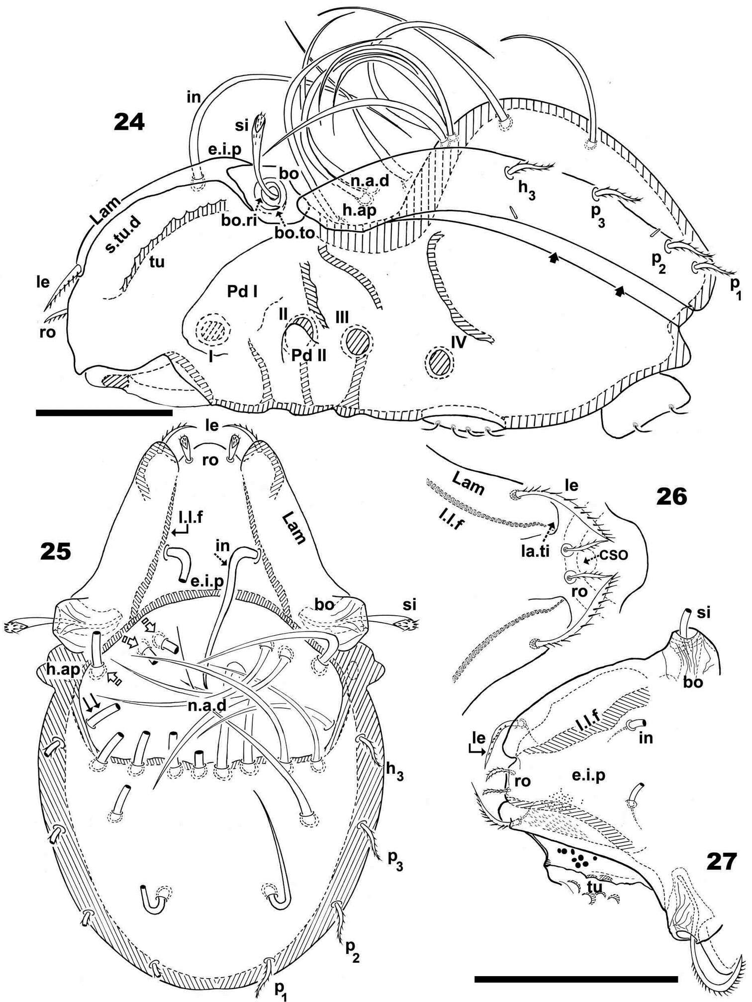
Figures 24–27.Malgasodes hungarorum Mahunka, 2000, adult. Optic observations. 24 lateral, slightly inclined view 25 dorsal view 26 prodorsum anterior part, dorsal view, inclined anteroposterior 27 prodorsum dorsal, inclined laterally. Abbreviations: see “Material and methods”. Scale bar: 24–25 = 60 μm; 26, 27 = 55 μm.
-
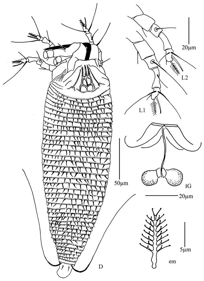
Xiao-Feng Xue, Hussein Sadeghi, Xiao-Yue Hong, Samira Sinaie
Zookeys
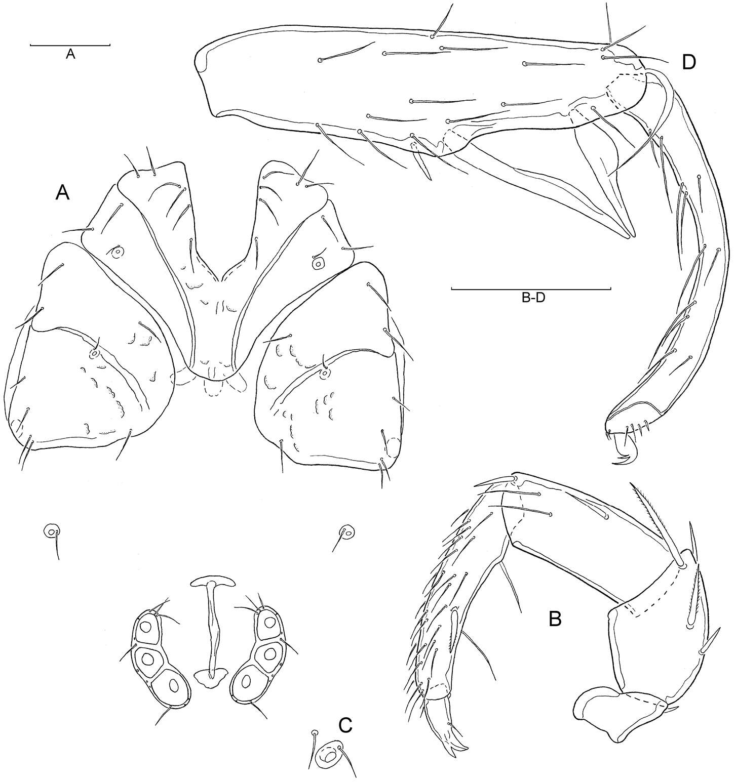
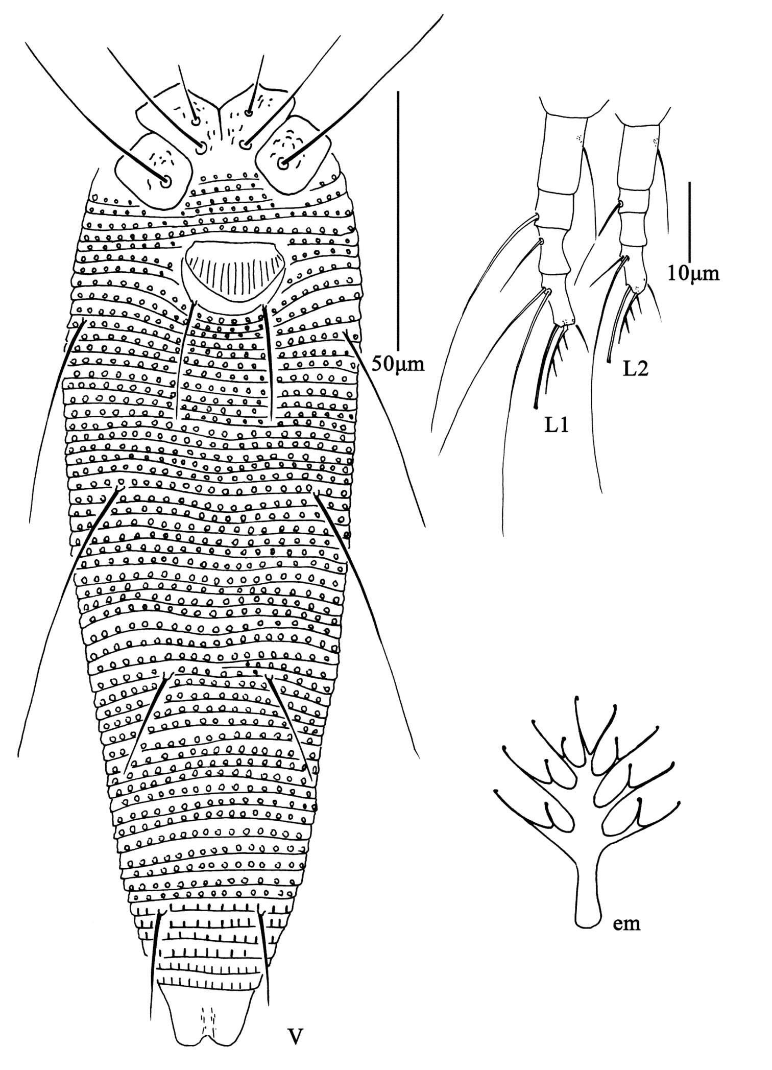
Figure 7. Aceria pulicaris sp. n. V ventral view of female em empodium L1 leg І L2 leg ІІ.
-
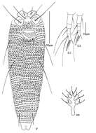
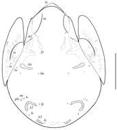
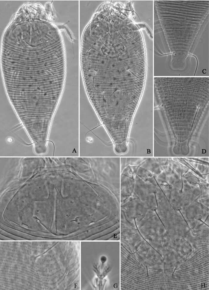
Figure 1.Conchogneta glabrisensillata sp. n. A Dorsal view of idiosoma B Ventral view of idiosoma C Prodorsum D Sensillus and bothridium, lateral view E Slight variation of sensillus, lateral view.
-
Hao-Sen Li, Xiao-Feng Xue, Xiao-Yue Hong
Zookeys
Figure 33.Diptacus mengdaensissp. n.: A dorsal view of female B ventral view of female C dorsal view of female posterior part D ventral view of female posterior part E prodorsal shield F lateral microtubercles G empodium H coxae and female genitalia.
-
Vladimir Pešić, Ksenia A. Semenchenko, Wonchoel Lee
Zookeys
Figure 6.Torrenticola nipponica (Enami, 1940), female, Tigrovaya River, Russia: A dorsal shield B ventral shield C palp, lateral view. Scale bars = 100 μm (A–B), 25 μm (C).
-
Michael J. Skvarla, J. Ray Fisher, Ashley P. G. Dowling
Zookeys
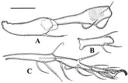
Figures 45.Scirula key illustrations. 45a Scirula impressa 45b Scirula papillata.
-
Ioana Cristina Constantinescu, Gabriel Chişamera, D. Khlur B. Mukhim, Costică Adam
Zookeys
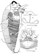
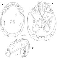
Figure 1.Pedanodectes angustilobus sp. n., male holotype: A dorsal view of idiosoma B ventral view of idiosoma.
-
Sergey G. Ermilov, Olman Alvarado-Rodríguez, Axel P. Retana-Salazar
Zookeys
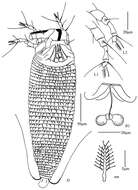
Figure 1.Pergalumna elongatiporosa sp. n.: dorsal view. Scale bar 100 μm.
-
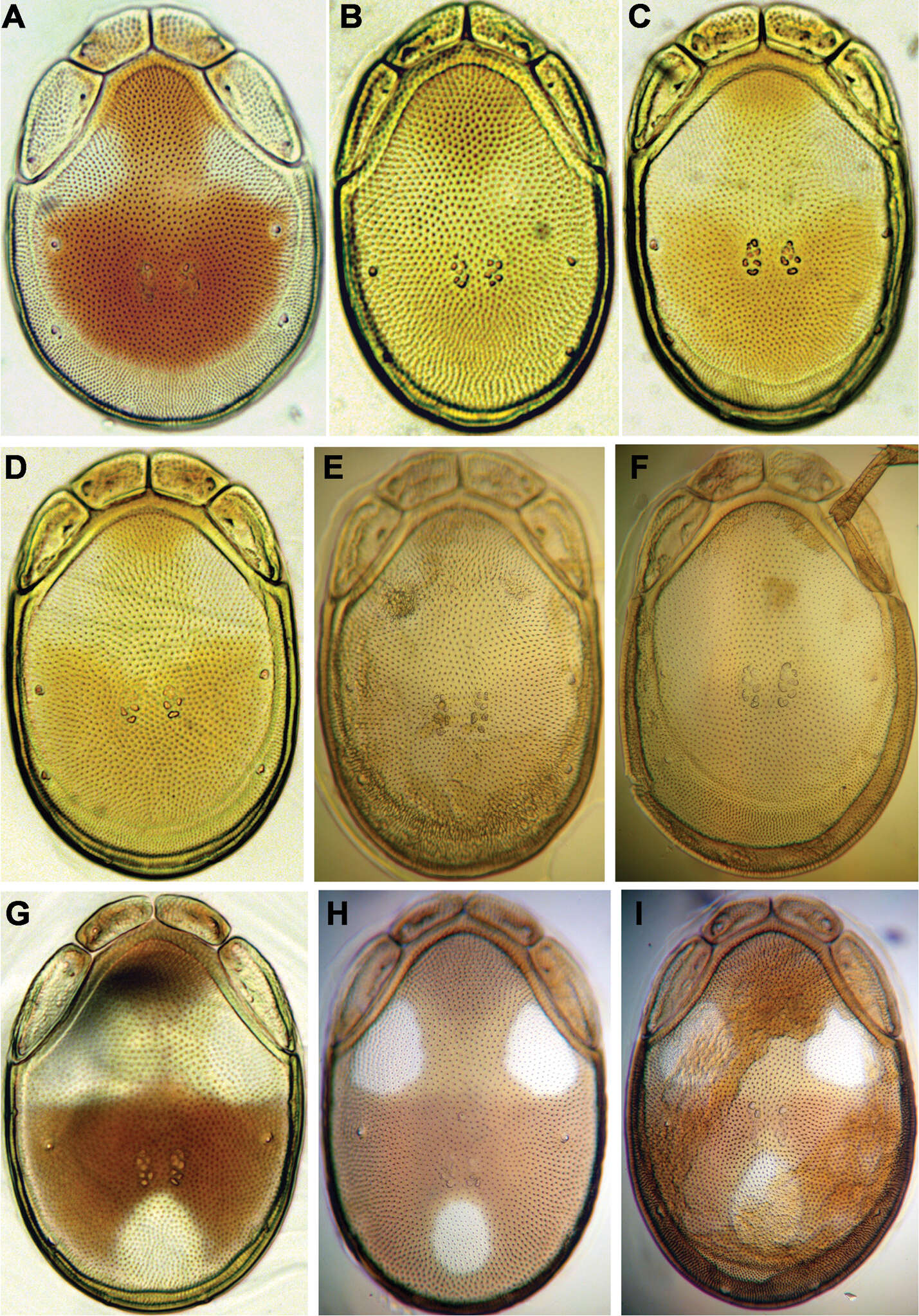
Xiao-Feng Xue, Hussein Sadeghi, Xiao-Yue Hong, Samira Sinaie
Zookeys
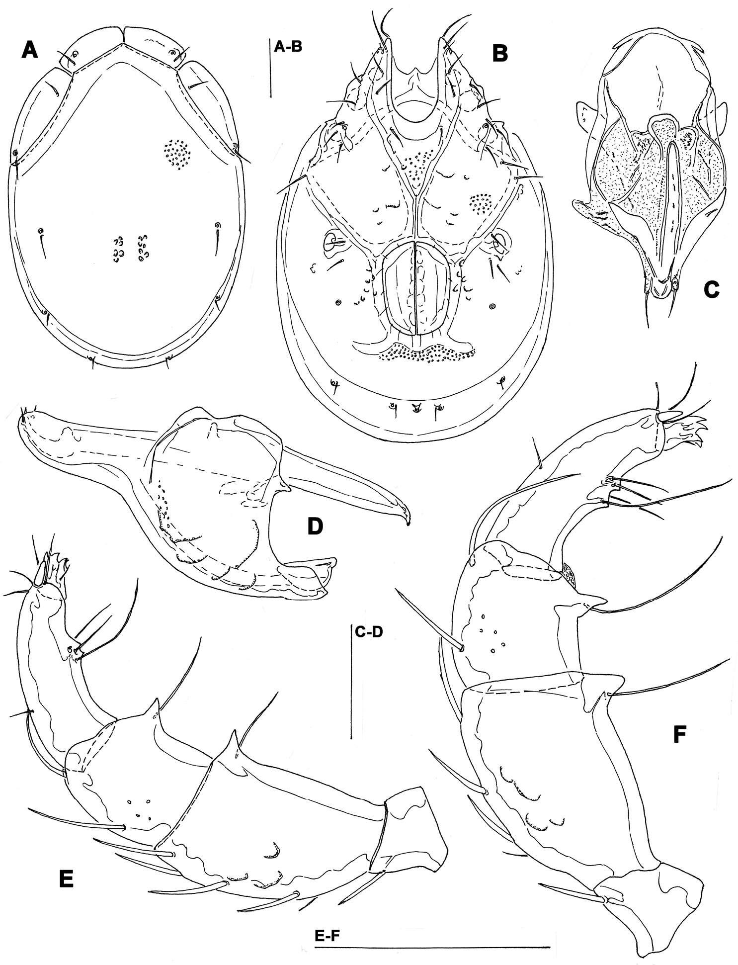
Figure 8. Aceria pulicaris sp. n. A dorsal view of female B ventral view of female C prodorsal shield D coxae and female genitalia.
-
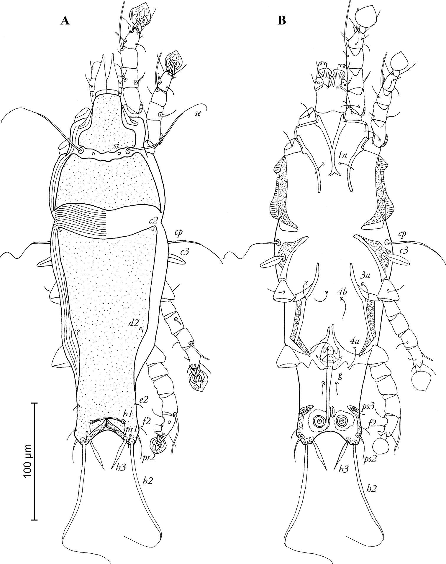
Figure 2.Conchogneta glabrisensillata sp. n. A Lateral view of prodorsum and anterior part of notogaster B Humeral region, showing tubercles Ha and Hp C Palp, right, antiaxial view D Leg I, right, antiaxial view E Genu and tibia of leg II, right, antiaxial view F Genu and tibia of leg III, right, antiaxial view G Leg IV, right, antiaxial view.
-
Hao-Sen Li, Xiao-Feng Xue, Xiao-Yue Hong
Zookeys
Figure 34.Rhyncaphytoptus spinus sp. n.: D dorsal view of female L1 leg I L2 leg II IG female internal genitalia em empodium.
-
Vladimir Pešić, Ksenia A. Semenchenko, Wonchoel Lee
Zookeys
Figure 7.Photographs of dorsal shield: A Torrenticola brevirostris (Halbert, 1911), female, stream in Naebyeansan NP, Korea B Torrenticola dentifera Wiles, 1991, male, stream in Naebyeansan NP, Korea: C–F Torrenticola kimichungi sp. n. (C–D specimens from stream in Seoraksan NP, Korea, E–F specimens from Tigrovaya River, Russia): C maleholotype D–E maleparatypes F femaleparatype G–I Torrenticola nipponica (Enami, 1940) (G specimen from Dobong stream, Korea H–I specimens from Tigrovaya River, Russia): G–H male I female. Photos. V. Pešić (Figs A–D, G), K. Semenchenko (Figs E–F, H–I).
-
Michael J. Skvarla, J. Ray Fisher, Ashley P. G. Dowling
Zookeys
Figures 49–53.Armascirus key illustrations. 49–51 Dorsal idiosoma 49a–e Hysterosomal shield complemented with setae 50a–d Hysterosomal shield small, not complemented with setae 51a–c Hysterosomal shield absent 52a, b Pedipalp tibiotarsal claw 52a Single claw 52b Bifid claw 53a Hysterosomal plate concave on lateral edges 53b Hysterosomal plate not concave on lateral edges.
-
Ioana Cristina Constantinescu, Gabriel Chişamera, D. Khlur B. Mukhim, Costică Adam
Zookeys
Figure 2.Pedanodectes angustilobus sp. n., female paratype: A dorsal view of idiosoma B ventral view of idiosoma.