-
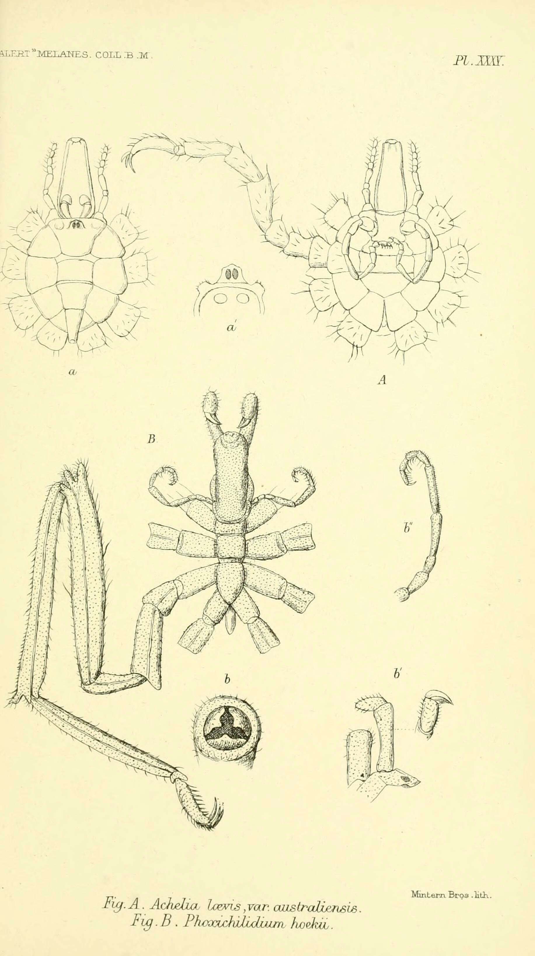
Report on the zoological collections made in the Indo-Pacific Ocean during the voyage of H.M.S. 'Alert' 1881-2.London :Printed by order of the Trustees,1884.
biodiversitylibrary.org/page/12067752
-
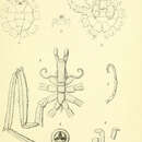
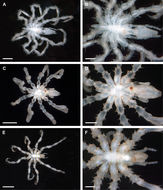
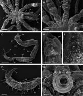
Figure 2.Ammotheidae 1; A, B: Achelia echinata, male, dorsal view; scales 500 µm and 250 µm, respectively; C, D: Achelia langi, male, dorsal view; scales 1 mm and 500 µm, respectively; E, F: Achelia vulgaris, male, dorsal view; scales 1 mm and 250 µm, respectively.
-
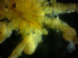
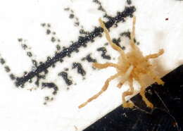
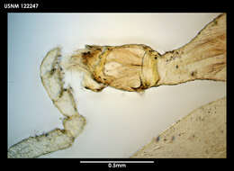
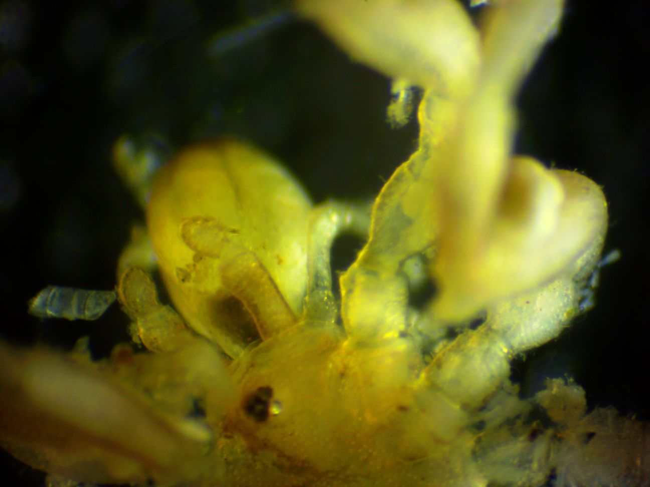
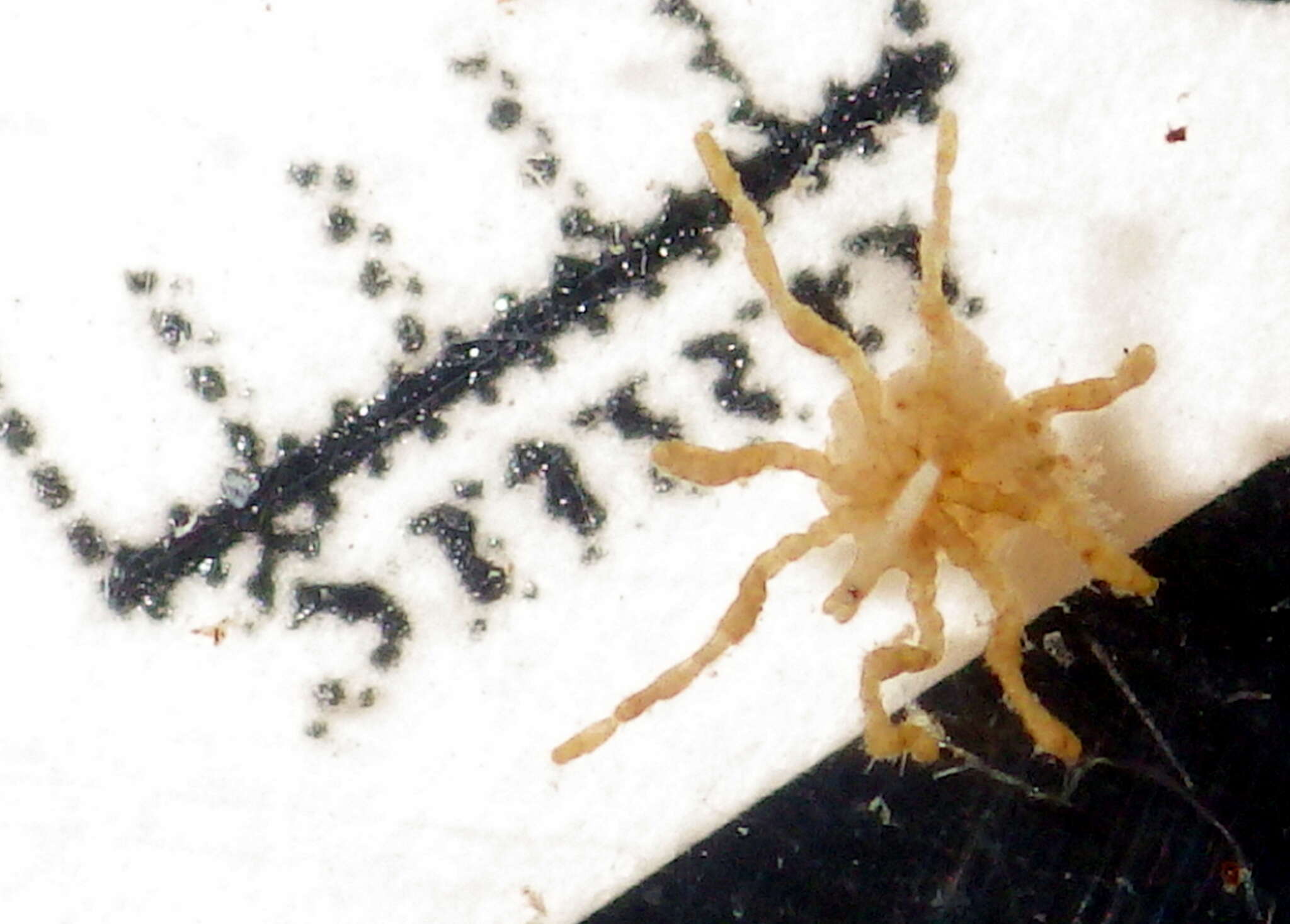
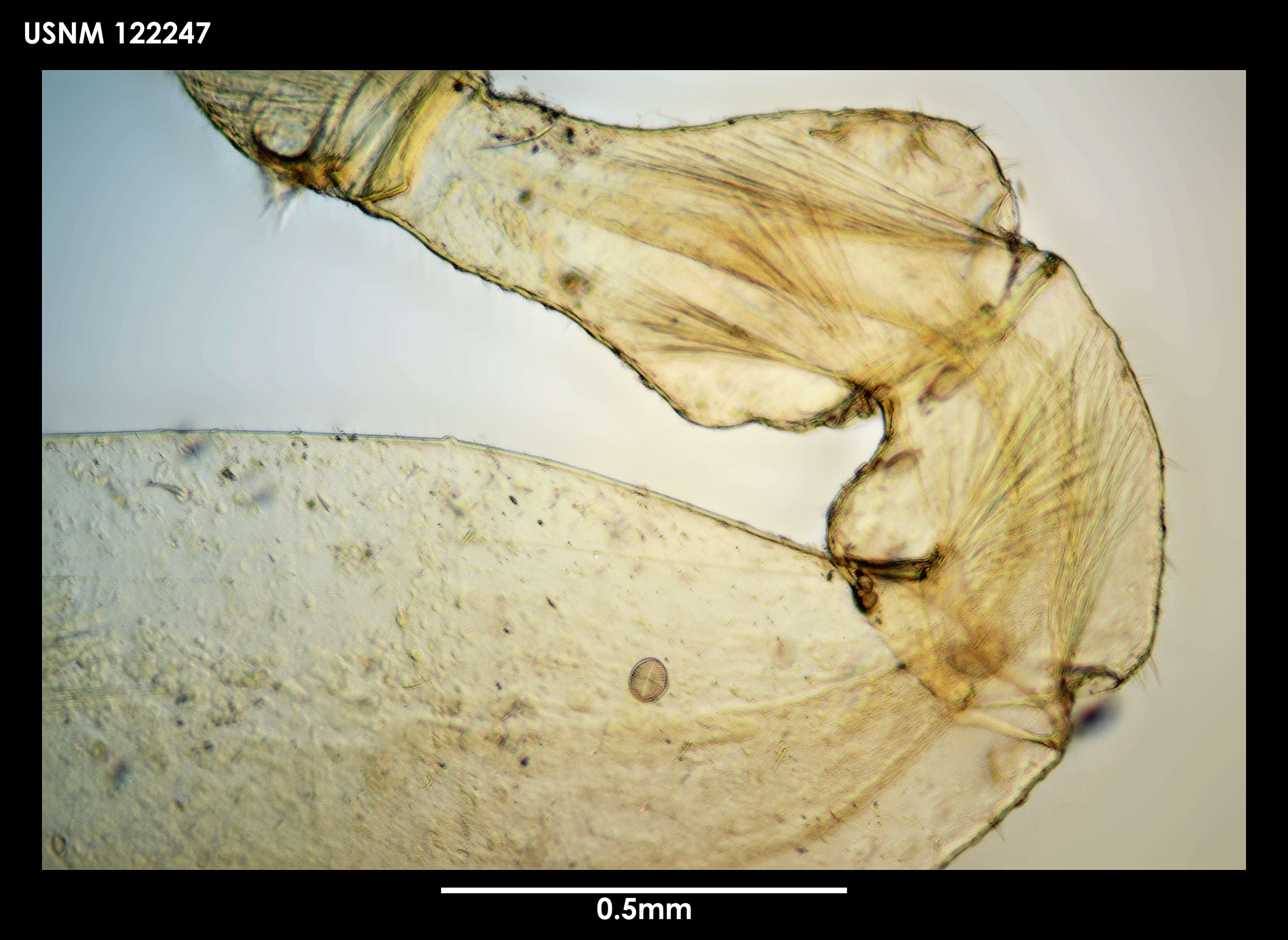
This closeup of the head shows the eye turret, the proboscis (top left), the chelicerae (chelifores) next to the proboscis which are two-segmented and end with a knob rather than a chela, the basal segments of the palps, and the coxae of the first several legs. Note that there is no finger-like projection on the dorsodistal part of the coxa of the first leg.
-
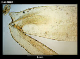
In this closeup of the posterior body, the stub-like abdomen projecting from the thorax can be seen. Several algal cells are adhered to the leg in the lower right.
-
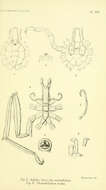
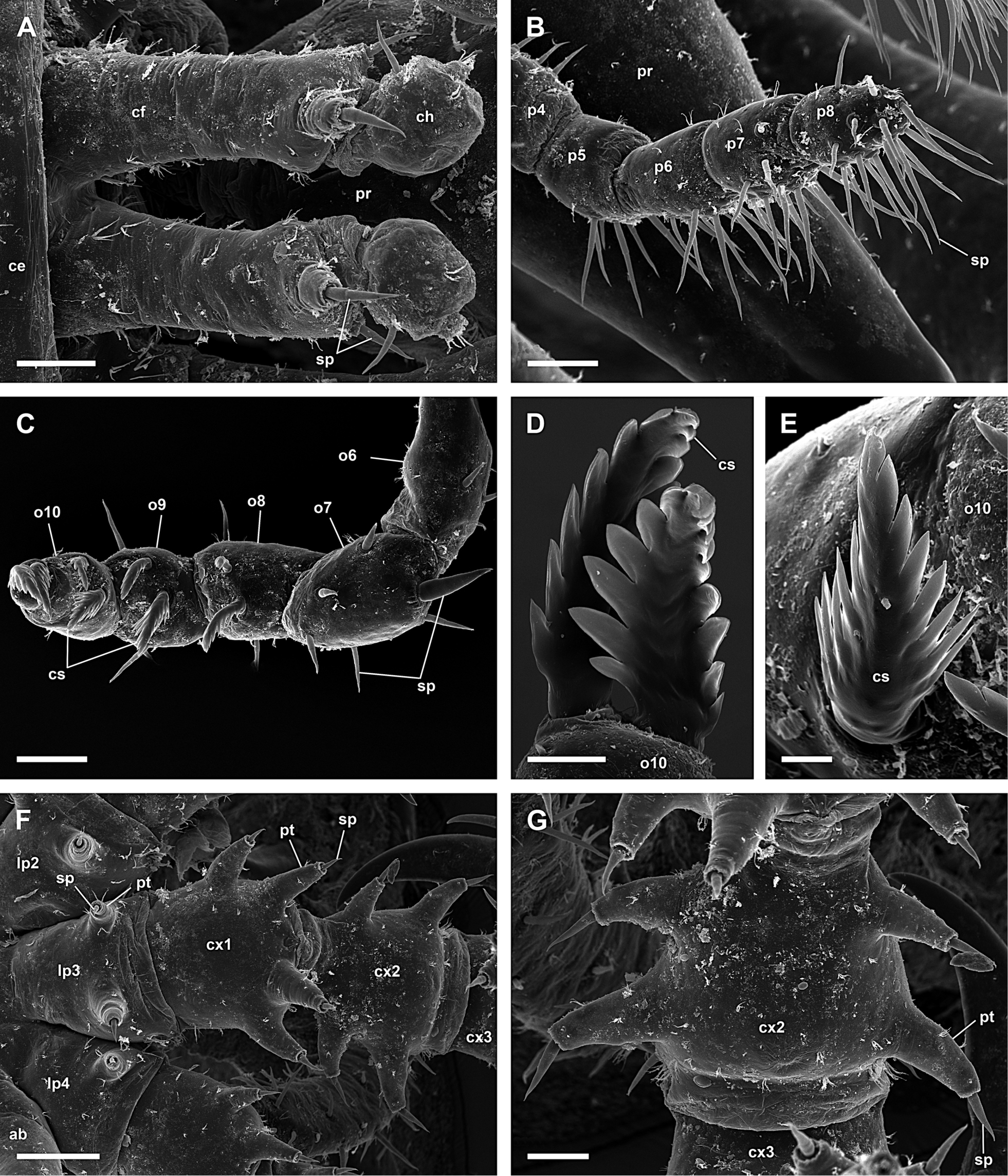
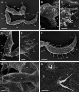
Figure 6.Achelia echinata, male; A: Chelifores with reduced chela; scale 40 µm; B: Distal articles of right 8-articled palp; scale 40 µm; C: Distal articles of 10-articled oviger; scale 40 µm; D, E: Compound spines on last oviger-article; scales 10 µm and 5 µm, respectively; F: Lateral process, coxa 1 and 2 of right 3rd leg, 2 protuberances with spine on each side of coxa 2; scale 100 µm; G: Coxa 2 with 2 protuberances with spine on each side (right 3rd leg); scale 40 µm.
-
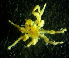
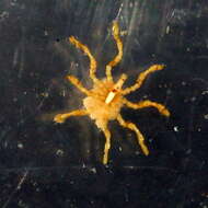
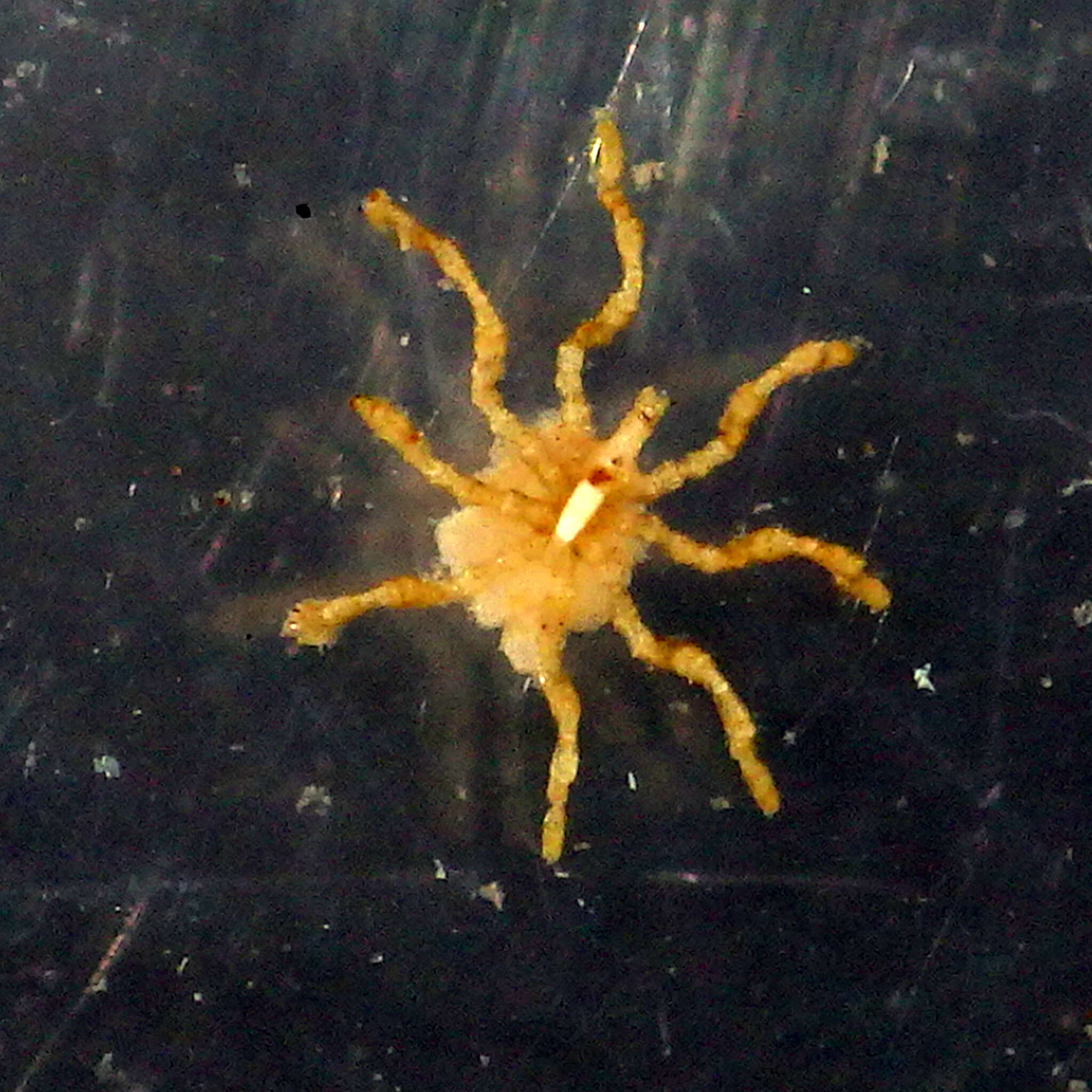
Achelia latifrons found on an Epiactis prolifera anemone at Cape Flattery. The distance from the tip of the proboscis (top left) to the tip of the abdomen (bottom right) is 3 mm. Total leg span is 11 mm. (Photo by: Dave Cowles, July 2014)
-
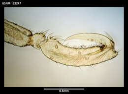
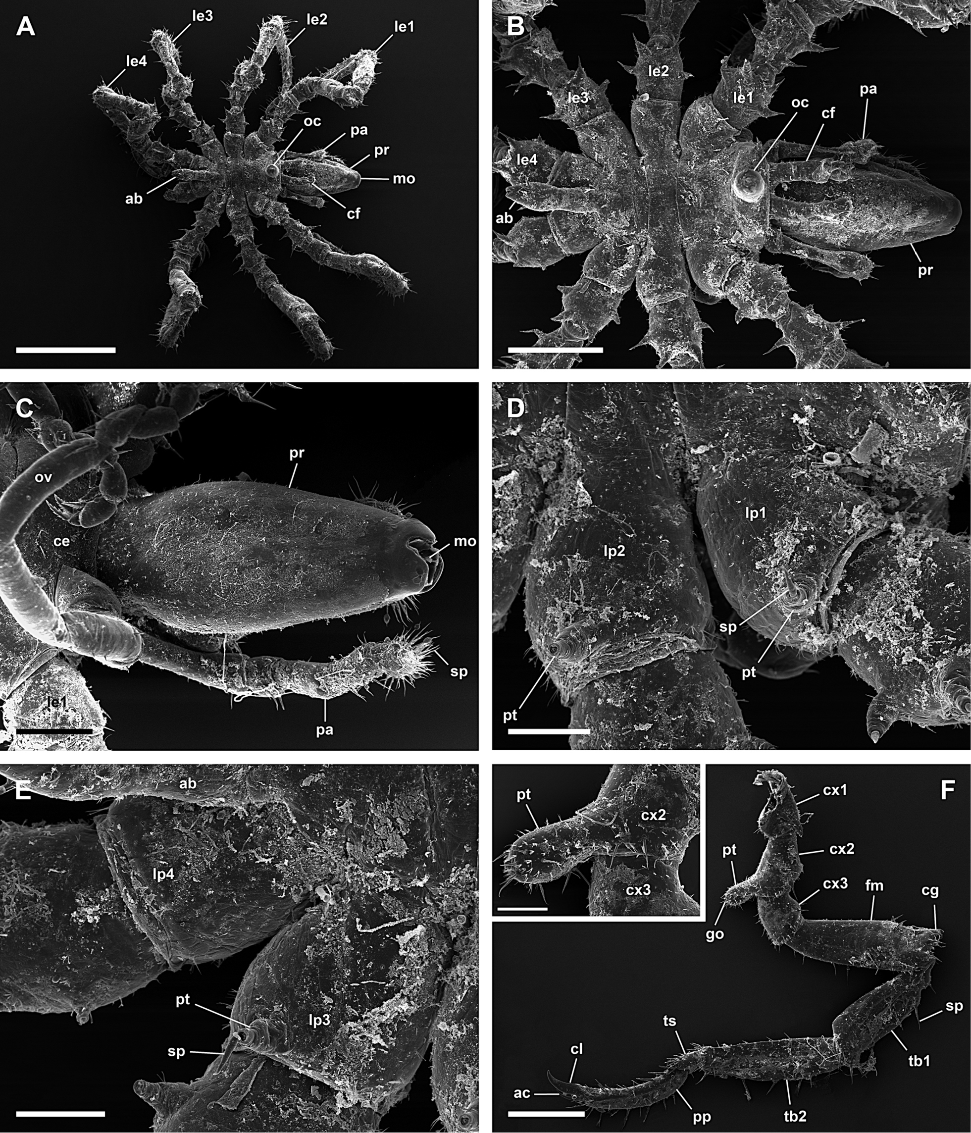
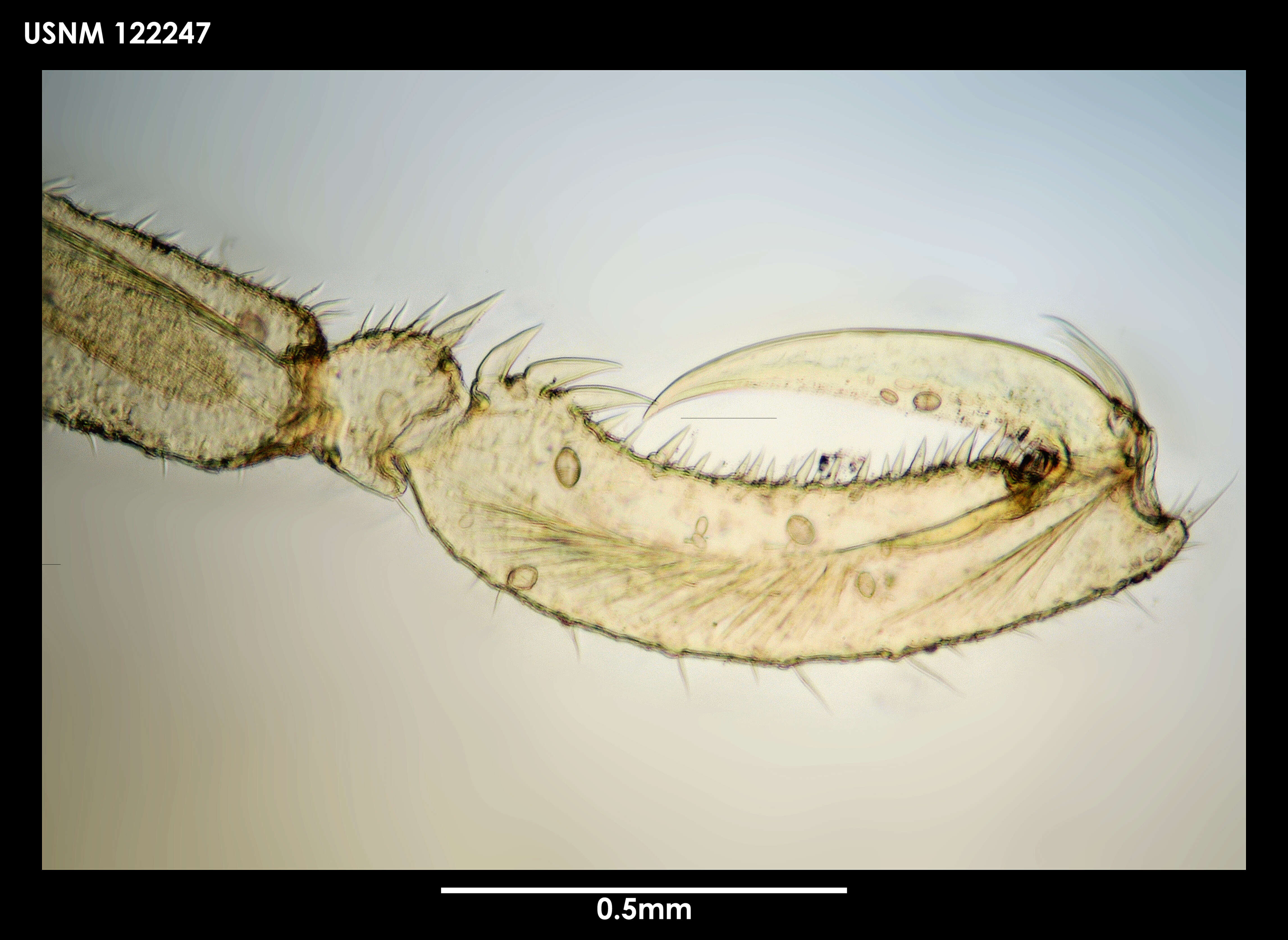
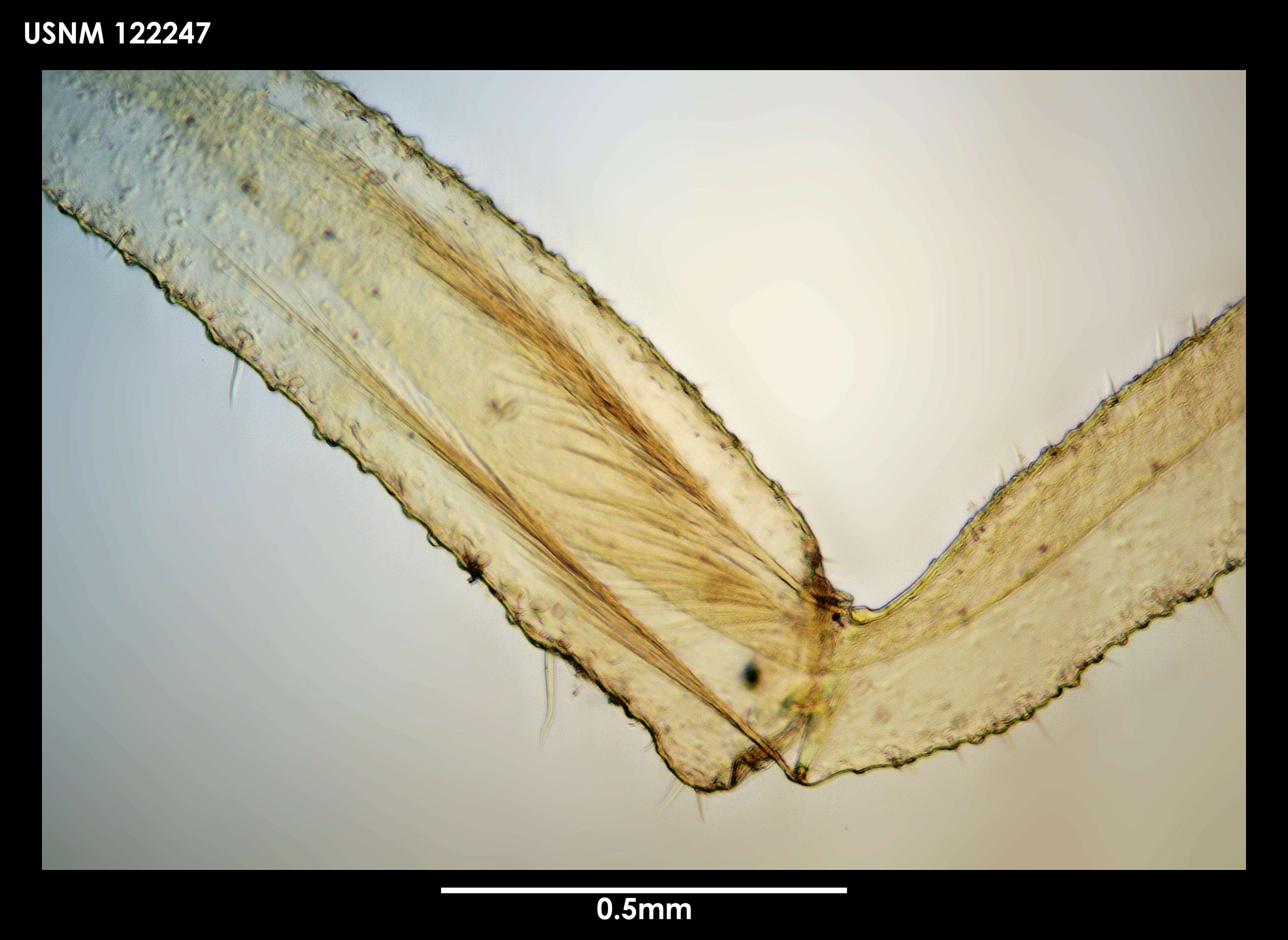
Figure 7.Achelia echinata, male; A: Left 3rd leg; scale 200 µm; B: Lateral view of coxa 2 with genital protuberance (right 4th leg); scale 40 µm; C: Genital opening (right 3rd leg); scale 20 µm; D: Lateral view of femur with cement gland on distal part (left 3rd leg); scale 100 µm; E: Cement gland (right 4th leg); scale 40 µm; F: Tarsus, propodus, and claw, auxiliary claws about half as long as claw (left 3rd leg); scale 100 µm; G: Abdomen; scale 40 µm; H: Hair and slit organ on dorsal side of trunk; scale 5 µm.
-
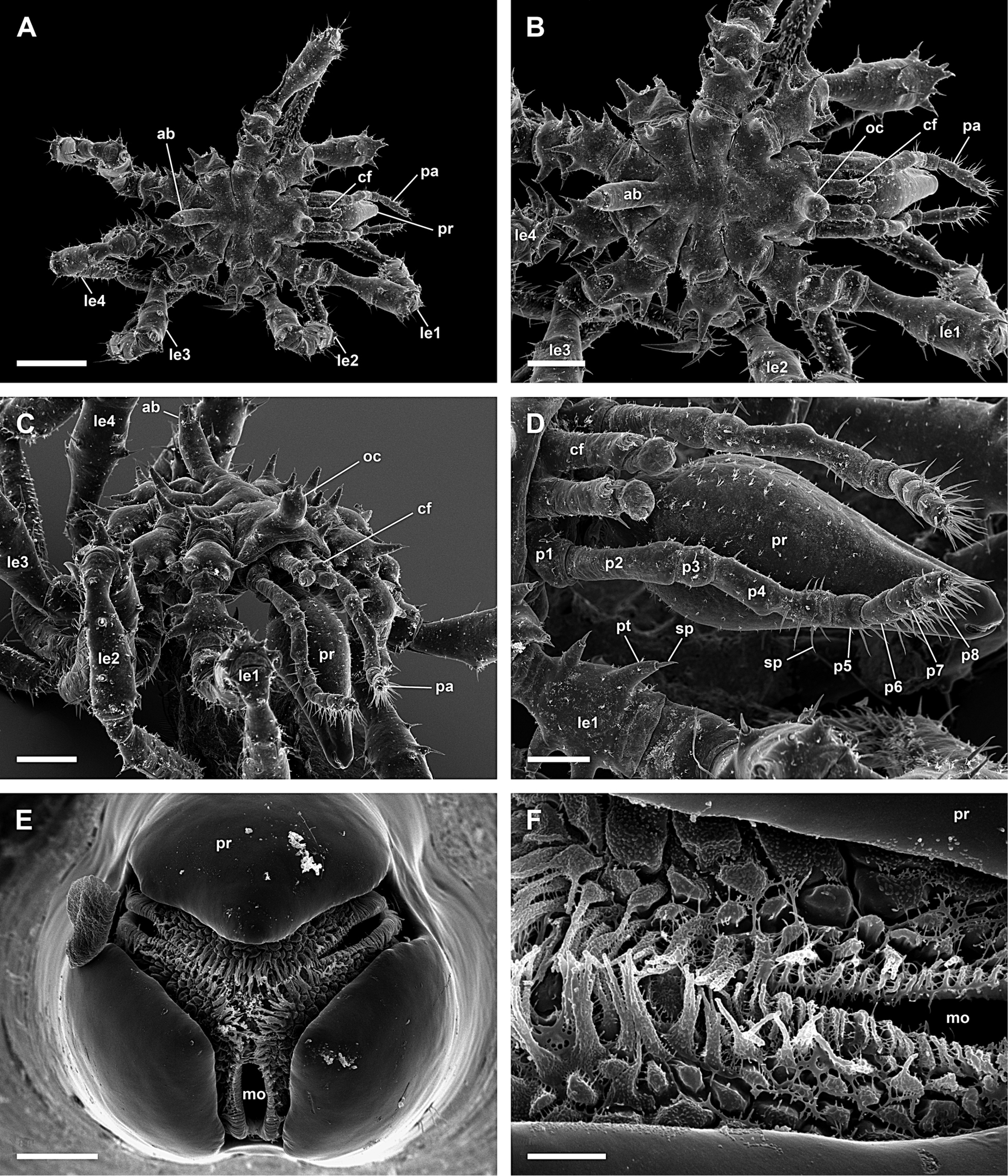
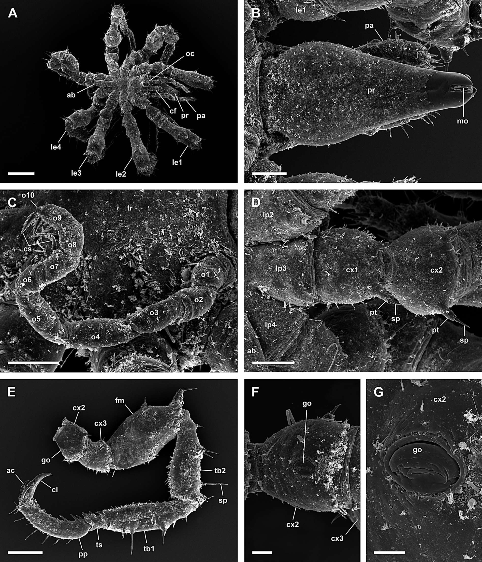
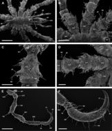
Figure 5.Achelia echinata, male; A: Dorsal view; scale 400 µm; B: Dorsal view of trunk, lateral processes touch each other; scale 200 µm; C: Frontal view of trunk, protuberances with spines on lateral process; scale 200 µm; D: Lateral view of proboscis, palp and chelifores with reduced chela; scale 100 µm; E: Mouth opening, dorsal is up; scale 20 µm; F: Mouth opening; scale 5 µm.
-
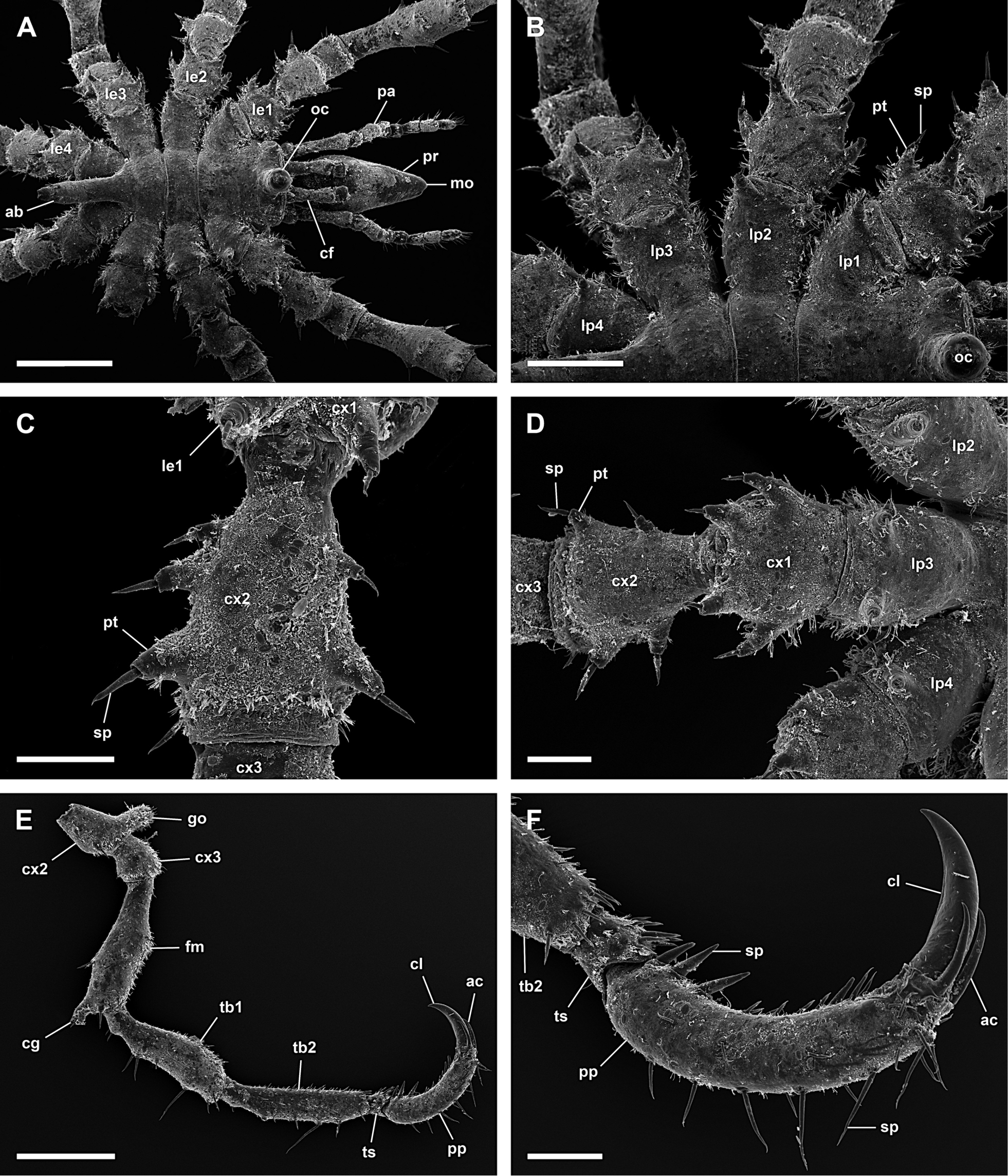
Figure 8.Achelia echinata, female; A: Dorsal view; scale 400 µm; B: Ventral view of proboscis; scale 100 µm; C: Left 10-articled oviger; scale 100 µm; D: Lateral process, coxa 1 and 2 of right 3rd leg, lateral processes touch each other; scale 100 µm; E: Left 3rd leg; scale 200 µm; F: Ventral view of coxa 2 with genital opening, distal is right (left 3rd leg); scale 40 µm; G: Genital opening; scale 20 µm.
-
Figure 2.Ammotheidae 1; A, B: Achelia echinata, male, dorsal view; scales 500 µm and 250 µm, respectively; C, D: Achelia langi, male, dorsal view; scales 1 mm and 500 µm, respectively; E, F: Achelia vulgaris, male, dorsal view; scales 1 mm and 250 µm, respectively.
-
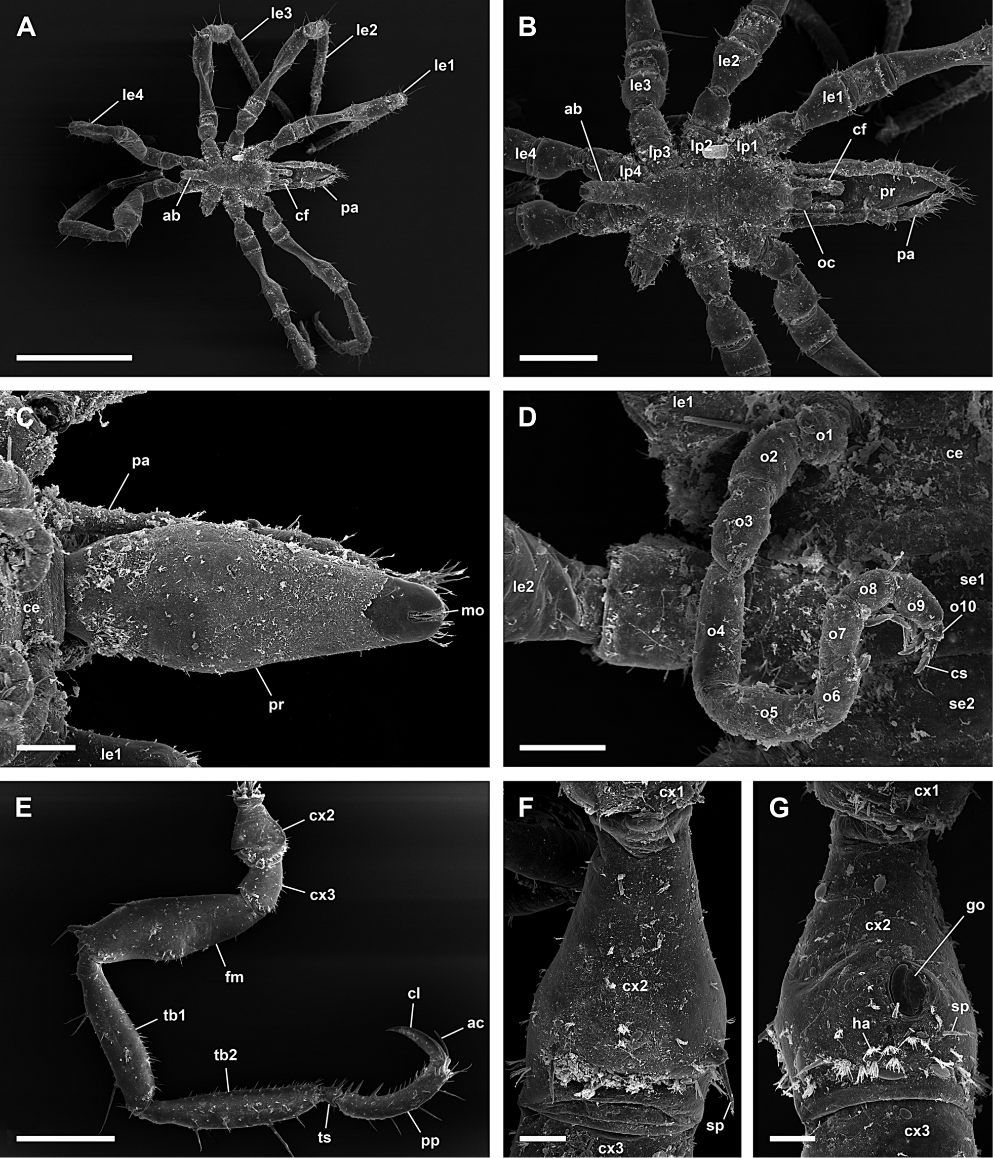
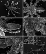
Figure 9.Achelia langi, male; A: Dorsal view; scale 1 mm; B: Dorsal view of trunk; scale 400 µm; C: Ventral view of proboscis; scale 200 µm; D: Dorsal view of right lateral processes 1 and 2, 2 protuberances with spine on lateral process 1 and 1 on lateral process 2; scale 100 µm; E: Dorsal view of right lateral processes 3 and 4, 1 protuberance with spine on lateral process 3 and lateral process 4 without protuberance or spine; scale 100 µm; F: Left 3rd leg; scale 400 µm; insert: Genital protuberance; scale 100 µm.
-
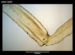
Figure 10.Achelia langi, female; A: Dorsal view of trunk; scale 400 µm; B: Dorsal view of left lateral processes, 1 protuberance with spine on lateral process 1-3, lateral process 4 without protuberance or spine; scale 200 µm; C: Right 3rd leg; scale 400 µm; D: Ventral view of coxa 2 with genital opening, distal is down (right 3rd leg); scale 100 µm; E: Genital opening; scale 20 µm; F: Tarsus, propodus, and claw, auxiliary claws about 2/3 of length of claw; scale 100 µm; G: Abdomen with anus; scale 40 µm.
-
Figure 2.Ammotheidae 1; A, B: Achelia echinata, male, dorsal view; scales 500 µm and 250 µm, respectively; C, D: Achelia langi, male, dorsal view; scales 1 mm and 500 µm, respectively; E, F: Achelia vulgaris, male, dorsal view; scales 1 mm and 250 µm, respectively.
-
Figure 11.Achelia vulgaris, male; A: Dorsal view of trunk; scale 400 µm; B: Dorsal view of left lateral processes, lateral processes do not touch each other; scale 200 µm; C: 2 protuberances with spine on right side and 3 on left side of coxa 2, distal is down (right 1st leg); scale 100 µm; D: Lateral process, coxa 1 and 2, 2 protuberances with spine on each side of coxa 2 (left 3rd leg); scale 100 µm; E: Right 3rd leg; scale 400 µm; F: Tarsus, propodus, and claw, auxiliary claws about half as long as claw; scale 100 µm.
-
Figure 12.Achelia vulgaris, female; A: Dorsal view; scale 1 mm; B: Dorsal view of trunk, lateral processes do not touch each other; scale 300 µm; C: Ventral view of proboscis; scale 100 µm; D: Right 10-articled oviger; scale 100 µm; E: Right 3rd leg; scale 400 µm; F: Dorsal view of coxa 2 (left 3rd leg); scale 40 µm; G: Ventral view of coxa 2 with genital opening (left 3rd leg); scale 40 µm.
-
-
-
-
-
-
-
-
-