-
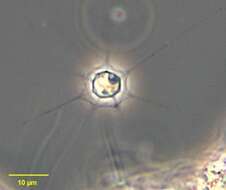
Clathrulina, one of the desmothoracid protists. Traditionally classified as the heliozoa because of the star-shaped appearance of the cell, but the heliozoa have always been a rag-bag of unrelated organisms. Desmothoracids may adopt one of several forms. A mature trophont (feeding form) is an amoeboid organism with stiffened arms. It lives in an spherical (ish) organic lorica which has large openings. The chamber is borned on a hollow stalk, and several stalked loricae may be joined end to end or in a branching arrangement. The stiffened pseudopodia extend out of the openings to capture food. Division leads to the formation of flagellated cells one or all progeny vacate the lorica, swim around and then settle as an amoeba. The amoeba secrete a stalk and mucus, and the mucus is shaped and accretes to form a lorica. The lorica strengthens and thickens with age. This image is of a cell in the lorica, pseudopodia can be seen. Differential interference contrast.Phase contrast micrograph.Phase contrast micrograph.
-
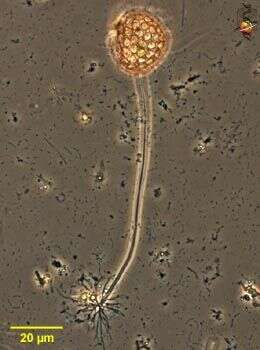
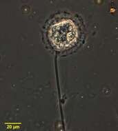
Clathrulina, one of the desmothoracid protists. Traditionally classified as the heliozoa because of the star-shaped appearance of the cell, but the heliozoa have always been a rag-bag of unrelated organisms. Desmothoracids may adopt one of several forms. A mature trophont (feeding form) is an amoeboid organism with stiffened arms. It lives in an spherical (ish) organic lorica which has large openings. The chamber is borned on a hollow stalk, and several stalked loricae may be joined end to end or in a branching arrangement. This image shows the rootlet-like secretrions which attach the stalk to the substrate. Phase contrast micrograph.
-
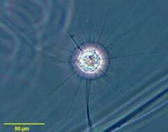
Clathrulina, one of the desmothoracid protists. Traditionally classified as the heliozoa because of the star-shaped appearance of the cell, but the heliozoa have always been a rag-bag of unrelated organisms. Desmothoracids may adopt one of several forms. A mature trophont (feeding form) is an amoeboid organism with stiffened arms. It lives in an spherical (ish) organic lorica which has large openings. The stiffened pseudopodia extend out of the openings to capture food. The central nucleus is also evident. Differential interference contrast.
-

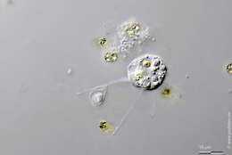
Clathrulina, one of the desmothoracid protists. Traditionally classified as the heliozoa because of the star-shaped appearance of the cell, but the heliozoa have always been a rag-bag of unrelated organisms. Desmothoracids may adopt one of several forms. A mature trophont (feeding form) is an amoeboid organism with stiffened arms. It lives in an spherical (ish) organic lorica which has large openings. The chamber is borned on a hollow stalk, and several stalked loricae may be joined end to end or in a branching arrangement. The stiffened pseudopodia extend out of the openings to capture food. Division leads to the formation of flagellated cells one or all progeny vacate the lorica, swim around and then settle as an amoeba. The amoeba secrete a stalk and mucus, and the mucus is shaped and accretes to form a lorica. The lorica strengthens and thickens with age. This image is of a young cell that is part way through the process of creating a lorica. Differential interference contrast.Phase contrast micrograph.
-
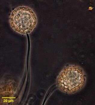
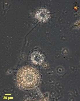
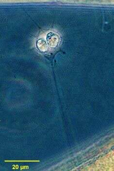
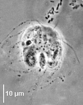
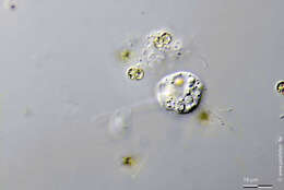
Clathrulina, one of the desmothoracid protists. Traditionally classified as the heliozoa because of the star-shaped appearance of the cell, but the heliozoa have always been a rag-bag of unrelated organisms. Desmothoracids may adopt one of several forms. A mature trophont (feeding form) is an amoeboid organism with stiffened arms. It lives in an spherical (ish) organic lorica which has large openings. The chamber is borned on a hollow stalk, and several stalked loricae may be joined end to end or in a branching arrangement. The stiffened pseudopodia extend out of the openings to capture food. Division leads to the formation of flagellated cells one or all progeny vacate the lorica, swim around and then settle as an amoeba. The amoeba secrete a stalk and mucus, and the mucus is shaped and accretes to form a lorica. The lorica strengthens and thickens with age. This image shows several cells at different stages of development. Differential interference contrast.
-
Clathrulina (cla-through-line-a), showing head region and included amoeboid cell. Differential interference contrast.
-
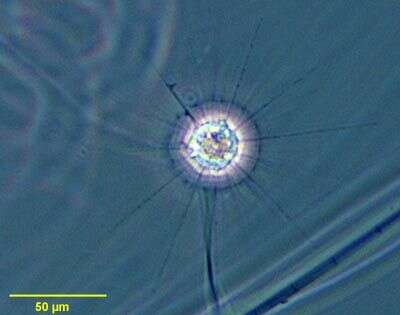
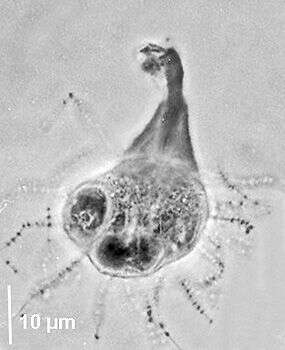
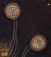
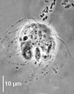
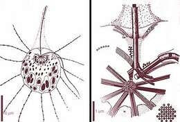
Hedriocystis undergoing division within faceted stalked capsule. Axopodia protrude from pores in capsule. Daughter cells become flagellated, escape the lorica then secrete their own stalk and lorica. Two of the five species in the genus have a spherical non-faceted lorica. Recently, a new genus, Penardiophrys has been erected for the faceted desmothoracid species such as this one. From freshwater pond near Boise, Idaho. Phase contrast. See Mikrjukov,K. Taxonomy and Phylogeny of Heliozoa I. Acta Protozool 39:81-97, 2000.
-
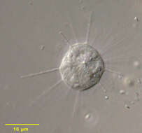
Hedriocystis - portrait of a species with smooth non-faceted spherical lorica. Phase contrast. From a freshwater pond near Boise, Idaho.
-
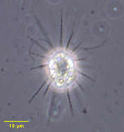
Hedriocystis species within faceted stalked siliceous capsule. Axopodia protrude from pores in capsule. Two of the five species in the genus have a spherical non-faceted lorica. Recently a new genus, Penardiophrys has been erected for the faceted desmothoracid species such as this one. From freshwater pond near Boise, Idaho. Phase contrast. See Mikrjukov,K. Taxonomy and Phylogeny of Heliozoa I. Acta Protozool 39:81-97, 2000.
-
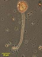
Hedriocystis, a small desmothoracid protist. The cell lives as an amoeba with stiffened arms (axopodia) within a loose lorica that has large holes or perforations. The lorica is stalked. When the cell divides, it may produce flagellated cells which leave the lorica and set up home elsewhere. From Lake Donghu, China. Phase contrast micrograph.
-
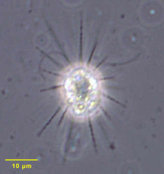
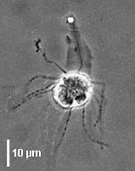
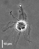
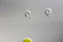
Portrait of Dimorpha mutans, a biflagellate helioflagellate of uncertain affinity. This is the type species of the genus. The two flagella can be seen at approximately the 11 o clock and one o clock positions in this image. The nucleus lies just above the central axoplast upon which the axonemes of the radiating axopodia terminate (seen more clearly in accompanying images). The axopodia bear granular extrusomes (visible in this image). Dimorpha elegans differs from D. mutans by having an axoplast, which is adjoined to the nucleus. Mikrjukov proposed that Dimorpha descends from a cercomonad ancestor (Acta Protozool (2000) 39:99-115). From a polysaprobic freshwater farm pond near Boise, Idaho. Phase contrast.
-
Portrait of Dimorpha mutans, a biflagellated helioflagellate of uncertain affinity. This is the type species of the genus. The central axoplast upon which the axonemes of the radiating axopodia terminate is well seen in this image. The axopodia bear granular extrusomes. Dimorpha elegans differs from D. mutans by having an axoplast, which is adjoined to the nucleus. Mikrjukov proposed that Dimorpha descends from a cercomonad ancestor (Acta Protozool (2000) 39:99-115). From a polysaprobic freshwater farm pond near Boise, Idaho.
-
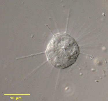
Dimorpha mutans has two anterior flagella one is anteriorly directed and bears microfibrils which form a sleeve around it, the second flagellum is lateral. The nucleus is situated in the anterior part. The cell body of about 20 µm bears axopods with extrusomes and are deflected posteriorly. Flagella and axopods arise from a centroplast located on the anterior face of the nucleus. It is a freshwater and free-living species that phagocytoses prey from the environment. Dimorpha mutans with posterior axopods, two anterior flagella are visible one is deflected backwards (osmium impregnated cell, phase contrast).
-
imorpha mutans has two anterior flagella one is anteriorly directed and bears microfibrils which form a sleeve around it, the second flagellum is lateral. The nucleus is situated in the anterior part. The cell body of about 20 µm bears axopods with extrusomes and are deflected posteriorly. Flagella and axopods arise from a centroplast located on the anterior face of the nucleus. It is a freshwater and free-living species that phagocytoses prey from the environment. Dimorpha mutans osmium impregnated cell with an anterior flagellum surrounded by a microfbrillar gangue, posterior axopods (fixed cell, phase contrast).
-
Dimorpha mutans has two anterior flagella one is anteriorly directed and bears microfibrils which form a sleeve around it, the second flagellum is lateral. The nucleus is situated in the anterior part. The cell body of about 20 µm bears axopods with extrusomes and are deflected posteriorly. Flagella and axopods arise from a centroplast located on the anterior face of the nucleus. It is a freshwater and free-living species that phagocytoses prey from the environment. Dimorpha mutans osmium impregnated cell with an anterior flagellum surrounded by microfbrillar gangue, posterior axopods (preserved cell, phase contrast).
-
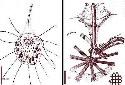
Diagrams based on and electron microscopy, the axoplast is located inside a concavity of the nucleus, axopodial microtubules arise from the axoplast and traverse the nucleus through channels, the microtubules forming the axopods are arranged in squared packed arrays, the two basal bodies/flagella are linked to the axoplast (from G. Brugerolle and J.-P. Mignot)
-

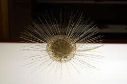
Summary.mw-parser-output table.commons-file-information-table,.mw-parser-output.fileinfotpl-type-information{border:1px solid #a2a9b1;background-color:#f8f9fa;padding:5px;font-size:95%;border-spacing:2px;box-sizing:border-box;margin:0;width:100%}.mw-parser-output table.commons-file-information-table>tbody>tr,.mw-parser-output.fileinfotpl-type-information>tbody>tr{vertical-align:top}.mw-parser-output table.commons-file-information-table>tbody>tr>td,.mw-parser-output table.commons-file-information-table>tbody>tr>th,.mw-parser-output.fileinfotpl-type-information>tbody>tr>td,.mw-parser-output.fileinfotpl-type-information>tbody>tr>th{padding:4px}.mw-parser-output.fileinfo-paramfield{background:#ccf;text-align:right;padding-right:0.4em;width:15%;font-weight:bold}.mw-parser-output.commons-file-information-table+table.commons-file-information-table,.mw-parser-output.commons-file-information-table+div.commons-file-information-table>table{border-top:0;padding-top:0;margin-top:-8px}@media only screen and (max-width:719px){.mw-parser-output table.commons-file-information-table,.mw-parser-output.commons-file-information-table.fileinfotpl-type-information{border-spacing:0;padding:0;word-break:break-word;width:100%!important}.mw-parser-output.commons-file-information-table>tbody,.mw-parser-output.fileinfotpl-type-information>tbody{display:block}.mw-parser-output.commons-file-information-table>tbody>tr>td,.mw-parser-output.commons-file-information-table>tbody>tr>th,.mw-parser-output.fileinfotpl-type-information>tbody>tr>td,.mw-parser-output.fileinfotpl-type-information>tbody>tr>th{padding:0.2em 0.4em;text-align:left;text-align:start}.mw-parser-output.commons-file-information-table>tbody>tr,.mw-parser-output.fileinfotpl-type-information>tbody>tr{display:flex;flex-direction:column}.mw-parser-output.commons-file-information-table+table.commons-file-information-table,.mw-parser-output.commons-file-information-table+div.commons-file-information-table>table{margin-top:-1px}.mw-parser-output.fileinfo-paramfield{box-sizing:border-box;flex:1 0 100%;width:100%}} Description: Français : Leopold et Rudolf Blaschka. Clathrulina elegans. Modèle en verre. Collection du Musée zoologique de Strasbourg. Exposition Laboratoire d'Europe Strasbourg, 1880-1930 au Musée d'art moderne et contemporain de Strasbourg. Date: 8 February 2018, 15:29:43. Source: Own work. Author:
Ji-Elle.
-
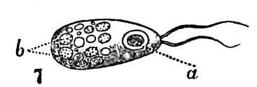
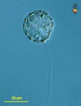
Summary.mw-parser-output table.commons-file-information-table,.mw-parser-output.fileinfotpl-type-information{border:1px solid #a2a9b1;background-color:#f8f9fa;padding:5px;font-size:95%;border-spacing:2px;box-sizing:border-box;margin:0;width:100%}.mw-parser-output table.commons-file-information-table>tbody>tr,.mw-parser-output.fileinfotpl-type-information>tbody>tr{vertical-align:top}.mw-parser-output table.commons-file-information-table>tbody>tr>td,.mw-parser-output table.commons-file-information-table>tbody>tr>th,.mw-parser-output.fileinfotpl-type-information>tbody>tr>td,.mw-parser-output.fileinfotpl-type-information>tbody>tr>th{padding:4px}.mw-parser-output.fileinfo-paramfield{background:#ccf;text-align:right;padding-right:0.4em;width:15%;font-weight:bold}.mw-parser-output.commons-file-information-table+table.commons-file-information-table,.mw-parser-output.commons-file-information-table+div.commons-file-information-table>table{border-top:0;padding-top:0;margin-top:-8px}@media only screen and (max-width:719px){.mw-parser-output table.commons-file-information-table,.mw-parser-output.commons-file-information-table.fileinfotpl-type-information{border-spacing:0;padding:0;word-break:break-word;width:100%!important}.mw-parser-output.commons-file-information-table>tbody,.mw-parser-output.fileinfotpl-type-information>tbody{display:block}.mw-parser-output.commons-file-information-table>tbody>tr>td,.mw-parser-output.commons-file-information-table>tbody>tr>th,.mw-parser-output.fileinfotpl-type-information>tbody>tr>td,.mw-parser-output.fileinfotpl-type-information>tbody>tr>th{padding:0.2em 0.4em;text-align:left;text-align:start}.mw-parser-output.commons-file-information-table>tbody>tr,.mw-parser-output.fileinfotpl-type-information>tbody>tr{display:flex;flex-direction:column}.mw-parser-output.commons-file-information-table+table.commons-file-information-table,.mw-parser-output.commons-file-information-table+div.commons-file-information-table>table{margin-top:-1px}.mw-parser-output.fileinfo-paramfield{box-sizing:border-box;flex:1 0 100%;width:100%}} Description: English: Bi-flagellate “flagellula” of Clathrulina elegans. a, nucleus; b, granules. Date: 1911. Source: Fig. 7 of
File:EB1911_Heliozoa_(1).jpg (extract). Original source: “Heliozoa,” Encyclopædia Britannica (11th ed.), v. 13, 1911, p. 232, fig. 1. Author: Marcus Manuel Hartog.
-
Summary.mw-parser-output table.commons-file-information-table,.mw-parser-output.fileinfotpl-type-information{border:1px solid #a2a9b1;background-color:#f8f9fa;padding:5px;font-size:95%;border-spacing:2px;box-sizing:border-box;margin:0;width:100%}.mw-parser-output table.commons-file-information-table>tbody>tr,.mw-parser-output.fileinfotpl-type-information>tbody>tr{vertical-align:top}.mw-parser-output table.commons-file-information-table>tbody>tr>td,.mw-parser-output table.commons-file-information-table>tbody>tr>th,.mw-parser-output.fileinfotpl-type-information>tbody>tr>td,.mw-parser-output.fileinfotpl-type-information>tbody>tr>th{padding:4px}.mw-parser-output.fileinfo-paramfield{background:#ccf;text-align:right;padding-right:0.4em;width:15%;font-weight:bold}.mw-parser-output.commons-file-information-table+table.commons-file-information-table,.mw-parser-output.commons-file-information-table+div.commons-file-information-table>table{border-top:0;padding-top:0;margin-top:-8px}@media only screen and (max-width:719px){.mw-parser-output table.commons-file-information-table,.mw-parser-output.commons-file-information-table.fileinfotpl-type-information{border-spacing:0;padding:0;word-break:break-word;width:100%!important}.mw-parser-output.commons-file-information-table>tbody,.mw-parser-output.fileinfotpl-type-information>tbody{display:block}.mw-parser-output.commons-file-information-table>tbody>tr>td,.mw-parser-output.commons-file-information-table>tbody>tr>th,.mw-parser-output.fileinfotpl-type-information>tbody>tr>td,.mw-parser-output.fileinfotpl-type-information>tbody>tr>th{padding:0.2em 0.4em;text-align:left;text-align:start}.mw-parser-output.commons-file-information-table>tbody>tr,.mw-parser-output.fileinfotpl-type-information>tbody>tr{display:flex;flex-direction:column}.mw-parser-output.commons-file-information-table+table.commons-file-information-table,.mw-parser-output.commons-file-information-table+div.commons-file-information-table>table{margin-top:-1px}.mw-parser-output.fileinfo-paramfield{box-sizing:border-box;flex:1 0 100%;width:100%}} Description: English: Blaschka glass model in the Muséum d'histoire naturelle de Genève : Clathrulina elegans. Date: 22 October 2014, 14:06:14. Source: Own work. Author:
Vassil.
-
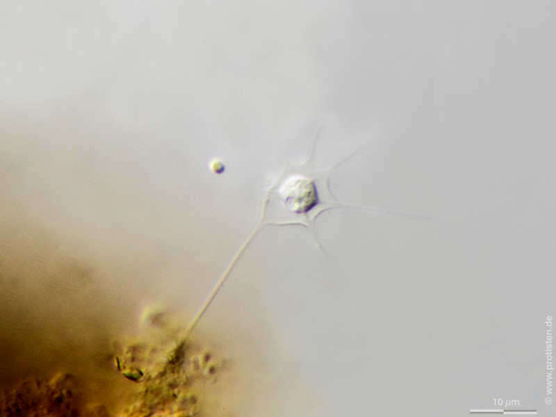
Hedriocystis pellucida Scale bar indicates 10 µm. Sample from a pond near Großostheim, Germany. Sampling date 04/2021. The image was built up using several photomicrographic frames with manual stacking technique. Images were taken using Zeiss Axioplan with Olympus OM-D M5 MKII. Image under Creative Commons License V 3.0 (CC BY-NC-SA). Place name: Pond near Großostheim (Germany) Latitude: 49.88482168 Longitude: 9.09980822 Multiebenen-Abbildung, manuell gestapelt. Der Messbalken markiert eine Länge von 10 µm. Probe aus einem Waldteich bei Großostheim. Datum der Aufsammlung: 04/2021. Mikrotechnik: Zeiss Axioplan, Kamera: Olympus OM-D M5 MKII. Creative Commons License V 3.0 (CC BY-NC-SA). For permission to use of (high-resolution) images please contact postmaster@protisten.de.
-
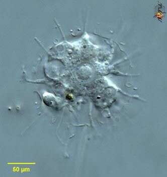
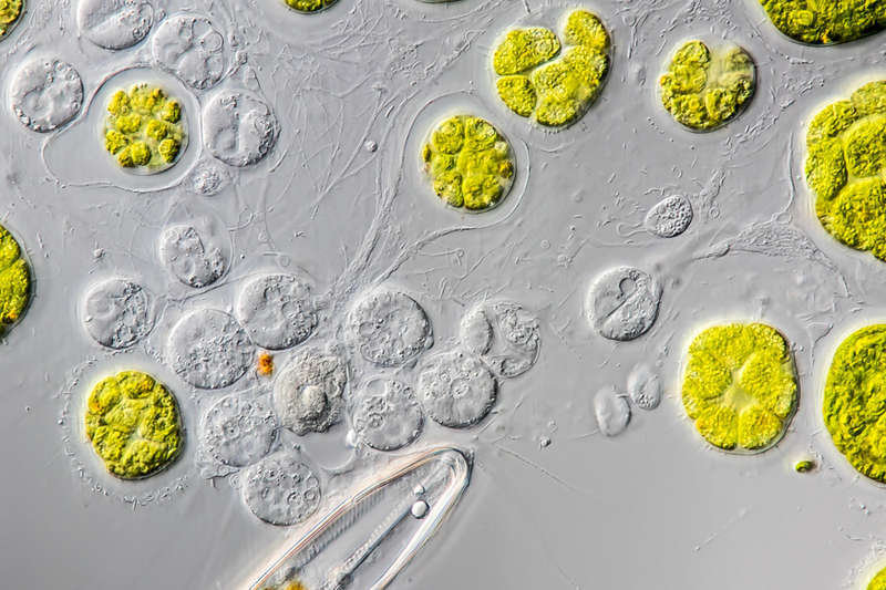
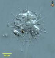
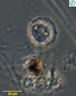
Microgromia socialis The specimen was gathered in the pond Birkensee near Rödelsee (Lower Franconia, Germany). Sampling date 7/2018.Copyright Dr. Rainer Meisch, Würzburg, Germany.Images were taken using Zeiss Axioplan with Canon DSLR Image under Creative Commons License V 3.0 (CC BY-NC-SA). Place name: Pond Birkensee near Rödelsee (Lower Franconia, Germany) Latitude: 49.71819841 Longitude: 10.27807474 Probe aus dem Birkensee bei Rödelsee (Unterfranken). Datum der Aufsammlung: 7/2018. Copyright Dr. Rainer Meisch, Würzburg. Mikrotechnik: Zeiss Axioplan, Kamera: Canon DSLR. Creative Commons License V 3.0 (CC BY-NC-SA). For permission to use of (high-resolution) images please contact postmaster@protisten.de.
-
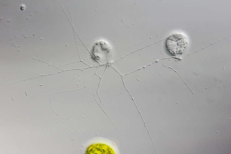
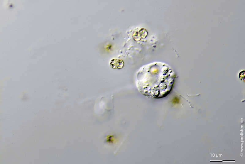
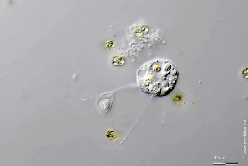
Heliomorpha mutans Synonym: Dimorpha mutans.Ingestion series. Scale bar indicates 10 µm. Sample from a pond in Lemkendorf on the isle Fehmarn (Baltic Sea). Images were taken using a Zeiss Axioplan Microscope with Canon EOS 70D.Image under Creative Commons License V 3.0 (CC BY-NC-SA). Place name: Pond in Lemkendorf, isle Fehmarn (Baltic Sea, Germany) Latitude: 54.472296 Longitude: 11.095662 Synonym: Dimorpha mutans.Bilderserie des Vorgangs der Nahrungsaufnahme. Der Messbalken markiert eine Länge von 10 µm. Probe aus einem Teich in Lemkendorf, Fehmarn. Mikrotechnik: Zeiss Axioplan, Kamera: Canon EOS 70D. Creative Commons License V 3.0 (CC BY-NC-SA). For permission to use of (high-resolution) images please contact postmaster@protisten.de.
-
Heliomorpha mutans Synonym: Dimorpha mutans.Ingestion series. Scale bar indicates 10 µm. Sample from a pond in Lemkendorf on the isle Fehmarn (Baltic Sea). Images were taken using a Zeiss Axioplan Microscope with Canon EOS 70D.Image under Creative Commons License V 3.0 (CC BY-NC-SA). Place name: Pond in Lemkendorf, isle Fehmarn (Baltic Sea, Germany) Latitude: 54.472296 Longitude: 11.095662 Synonym: Dimorpha mutans.Bilderserie des Vorgangs der Nahrungsaufnahme. Der Messbalken markiert eine Länge von 10 µm. Probe aus einem Teich in Lemkendorf, Fehmarn. Mikrotechnik: Zeiss Axioplan, Kamera: Canon EOS 70D. Creative Commons License V 3.0 (CC BY-NC-SA). For permission to use of (high-resolution) images please contact postmaster@protisten.de.


.jpg)