-
-
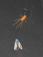
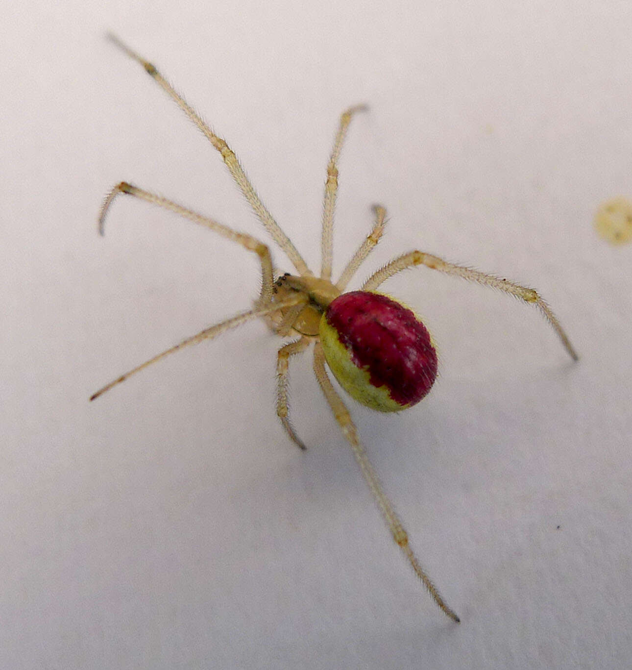
Figures 177–182.Habitats and natural history of some Selenops species. 177 Selenops arikok sp. n. from Aruba guarding egg sac with several spiderlings inside 178 Selenops geraldinae Corronca eating a fly and guarding her egg sac on a bromeliad, Gaspar Grande Island, Trinidad and Tobago 179 Selenops willinki Corronca on a tree trunk, Little Tobago, Trinidad and Tobago. Terminal setal tufts and festoon pattern are visible 180 Selenops micropalpus Muma on the trunk of Bursera simaruba, in a dry forest, St. Lucia. Terminal setal tufts and festoon pattern are visible 181 Selenops mexicanus Keyserling on a large tree trunk outside of Cueva Actun Kan, Guatemala. Note the alternating light and dark leg annulations 182 Egg sacs of Selenops bifurcatus Banks on rocks in a wash in dry forest and thornscrub, Zacatan, Guatemala. Selenops bifurcatus sometimes guards the yellowish egg sacs, and other times does not.
-
Jeremy Miller, Cahyo Rahmadi
Zookeys
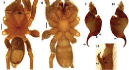
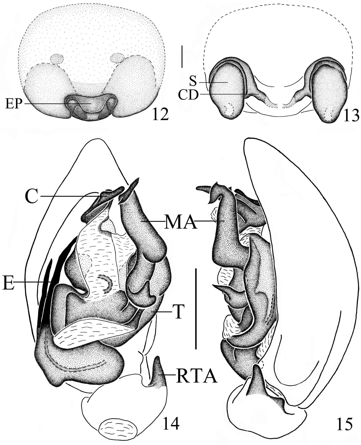
Figures 7–12.Amauropelma matakecil sp. n., female holotype (MZB.Aran.500) 7 Epigynum, ventral view. Note that the right epigynal tooth has broken off leaving a round hole; the tooth itself is lying unattached near the epigastric furrow 8 Vulva, dorsal view, left side, cleared, white arrow indicates fertilization duct 9 Right tarsus, leg I, prolateral view 10 Left tarsus, leg I, dorsal view, arrow indicates tarsal organ 11 Spinnerets, anal tubercle, and tracheal spiracle, posterior view, arrow indicates tracheal spiracle 12 Spinnerets, lateral view. ALS, anterior lateral spinneret; AT, anal tubercle; CD, copulatory duct; ET, epigynal tooth; PLS, posterior lateral spinneret; PMS, posterior median spinneret; S, spermatheca.
-
Jeremy A. Miller, Charles E. Griswold, Nikolaj Scharff, Milan Řezáč, Tamás Szűts, Mohammad Marhabaie
Zookeys
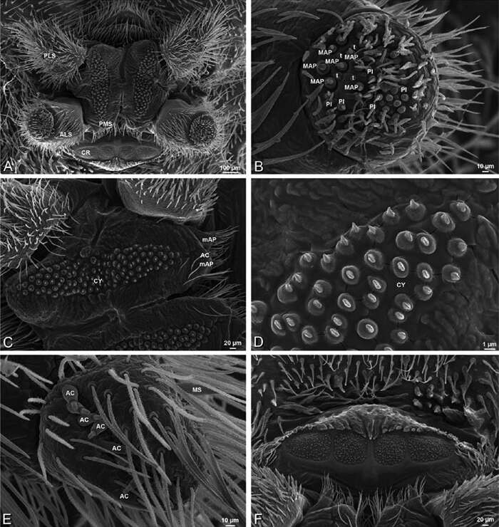
Figure 36.A–F Dresserus sp., female from Mazumbai, Tanzania (CASENT 9025747, CAS), scanning electron micrographs of spinnerets. A overview B left ALS C right PMS D detail, cylindrical gland spigots on right PMS E left PLS F cribellum. AC aciniform gland spigot ALS anterior lateral spinneret CR cribellum CY cylindrical gland spigot MAP major ampullate gland spigot mAP minor ampullate gland spigot MS modified spigot PI piriform gland spigot PLS posterior lateral spinneret PMS posterior median spinneret.
-
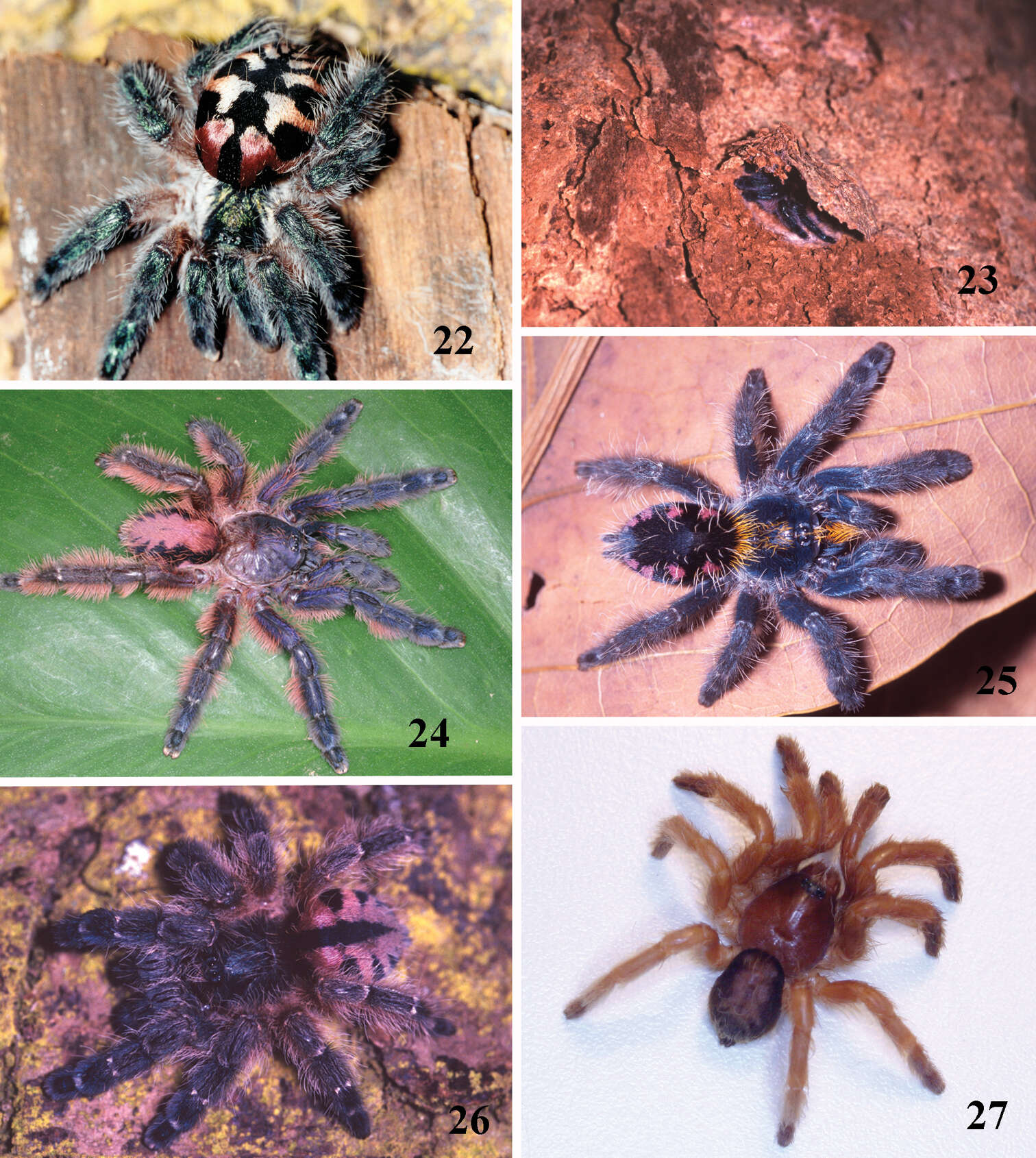
Figures 22–27.22–23 Typhochlaena seladonia C. L. Koch, 1841, habitus 22 female, Santa Luzia do Itanhy, state of Sergipe 23 immature inside its retreat in tree bark, same locality 24 Typhochlaena amma sp. n., female, Santa Teresa, state of Espirito Santo 25 Typhochlaena costae sp. n., female, Palmas, state of Tocantins 26 Typhochlena curumim sp. n., female, Areia, state of Paraiba 27 Typhochlaena paschoali sp. n., preserved female, Camacam, state of Bahia (holotype MNRJ 13723). Photos: R. Bertani.
-
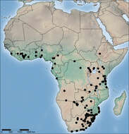
Figure 71.Distribution of Copa flavoplumosa Simon, 1885 in the Afrotropical Region.
-
Figures 12–15.Mallinella sphaerica sp. n., 12 epigyne, ventral view 13 vulva 14 left male palp, ventral view 15 same, retrolateral view. Scale bars: 0.2 mm (12–13); 0.5 mm (14–15).
-
Dan Quan, Jian Chen, Jie Liu
Zookeys
Figure 17.Collection localities of Sinopoda serrata (Wang, 1990) in China.
-
Peter Michalik, Luis Piacentini, Elisabeth Lipke, Martin J. Ramírez
Zookeys
Figure 4.Female genitalia. A Lateral view of the dome-shaped genital area (compare also to Fig. 3A) (MACN-Ar 30667) B Sagital section through genital area obtained by micro-CT (MACN-Ar 30667) C Posterior view of genital area (MACN-Ar 30667). Spermathecae in anterior (D) and ventral view (E) (ZIMG II/28128). Abbreviation: B bursa; EF epigastric furrow; GP genital pockets; Gl glands; PF postepigastric fold; Sp spermathecae; Ue uterus externus.
-
Yuri M. Marusik, Mikhail M. Omelko
Zookeys
Figures 6–12.Male palp of Cryptothele verrucosa (6–11) and Cryptothele alluaudi (12). 6 bulbus, ventral 7 bulbus, ventro-prolateral 8, 10 bulbus, retrolateral 9 bulbus, prolateral 11 palp with removed bulbus, retrolateral 12 palp, ventral (after Marusik and Omelko (2012)). Abbreviations: Co conductor; Ea terminal process of embolus; Eb embolus base; Em embolus; Ep posterior process of embolus; Sd seminal duct; Sp subtegular process; St subtegulum; Te triangle extesion of tegulum; Ts threads of subtegulum.
-
Jason E. Bond, Rebecca L. Godwin
Zookeys
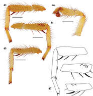
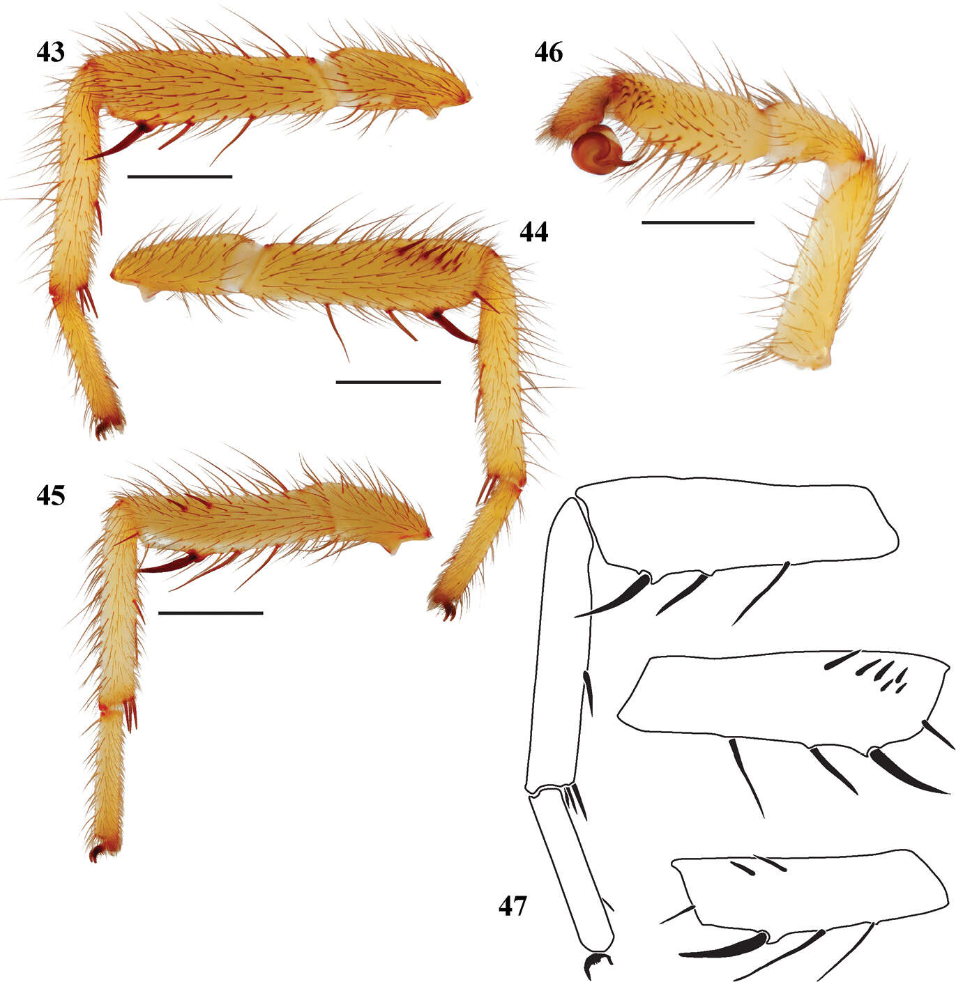
Figures 43–47.Eucteniza huasteca sp. n. from Nuevo Leon, Mexico male holotype 43 retrolateral aspect, leg I [832040] 44 prolateral aspect, leg I [832036] 45 retrolateral aspect, leg II [832042] 46 retrolateral aspect, pedipalp [832044] 47 line drawings, leg I retrolateral and prolateral (tibia) aspects; prolateral aspect tibia leg II.
-
Ning Sun, Yuri M. Marusik, Lihong Tu
Zookeys
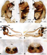
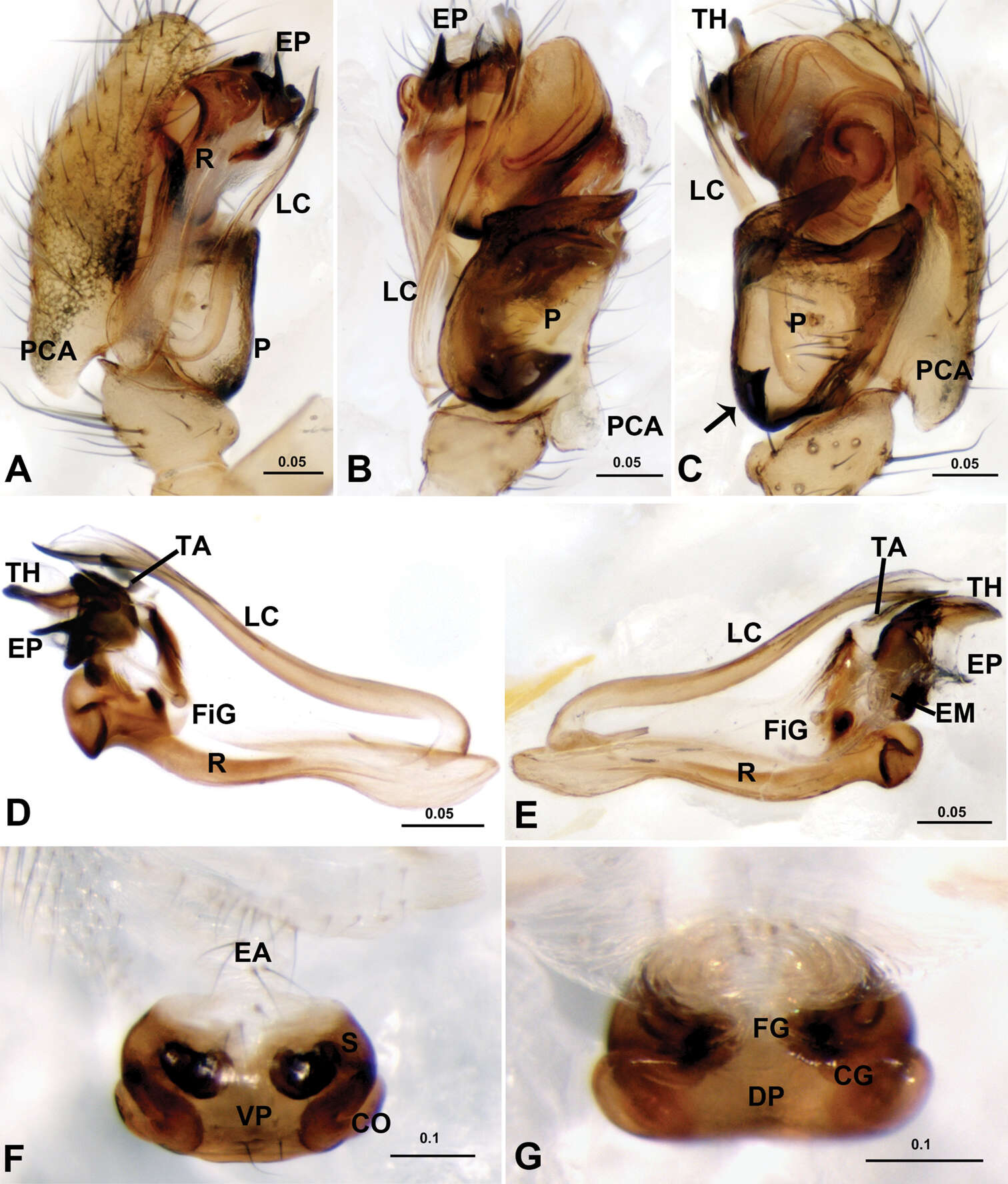
Figure 3.Acanoides hengshanensis. A male palp, prolateral B male palp, ventral C male palp, retrolateral, arrow indicates pointed tooth on posterolateral margin D embolic division, ventral E embolic division, dorsal F epigynum, ventral G epigynum, dorsal. CG copulatory groove; CO copulatory opening; DP dorsal plate; EA extensible area of epigynal basal part; EM embolic membrane; EP embolus proper; FG fertilization groove; FiG Fickert’s gland; LC lamella characteristica; P paracymbium; PCA proximal cymbial apophysis; R radix; S spermatheca; TA terminal apophysis; TH thumb of embolus; VP ventral plate. [Scale bars: mm].
-
Figure 2.Sinamma oxycera gen. n. & sp. n., male holotype. A, B Left palp C Left leg I D Left tibia I E Left metatarsus I and tarsus I. A prolateral view B, C retrolateral view D, E anterior view.
-
Figure 5.Xyphinus hwangi sp. n., female. A, C, E habitus, dorsal, lateral and ventral views B, D, F, G prosoma, dorsal, lateral, ventral and anterior views H, I abdomen, ventral and lateral views J, K genital area, ventral and dorsal views (cleared in lactic acid). Scale bars: A, C, E = 0.4 mm; B, D, F–I = 0.2 mm; J, K = 0.1 mm.
-
Carlos Perafán, Fernando Pérez-Miles
Zookeys
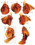
Figures 7–11.Melloleitaoina mutquina. 7–8 male holotype 7 dorsal view 8 ventral view 9–10 right palpal bulb 9 prolateral view 10 retrolateral view 11 right tibial apophysis. Arrow indicates apex widened. Scale bars = 1 mm.
-
Alejandro Valdez-Mondragón, Jorge I. Mendoza, Oscar F. Francke
Zookeys
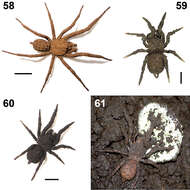
Figures 58–61.Paratropis tuxtlensis sp. n. Photographs of live specimens, kept in the laboratory 58 Adult male 59 Immature specimen 60 Adult female 61 Adult female protecting her egg sac. Scales: 1 mm (Figure 59), 4 mm (Figure 58), 6 mm (Figure 60).
-
Yuri M. Marusik, Alexander A. Fomichev, Mikhail M. Omelko
Zookeys
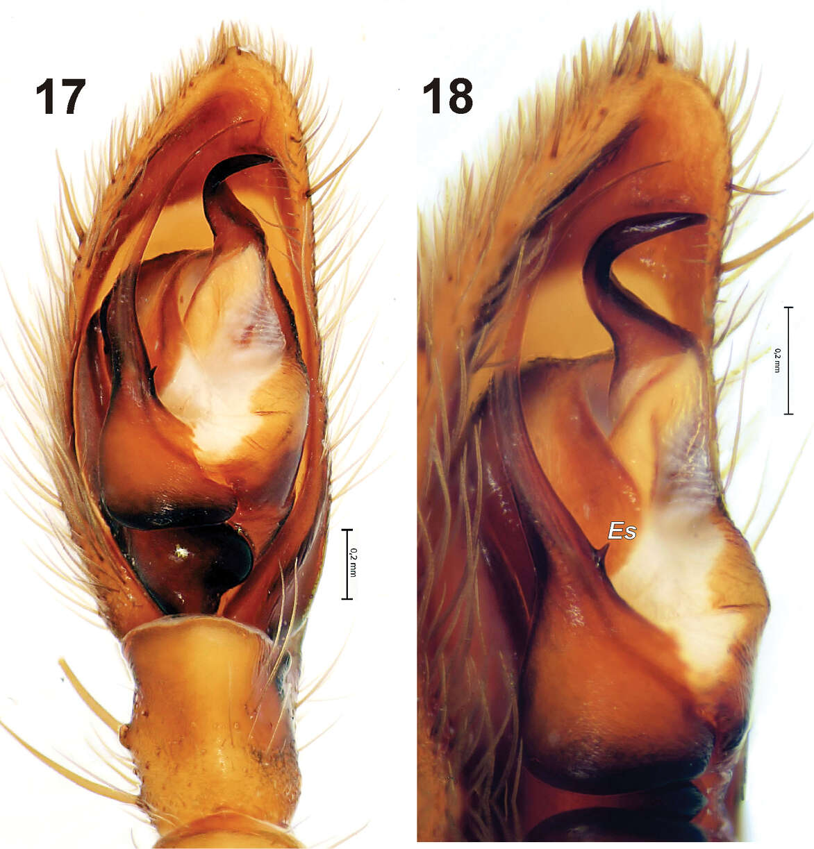
Figures 17–18.Male palp of Gnaphosa ustyuzhanini. 17 ventral 18 prolateral. Scale = 0.2 mm. Es – embolic spine.
-
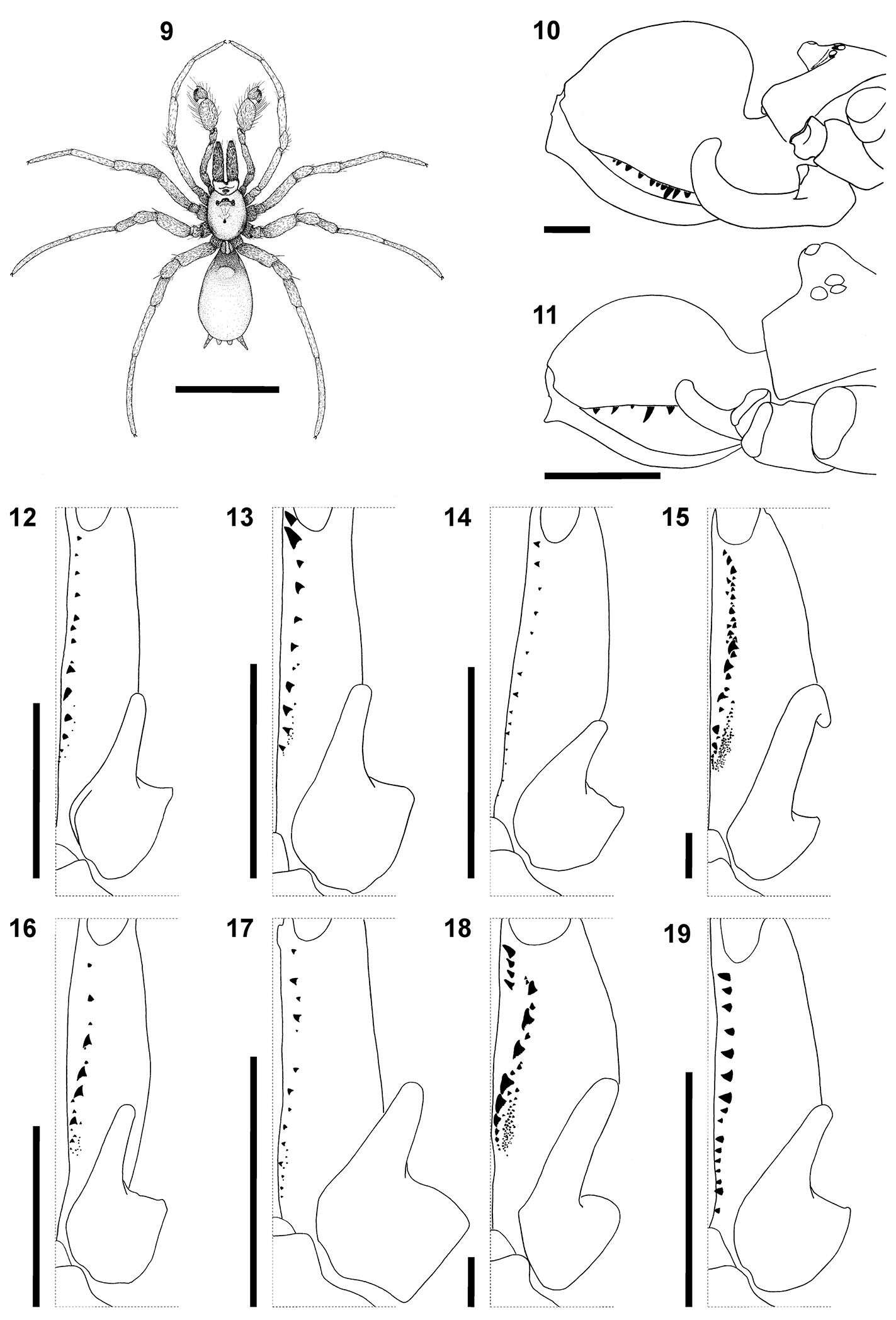
Figures 12–19.Heser vijayanagara sp. n. 12 Female allotype, dorsal view of body 13 Male palp, ventral, with conductor (C), embolus (E), and median apophysis (MA) indicated 14 Male palp, retrolateral 15 Epigyne, ventral 16 Female metatarsi III (left) and IV, with ventral terminal preening comb 17 Female cheliceral teeth 18 Vulva, ventral 19 Vulva, dorsal. Scale bars: 12: 1.0; 13–17: 0.5; 18–19: 0.1.
-
Fourie René, Haddad Charles R., Jocqué Rudy
Zookeys
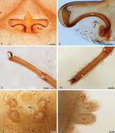
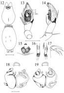
Figures 9–19.Somatic morphology of Calommata tibialis sp. n., male 9, 17 Calommata simoni Pocock, female 10, 15 and male 11, 16 Calommata megae sp. n., male 12 Calommata meridionalis sp. n., male 13 Calommata namibica sp. n., male 14and Calommata transvaalica Hewitt, female 18 and male 19: 9 dorsal habitus 10, 11 lateral view of chelicera, endites and anterior of carapace 12–19 left chelicera, ventral view, indicating dentition. Scale bars: 5mm (9), 1mm (10–19).
-
Cairns, Queensland, Australia
-
-
Mpumalanga, South Africa
-
Cradley, Malvern, Worcs.SO7347
-
Monteverde, Puntarenas, Costa Rica


