-
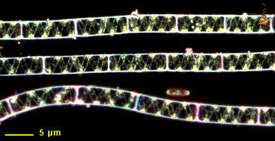
In Spyrogyra, the plastids (a type of chloroplast) are spiral around the interior of the cell. The nucleus is slightly to the right of the center. This alga was collected from the Gardner River near Sheepeater Cliffs.
-
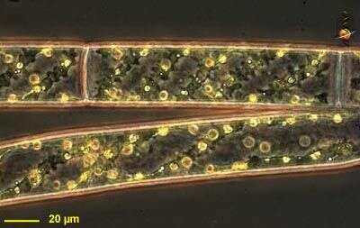
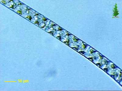
Spirogyra observed in freshwater sediments in the vicinity of Broome, Western Australia in September 2003. This image was taken using phase contrast optics. This work was supported by the Australian Biological Resources Study.
-
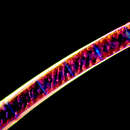
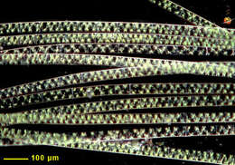
Dark ground illumination of this filamentous green alga. The chloroplasts are ribbon-like and spiral around the interior of the cell wall.
-
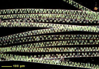
Small clump of filaments with spiral plastids viewed using dark-ground illumination.
-
Collected from Cumloden Swamp on October 7, 2002.
-
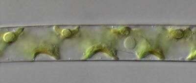
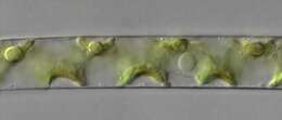
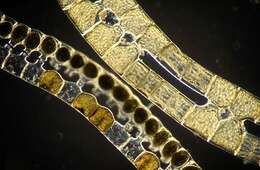
Dark ground image of preserved filaments showing various stages of the conjugation process. Paired filaments caught during conjugation are above, filaments with zygotes are below.
-
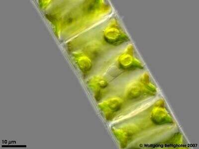
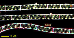
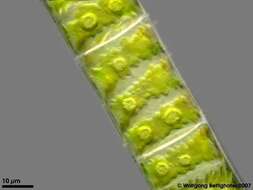
Part of a filament showing the shape of the ribbon like chloroplast with pyrenoids. Multi layer image (DOF) using 5 frames generating depth of focus, stacked manually using Corel Photopaint. Aufwuchs on roots dangling in a creek´s water. This image was taken using Zeiss Universal with Olympus C7070 CCD camera.
-
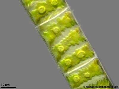
Part of a filament showing the nucleus and chloroplast´s pyrenoids. Multi layer image (DOF) using 3 frames generating depth of focus, stacked manually using Corel Photopaint. Aufwuchs on roots dangling in a creek´s water. This image was taken using Zeiss Universal with Olympus C7070 CCD camera.
-
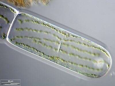
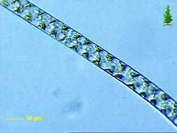
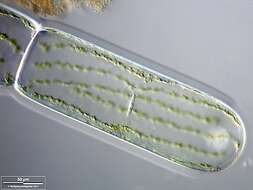
Scale bar indicates 50 µm.Sample from the pond Hegne Moor situated in the vicinity of Lake Constance. The image was built up using several photomicrographic frames with manual stacking technique. Images were taken using Zeiss Universal with Olympus C7070 CCD camera.Image under Creative Commons License V 3.0 (CC BY-NC-SA).
-
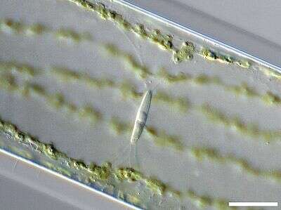
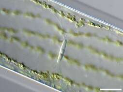
Zoom-in. The nucleus with its central nucleolus is visible. The suspension of the nucleus with fine cytoplasmic filaments indicates that almost the whole cell is filled with a large water vacuole. The parietal chloroplast is also lying surrounded by cytoplasm. Scale bar indicates 25 µm.Sample from the pond Hegne Moor situated in the vicinity of Lake Constance. The image was built up using several photomicrographic frames with manual stacking technique. Images were taken using Zeiss Universal with Olympus C7070 CCD camera.Image under Creative Commons License V 3.0 (CC BY-NC-SA).