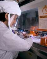
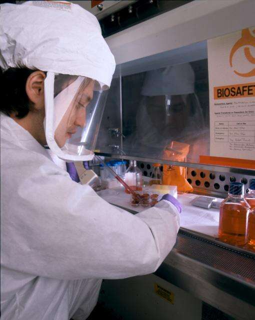
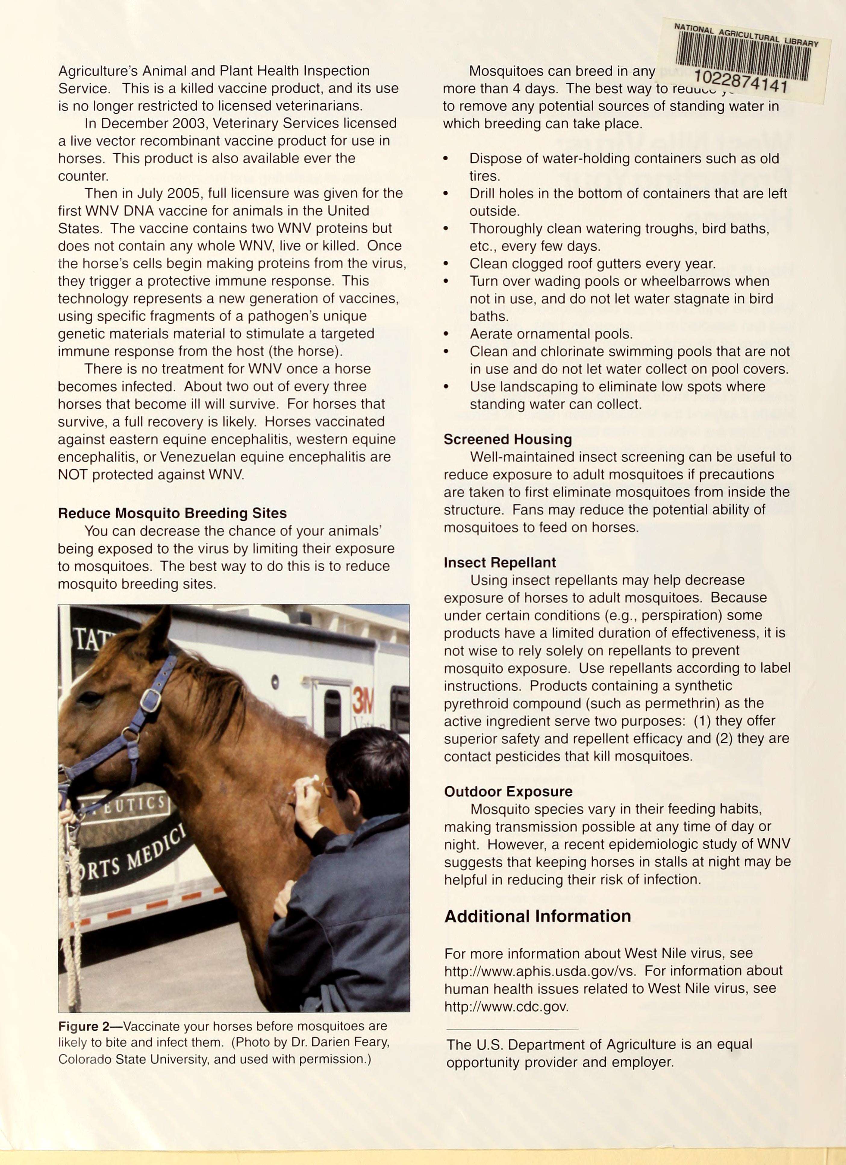
Description: NIAID scientist wears BSL-3 gear to demonstrate preparation of tissue culture plates infected with a chimera of West Nile virus and the dengue virus. The lab is developing a live, attenuated virus vaccine for West Nile virus. Credit: NIAID. Date: Taken on 27 April 2011, 12:50. Source: West Nile Vaccine Research in BSL-3 Lab. Author: NIAID. Camera location39° 00′ 03.27″ N, 77° 05′ 57.49″ WView all coordinates using: OpenStreetMap 39.000909; -77.099304.
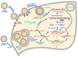
All images in this article were uploaded in the JPEG format even though it consists of non-photographic data. This information could be stored more efficiently or accurately in the PNG or SVG format. If possible, please upload a PNG or SVG version of this image without compression artifacts, derived from a non-JPEG source (or with existing artifacts removed). After doing so, please tag the JPEG version with {{Superseded|NewImage.ext}} and remove this tag. This tag should not be applied to photographs or scans. For more information, see {{BadJPEG}}. Description: English: West Nile virus life cycle. After binding and uptake, the virion envelope fuses with cellular membranes, followed by uncoating of the nucleocapsid and release of the RNA genome into the cytoplasm. The viral genome serves as messenger RNA (mRNA) for translation of all viral proteins and as template during RNA replication. Copies are subsequently packaged within new virus particles which are transported in vesicles to the cell membrane. Date: 26 May 2013, 21:37:46. Source: https://www.ncbi.nlm.nih.gov/pmc/articles/PMC3311072/. Author: De Filette, et al.
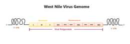
Description: English: Depicts the RNA genome of the West Nile Virus including all of its genes and the 5' and 3' untranslated regions. Date: 10 October 2017. Source: Made on Microsoft Publisher. Author: Puthuveetilnp.
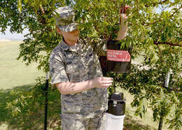
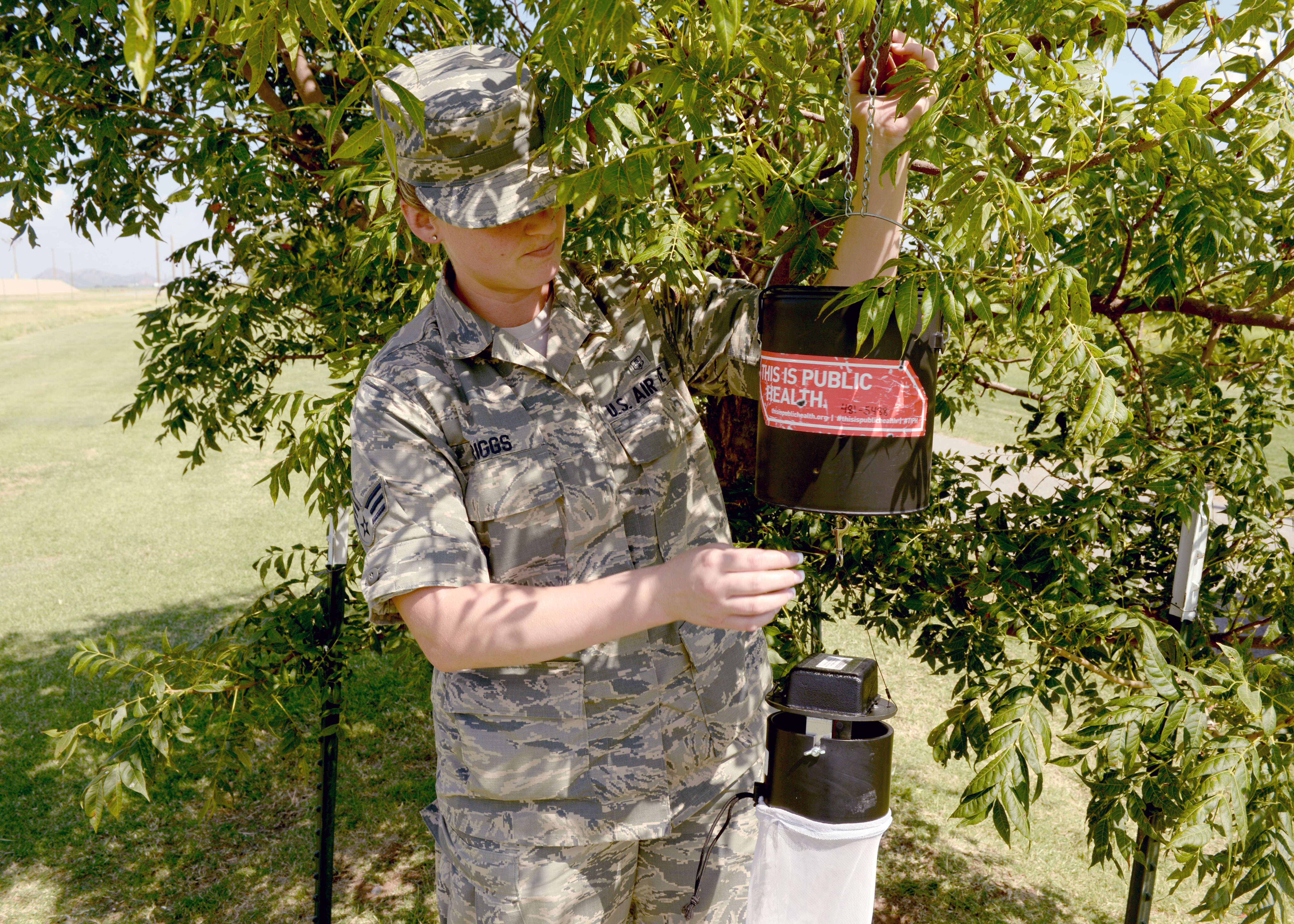
Description: English: U.S. Air Force Senior Airman Brandi Spriggs, 97th Medical Operations Squadron health technician, sets up a dry ice trap to catch mosquitoes, Aug. 11, 2016, at Altus Air Force Base, Okla. The Altus AFB Public Health Office is actively testing mosquitoes for viruses and conducting surveillance on mosquito populated areas. (U.S. Air Force photo by Airman 1st Class Cody Dowell/Released) Unit: 97th Air Mobility Wing, Public Affairs DVIDS Tags: medical; virus; zika; pest mangment. Date: 11 August 2016. Source: https://www.dvidshub.net/image/2811225/altus-staying-top-zika. Author: Airman 1st Class Cody Dowell. LocationALTUS AFB, OK, US. VIRIN : This Image was released by the United States Air Force with the ID 161811-F-LH697-0025 (next). This tag does not indicate the copyright status of the attached work. A normal copyright tag is still required. See Commons:Licensing. العربية | বাংলা | Deutsch | English | español | euskara | فارسی | français | italiano | 日本語 | 한국어 | македонски | മലയാളം | Plattdüütsch | Nederlands | polski | پښتو | português | svenska | Türkçe | українська | 中文 | 中文(简体) | +/− :. Search DVIDs. Posted23 August 2016, 13:08. Archive linkarchive copy.
Description: English: Susan Blumenthal, MD, Senior Fellow in Health Policy, New America. Date: 12 July 2016, 23:28:14. Source: https://www.flickr.com/photos/newamerica/28252124006/. Author: New America. Other versions: This file has multiple extracted images: [[|thumb|none|320x160px|[[:|Susan Blumenthal in 2016]]]] Susan Blumenthal in 2016.jpg.
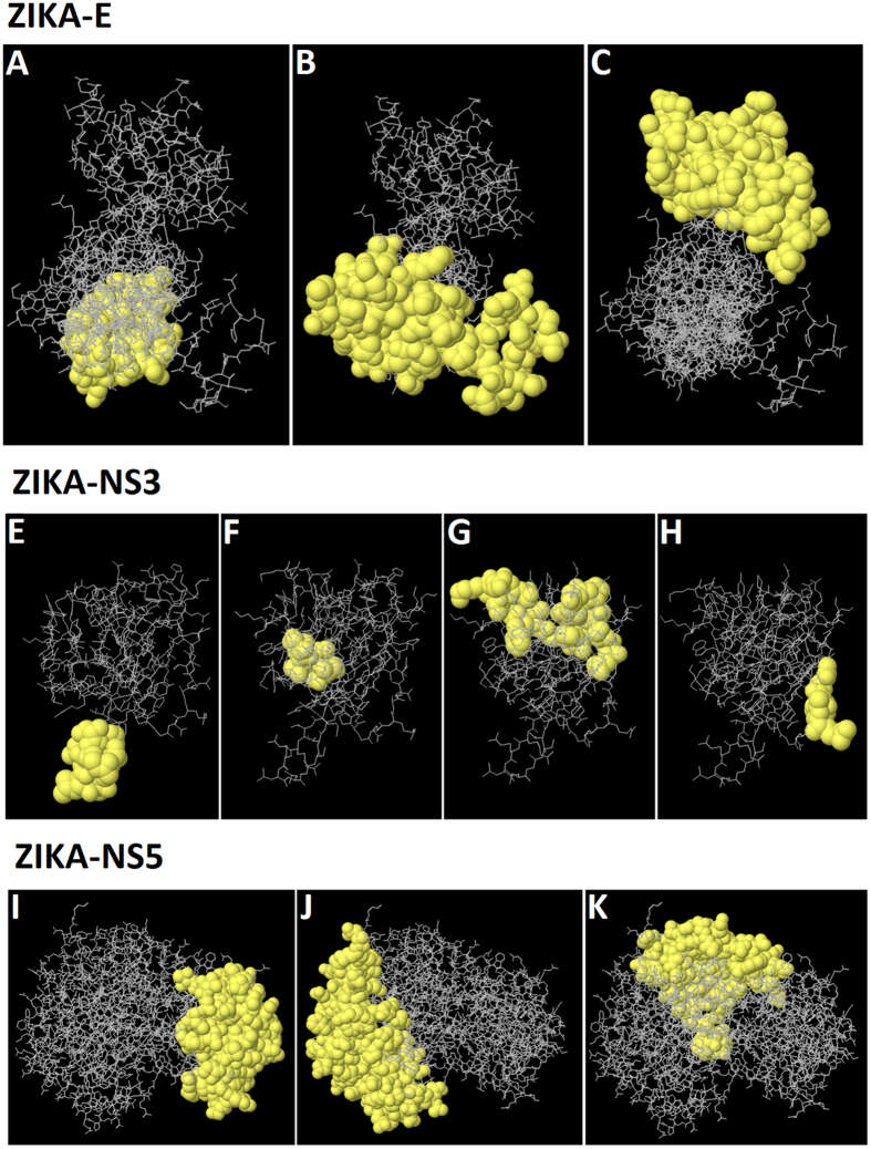
Muhammad Usman Mirza, Shazia Rafique, Amjad Ali, Mobeen Munir, Nazia Ikram, Abdul Manan, Outi M. H. Salo-Ahen, Muhammad Idrees
Wikimedia Commons
Description: English: 3D Representation of the predicted discontinuous epitopes ( A to C ) of Zika-E protein, ( E to H ) of Zika-NS3 protein and ( I to K ) of Zika-NS5 protein. Date: 9 December 2016. Source: Muhammad Usman Mirza et. al. "Towards peptide vaccines against Zika virus: Immunoinformatics combined with molecular dynamics simulations to predict antigenic epitopes of Zika viral proteins", Scientific Reports doi:10.1038/srep37313. Author: Muhammad Usman Mirza, Shazia Rafique, Amjad Ali, Mobeen Munir, Nazia Ikram, Abdul Manan, Outi M. H. Salo-Ahen, Muhammad Idrees. Permission(Reusing this file): https://creativecommons.org/licenses/by/4.0/ This work is licensed under a Creative Commons Attribution 4.0 International License. The images or other third party material in this article are included in the article’s Creative Commons license, unless indicated otherwise in the credit line; if the material is not included under the Creative Commons license, users will need to obtain permission from the license holder to reproduce the material. To view a copy of this license, visit https://creativecommons.org/licenses/by/4.0/.
Description: English: Tom Frieden, MD, MPH, Director, Centers for Disease Control and Prevention (CDC); Susan Blumenthal, MD, Senior Fellow in Health Policy, New America. Date: 12 July 2016, 23:29:47. Source: https://www.flickr.com/photos/newamerica/28207976511/. Author: New America.
Description: A Zika virus researcher at the NIAID Vaccine Research Center pipets samples. Credit: NIAID. Date: 19 April 2016, 11:07. Source: Zika Virus Research. Author: NIAID.
Description: A Zika virus researcher at the NIAID Vaccine Research Center loads samples into a microcentrifuge. Credit: NIAID. Date: 19 April 2016, 11:29. Source: Zika Virus Research. Author: NIAID.
Description: English: Mosquitoes lie in a petri dish at Altus Air Force Base, Okla. to be sent to the lab at Fort Sam Houston, Texas, to be tested for viruses, Aug. 12, 2016. The Altus AFB Public Health Office is actively testing mosquitoes for viruses and conducting surveillance on mosquito populated areas. (U.S. Air Force photo by Airman 1st Class Cody Dowell/Released) Unit: 97th Air Mobility Wing, Public Affairs DVIDS Tags: medical; virus; zika; pest managment. Date: Taken on 12 August 2016. Source: https://www.dvidshub.net/image/2811228/altus-staying-top-zika. Author: Airman 1st Class Cody Dowell. LocationInfoFieldALTUS AFB, OK, US. VIRINInfoField : This image was released by the United States Air Force with the ID 160812-F-LH697-034 (next). This tag does not indicate the copyright status of the attached work. A normal copyright tag is still required. See Commons:Licensing. العربية | বাংলা | Deutsch | Deutsch (Sie-Form) | English | español | euskara | فارسی | français | italiano | 日本語 | 한국어 | македонски | മലയാളം | Plattdüütsch | Nederlands | polski | پښتو | português | svenska | Türkçe | українська | 中文 | 中文(简体) | 中文(繁體) | +/− :. Search DVIDs. PostedInfoField23 August 2016, 13:08. Archive linkInfoFieldarchive copy.
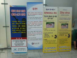
Description: English: Warning signs about Zika virus at the Incheon Airport, Korea. Date: Taken on 5 July 2016 05:28:44. Source: Own work. Author: Piotrus.
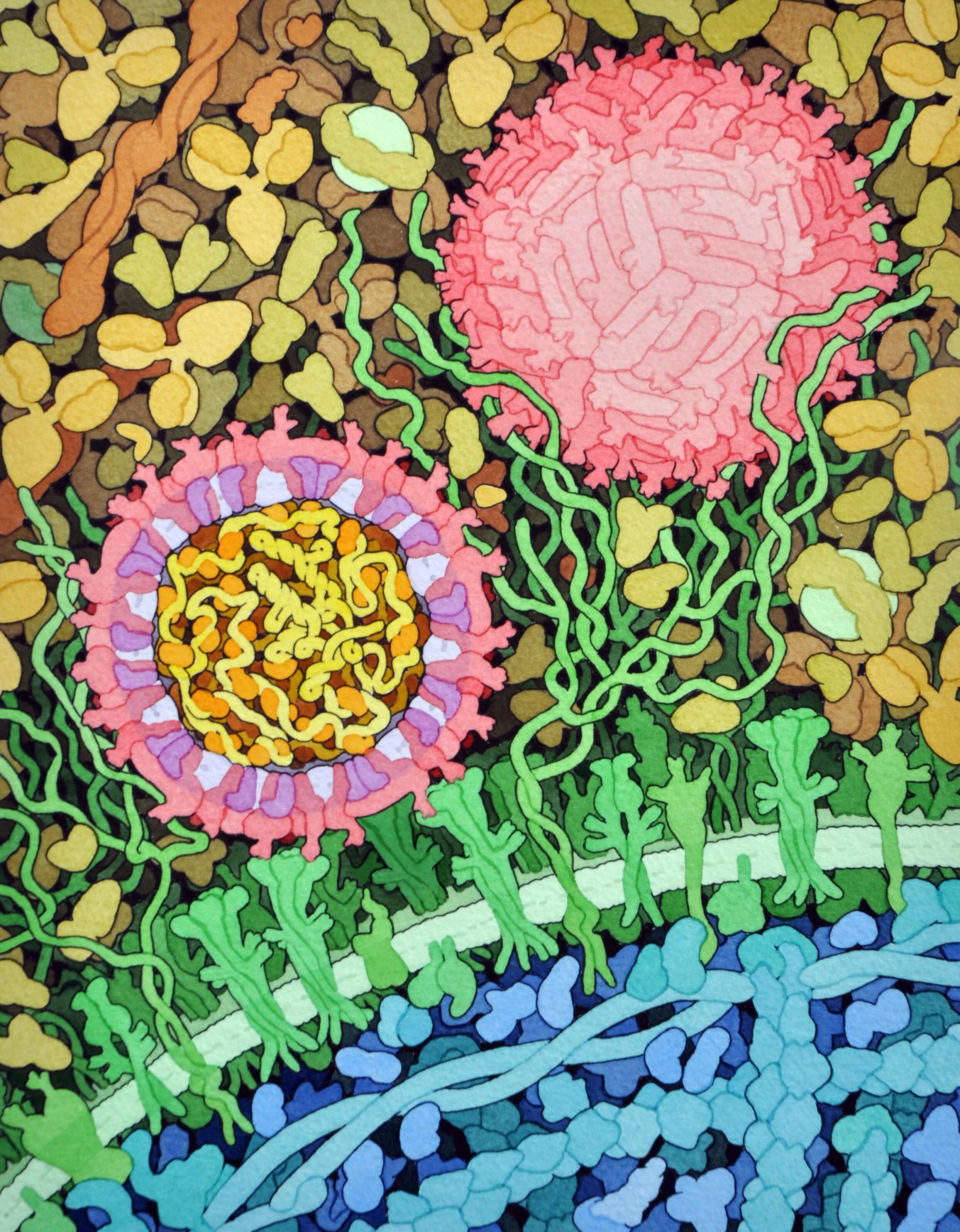
Description: English: Space-fill drawing of the outside of one Zika virus particle, and a cross-section through another as it interacts with a cell. The two main proteins of the viral envelope, the envelope proteins and membrane proteins, are shown in red and purple respectively. The lipid membrane of the envelope is shown in light lavender.The capsid proteins, in orange, are shown interacting with the RNA genome, in yellow, at the center of the virus. The cell-surface receptor proteins are in green, the cytoskeleton in blue, and blood plasma proteins in gold. Drawn by David Goodsell. Date: 1 June 2016. Source: RCSB Molecule of the Month 197, June 2016. Author: David Goodsell.
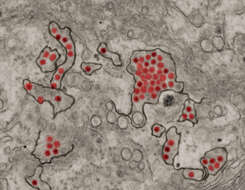
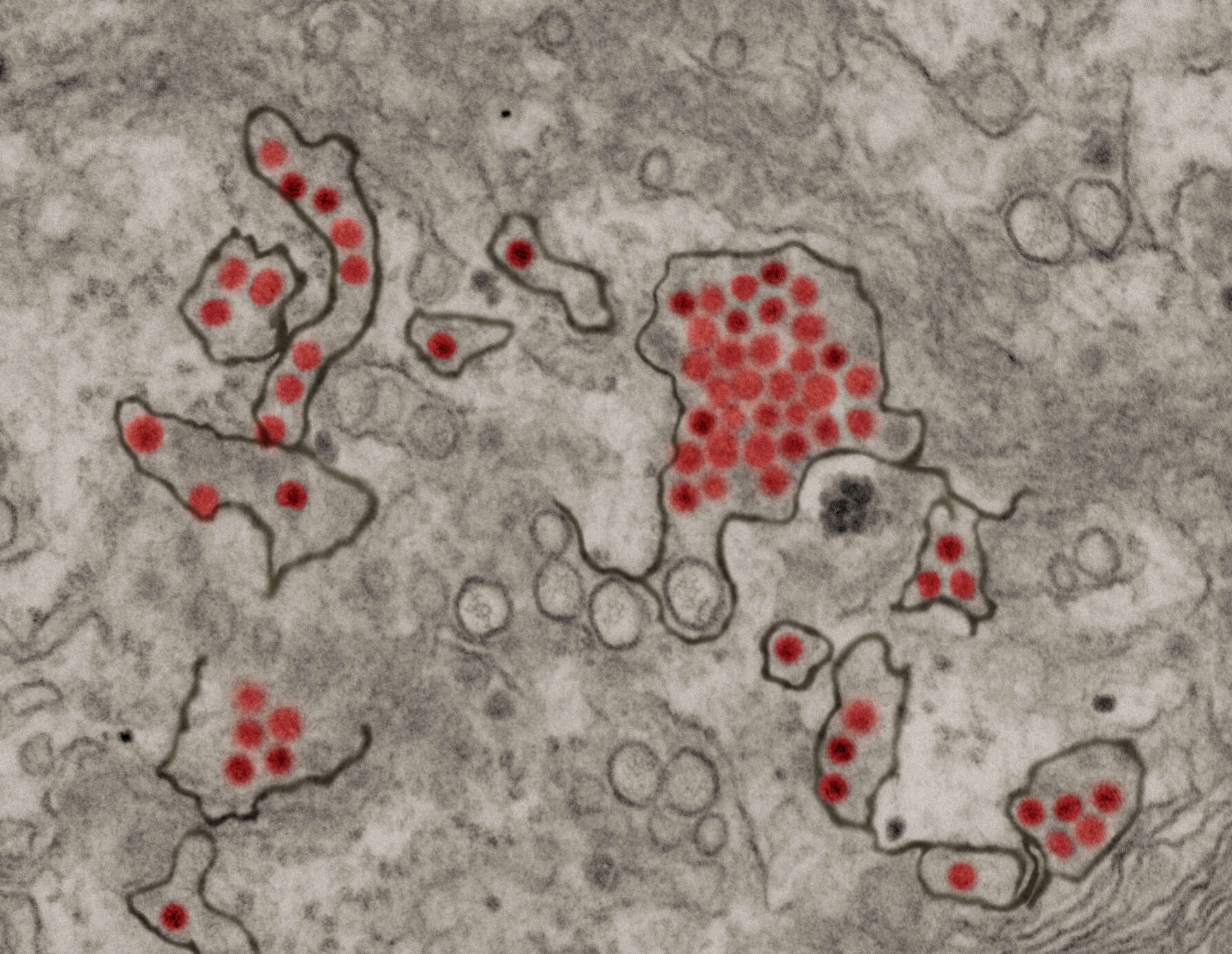
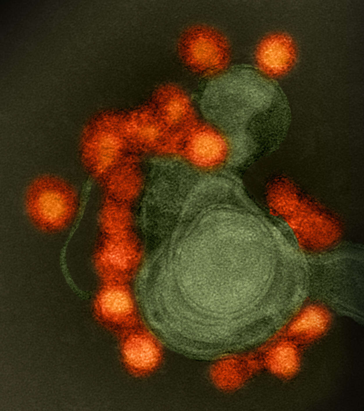
Description: Transmission electron microscope image of negative-stained, Fortaleza-strain Zika virus (red), isolated from a microcephaly case in Brazil. Credit: NIAID. Date: 19 May 2016, 17:04. Source: Zika Virus. Author: NIAID.
Description: English: Tom Frieden, MD, MPH, Director, Centers for Disease Control and Prevention (CDC); Susan Blumenthal, MD, Senior Fellow in Health Policy, New America. Date: 12 July 2016, 23:30:50. Source: https://www.flickr.com/photos/newamerica/27670841583/. Author: New America.
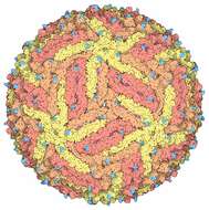
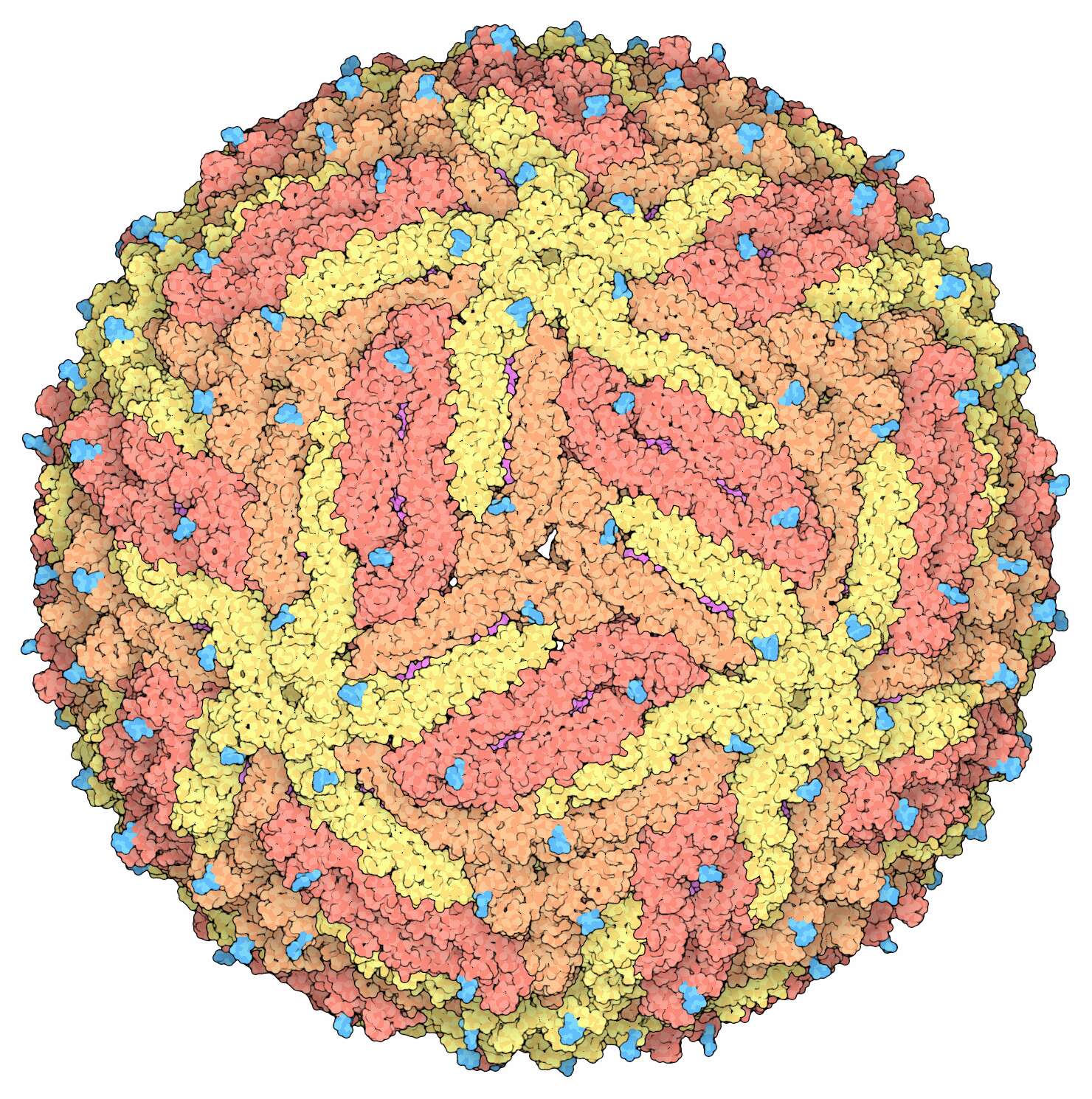
Description: English: Space-fill drawing of the Zika virus capsid, with the capsid proteins in shades of yellow and orange to show the icosahedral symmetry. The membrane proteins in the under layer (magenta) show through in some places, and the cyan protrusions are attached carbohydrate chains. Drawn by David Goodsell from the cryoEM structure 5ire. Date: 1 June 2016. Source: RCSB Molecule of the Month 197, June 2016. Author: David Goodwill.


