-
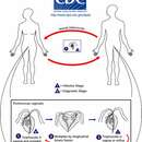
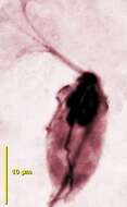
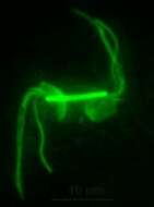
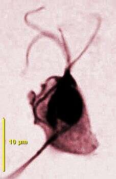
The genus Trichomonas includes polymorphic parabasalid flagellates (5-30 µm) with four free anteriorly directed flagella and a short recurrent flagellum associated with an undulating membrane shorter than the body; no free posterior recurrent flagellum. Costa relatively slender, axostylar trunk protruding posteriorly; parabasal body V-shaped with one or two long parabasal filaments. Amoeboid and polymastigotes forms present in natural or culture conditions. Several species living in the genitor-urinary tract of humans such as T. vaginalis, the mouth such as T. tenax or in the instestine of birds such as T. gallinae. Image of Trichomonas vaginalis with four anterior flagella, a recurrent flagellum associated with an undulating membrane, axostyle (Immunofluorescence with an anti-tubulin antibody).
-
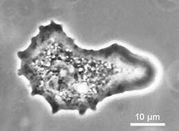
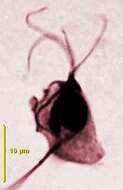
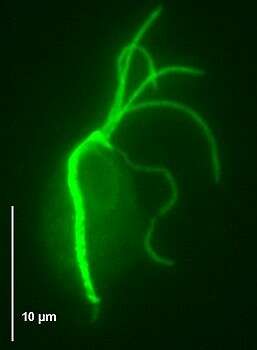
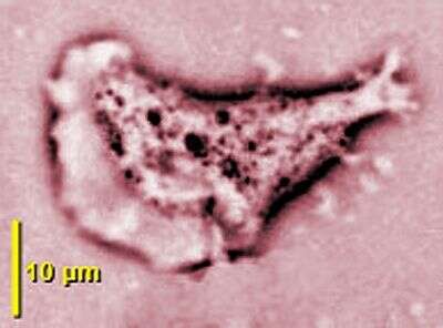
The genus Trichomonas includes polymorphic parabasalid flagellates (5-30 µm) with four free anteriorly directed flagella and a short recurrent flagellum associated with an undulating membrane shorter than the body; no free posterior recurrent flagellum. Costa relatively slender, axostylar trunk protruding posteriorly; parabasal body V-shaped with one or two long parabasal filaments. Amoeboid and polymastigotes forms present in natural or culture conditions. Several species living in the genitor-urinary tract of humans such as T. vaginalis, the mouth such as T. tenax or in the instestine of birds such as T. gallinae. Image of Trichomonas vaginalis giant adhering amoeboid cell (phase contrast).
-
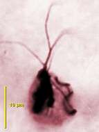
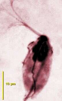
Cell showing the four anterior flagella, the short undulating membrane, the nucleus, the parabasal apparatus and the axostyle.
-
Cell stained by protargol, cell showing the four anterior flagella and the short undulating membrane and the subjacent costa.
-
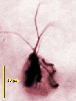
Cell showing the four anterior flagella, the short undulating membrane, the nucleus, the parabasal apparatus with the long parabasal fibre and the axostyle (protargol staining).
-
Cell stained by protargol, showing the four anterior flagella, the short undulating membrane and the subjacent costa.
-
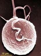
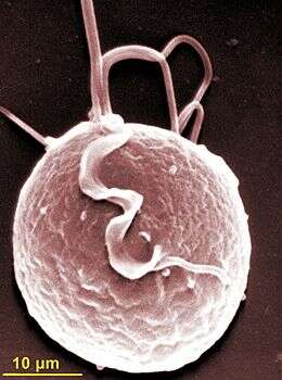
Scanning electron micrograph showing anterior flagella and the recurrent flagellum adhering to the short lamellar undulating membrane.
-
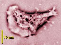
Adherent amoeboid cell by phase contrast microscopy.
-
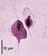
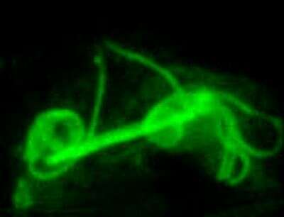
Division stages by immunofluorescence showing the two sets of flagella separated by a thick paradesmose.
-
Division, by immunofluorescence, showing the two sets of flagella separated by a thick paradesmose.