-
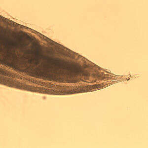
Öesophagostomum sp. adult femalePosterior end of a female Oesophagostomum sp., showing the pointed tail.From
CDC DPDx website
-
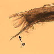
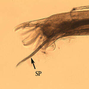
Male Oesophagostomum sp. Posterior end of male Oesophagostomum sp. showing the bursa. Note the spicule (SP).From
CDC DPDx Website
-
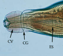
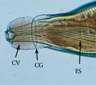
Öesophagostomum adultMagnification of the anterior end. Note the presence of the cephalic vesicle (CV), cephalic groove (CG) and esophagus (ES).From
CDC DPDx website
-
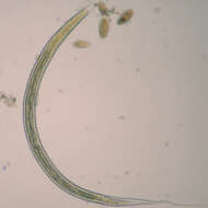
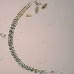
OesophagostomumL3 larva of Oesophagostomum sp., obtained via coproculture from the feces of a baboon (Papio ursinus) in South Africa. Note the long, thin, pointed tail. Image courtesy of the UTC Baboon Research Unit, University of Cape Town, South Africa.The definitive hosts of Oesophagostomum spp. become infected from the ingestion of infective L3 larvae, which develop in the environment. L3 larvae are longer than the L3 larvae of hookworms, measuring 710-950 µm in length. The intestine is made-up of alternating triangular-shaped cells (zigzag gut cells). The tail end has a long, thin, tapered sheath, with a considerable gap between the end of the tail and the end of the sheath.From
CDC-DPDx
-
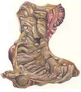
ÖesophagostomumPost-Mortem Pathology Plate borrowed from Brumpti and Thomas. "The Pathological Report of Oesophagostomiasis in Man."Colon and small intestine tissue samples from patient infected with Oesophagostomum. Important to note are the nodules lining the lumen of the colon (right).From:
"Introduction to the parasite family Oesophagostomum" (Stanford University Human Biology 103 class)
-
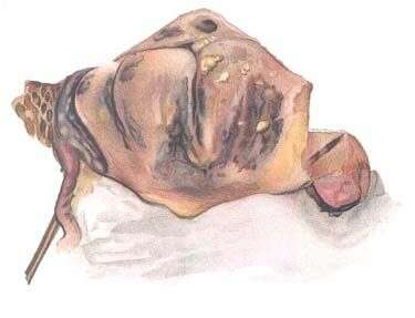
ÖesophagostomumPost-Mortem Pathology Plate borrowed from Brumpti and Thomas. "The Pathological Report of Oesophagostomiasis in Man." Worm protruding from nodule on the lining of the colon. From:
"Introduction to the parasite family Oesophagostomum" (Stanford University Human Biology 103 class)
-
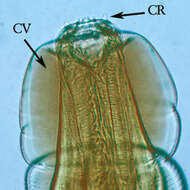
Öesophagostomum adultHigher magnification of the anterior end. Note the presence of the cephalic vesicle (CV) and corona radiata (CR).
-
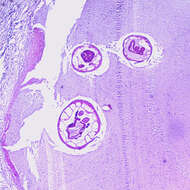
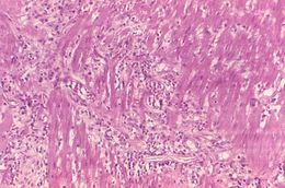
Öesophagostomum sp. adult in host tissueCross-section of an adult of Oesophagostomum sp. in a colon biopsy specimen from a patient from Africa, stained with hematoxylin and eosin (H&E). Image taken at 40x magnification.From
CDC DPDx
-
ÖesophagostomumPost-Mortem Pathology Plate borrowed from Brumpti and Thomas. "The Pathological Report of Oesophagostomiasis in Man." Outside covering of encysted worm. From:
"Introduction to the parasite family Oesophagostomum" (Stanford University Human Biology 103 class)
-
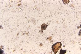
Magnified 125X, this photomicrograph revealed the presence of a fertile Ascaris sp. egg that was found in an unstained formalin-preserved stool sample. See PHIL 411 for an example of an unfertilized Ascaris lumbricoides egg.Geographic Distribution:The most common human helminthic infection, Ascaris sp. have a worldwide distribution. Their highest prevalence is in tropical and subtropical regions, and areas with inadequate sanitation. Ascariasis occurs in rural areas of the southeastern United States.Created: 1973
-
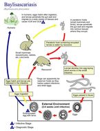
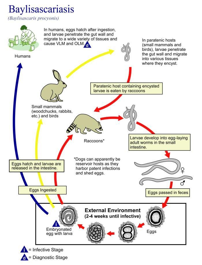
This illustration depicts the life cycle of Baylisascaris procyonis, the causal agent of Baylisascariasis.Created: 2002
-
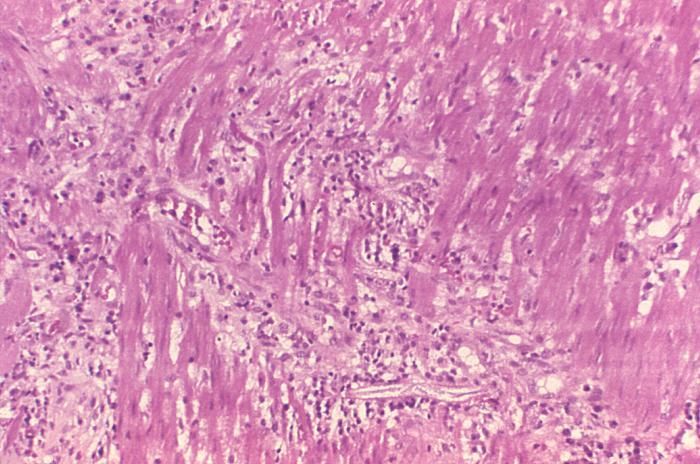
This photomicrograph reveals histopathologic changes indicative of the presence of the intestinal parasitic nematode (roundworm), Strongyloides stercoralis.Clinical Features:Frequently asymptomatic. Gastrointestinal symptoms include abdominal pain and diarrhea. Pulmonary symptoms (including Loefflers syndrome) can occur during pulmonary migration of the filariform larvae. Dermatologic manifestations include urticarial rashes in the buttocks and waist areas. Disseminated strongyloidiasis occurs in immunosuppressed patients, can present with abdominal pain, distension, shock, pulmonary and neurologic complications and septicemia, and is potentially fatal. Blood eosinophilia is generally present during the acute and chronic stages, but may be absent with dissemination.Created: 1972
-
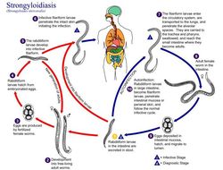
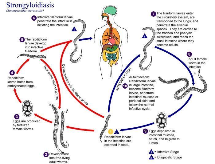
This is an illustration of the life cycle of Strongyloides stercoralis, the causal agent of Strongyloidiasis.Created: 2002
-
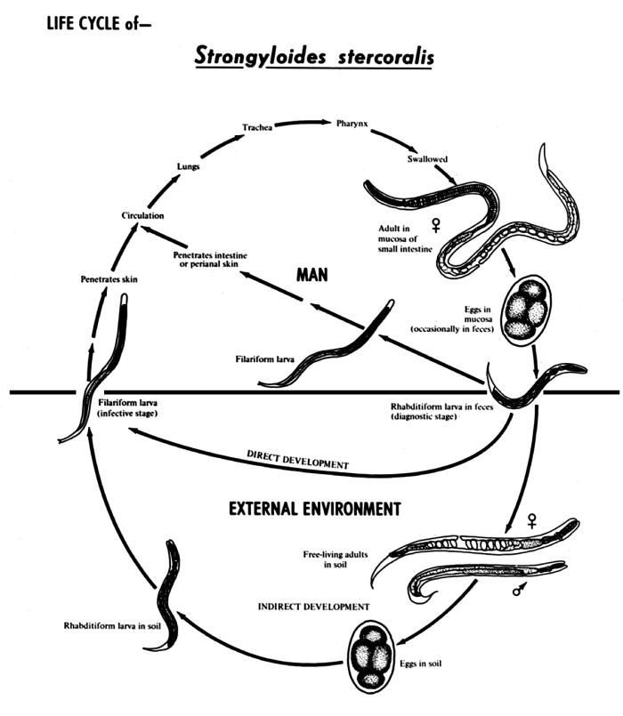
This diagram depicts the various stages in the life cycle of the Strongyloides stercoralis nematode.Created: 1982
-
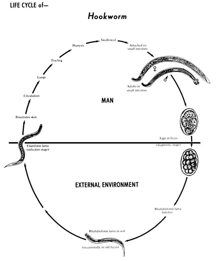
The human hookworms include two nematode (roundworm) species, Ancylostoma duodenale and Necator americanus.Created: 1982
-
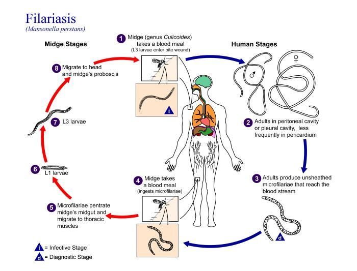
This is an illustration of the life cycle of Mansonella perstans, one of the causal agents of Filariasis.Created: 2002
-
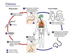
This is an illustration of the life cycle of Mansonella streptocerca, one of the causal agents of Filariasis.Created: 2002
-
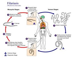
This is an illustration of the life cycle of Wuchereria bancrofti, one the causal agents of Filariasis.Created: 2003
-
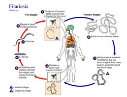
This is an illustration of the life cycle of Loa loa, one of the causal agents of Filariasis.Created: 2003
-
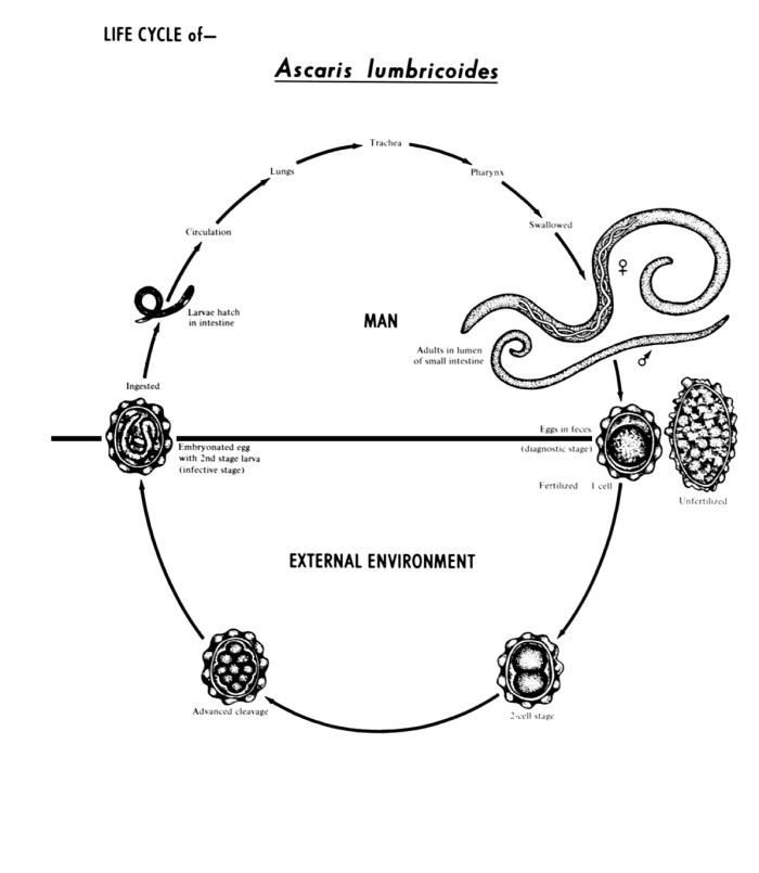
This diagram depicts the various stages in the life cycle of the intestinal roundworm nematode Ascaris lumbricoides.Created: 1982
-
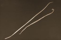
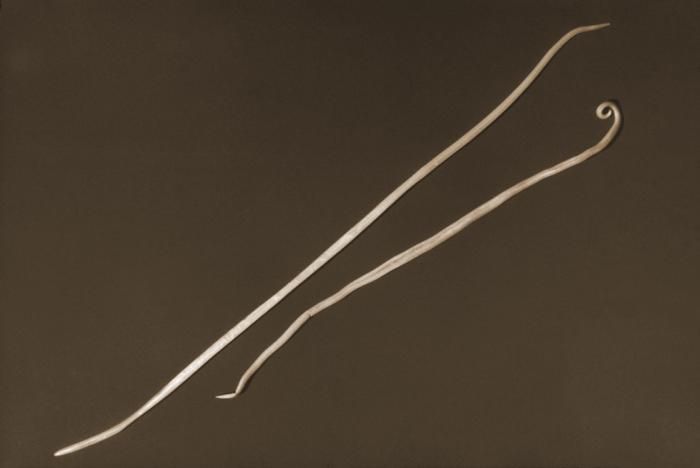
Depicted in this 1960 photograph were two Ascaris lumbricoides nematods, i.e., roundworms. The larger of the two was the female of the species, while the normally smaller male was on the right. Adult female worms can grow over 12 inches in length.Created: 1960
-
This 2007 photograph depicted Center for Disease Control/ NCZVED/DPD laboratory technician, Henry Bishop holding a mass of Ascaris lumbricoides worms, which had been passed by a child in Kenya, Africa. This nematode parasitizes the human small intestine, and is spread from human to human by the fecal-oral route. Children seem to be infected more often than adults, and though the organisms depicted here originated in Africa, the disease can be acquired in the southeastern United States.Created: 2007
-
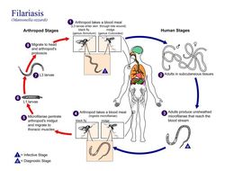
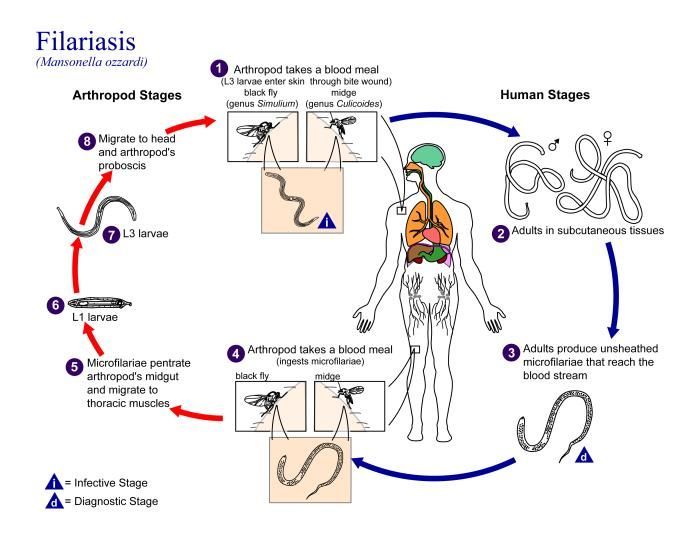
This is an illustration of the life cycle of Mansonella ozzardi, one of the causal agents of Filariasis.Created: 2002
-
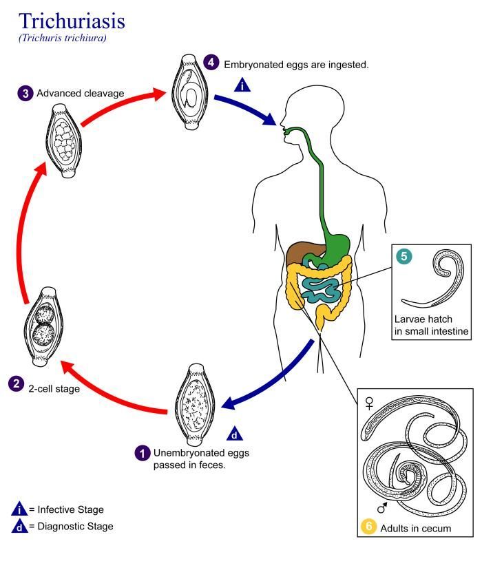
This is an illustration of the life cycle of Trichuris trichiura, the causal agent of Trichuriasis.Created: 2002