Masanori Okanishi, Timothy D. O’Hara, Toshihiko Fujita
Zookeys
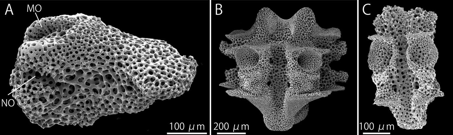
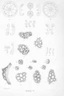
Figure 7.Squamophis albozosteres sp. n., paratype(MV F162658). SEM photographs of internal ossicles. A lateral arm plate from middle portion of the arm B, C vertebrae from middle (B) and distal (C) portion of the arm, oral views. Abbreviations: MO - muscle opening; NO - nerve opening.
Anne I. Gondim, Carmen Alonso, Thelma L. P. Dias, Cynthia L. C. Manso, Martin L. Christoffersen
Zookeys
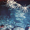
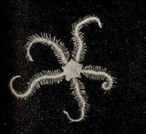
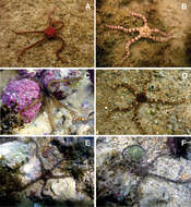
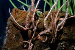
Figure 14.Some ophiurans in their natural habitat. A Ophiomyxa flaccida B Ophiolepis impressa C Ophiothrix angulata D Ophiocoma echinata E Ophioderma cinerea F Ophioderma appressa. Photos by Thelma L. P. Dias.
Rebeca Granja–Fernández, María D. Herrero-Pérezrul, Ramón A. López-Pérez, Luis Hernández, Fabián A. Rodríguez-Zaragoza, Robert Wallace Jones, Rubén Pineda-López
Zookeys
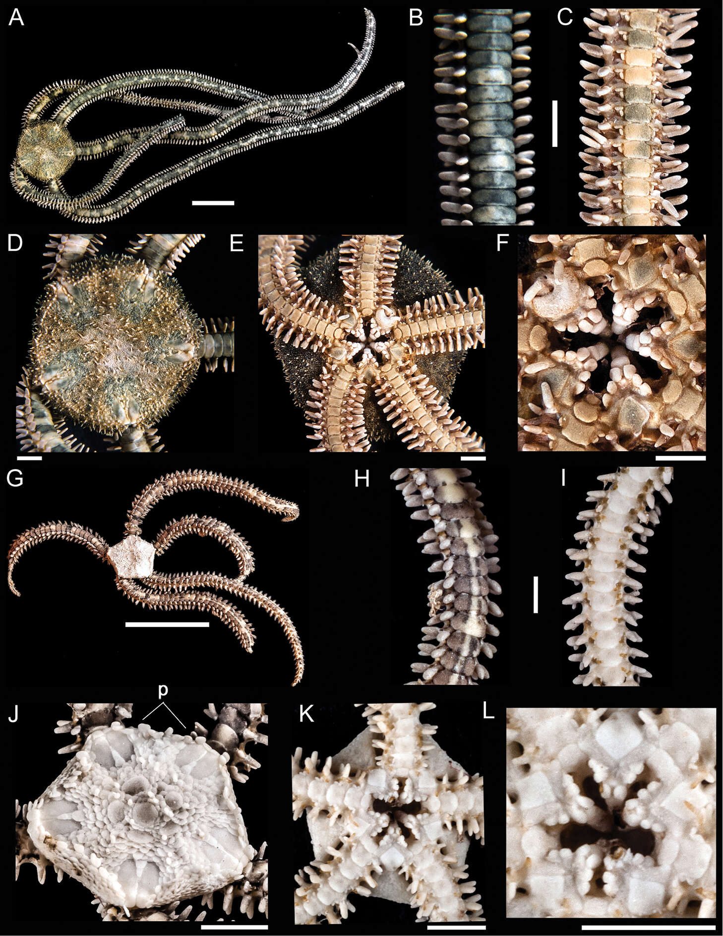
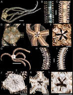
Figure 1.Ophiocnida hispida. A dorsal view. Scale bar = 5 mm B dorsal view of the arm C ventral view of the arm D dorsal view of the disk E ventral view of the disk F jaw. Scale bar = 1 mm. Ophiophragmus papillatus G dorsal view. Scale bar = 5 mm H dorsal view of the arm I ventral view of the arm J dorsal view of the disk (p = papillae around the margin of the disk) K ventral view of the disk L jaw. Scale bar = 1 mm.