Plate 7
Опис:
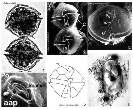
Plate 7. Alexandrium tamarense. Fig. 1. LM. Two cell chain: cells small to medium; slightly longer than wide, nearly spherical. Cingulum (C) deeply escavated and lipped. Left hypothcal lobe slightly larger than right. Nucleus (n) visible. Figs. 2-4. SEM. Fig. 2. Two cell chain: cingulum displaced 1X its width. Deep sulcus (s) widens posteriorly. Fig. 3. Epitheca: apical view. Apical pore plate (Po) rectangular; narrows ventrally. Po and first apical plate (1') in direct contact. Small ventral pore present on 1' plate. Fig. 4. Apical pore complex (APC): foramen large and fishhook shaped. Small round anterior attachment pore (aap) present (Hallegraeff 1991). Fig. 5. Line drawing. Fig. 6. LM. Oblong resting cyst with rounded ends, reddish lipid bodies; covered in mucilage.
Се јавува на следниве страници:
- Life
- Cellular
- Eukaryota (еукариот)
- SAR (Stramenopiles, Alveolates, Rhizaria)
- Alveolata
- Dinophyceae (светлечки камшичари)
- Gonyaulacales
- Gonyaulacaceae
- Alexandrium
- Alexandrium tamarense
- Dinoflagellata
Сликата ја нема во ниедна збирка.
Информации за изворот
- лиценца
- cc-publicdomain
- библиографски навод
- Faust, Maria A. and Rose A. Gulledge. Identifying Harmful Marine Dinoflagellates. Smithsonian Contributions from the United States National Herbarium, volume 42: 1-144 (including 48 plates, 1 figure and 1 table).
- изворно
- изворна податотека
- посети извор
- соработничко мреж. место
- NMNH Marine Dinoflagellates
- ID