Ventral infraciliature
Опис:
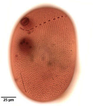
Ventral infraciliature of the large nassulid ciliate Obertrumia aurea (Ehrenberg, 1833; Foissner 1987). Synonym of Nassula aurea. Obertrumia is distinguished by it's bipartite "hypostomial frange",linear arrays of ciliary tufts. A sigmoid ventrolateral frange begins posterior to the cytostome running anteriorly and to the left (viewer's right) around to the dorsal surface. The dorsal termination is not seen in this image. The ventrolateral part of the frange terminates at the left end of the horizontal line of ciliary tufts on the dorsal surface. The uniform longitudinal somatic kineties, interrupted by the hypostomial frange, are well seen here. The multiple darkly stained micronuclei are seen overlying the spherical macronucleus in this image.The small circular structure to the viewer's left just posterior to the macronucleus is the excretory pore of the contractile vacuole. The thin dark longitudinal line posterior to this is the cytopyge (cell anus).In vivo the cell appears brightly colored (orange, green or violet) due to multiple food vacuoles containing ingested cyanobacteria. Numerous small mucocysts give the cortex a roughly granular appearance. O. aurea feeds mainly on cyanobacteria. Silver carbonate stain (see Foissner, W.Europ. J. Protistol.27,313-330;1991). Collected from a freshwater aquaculture pond near Boise, Idaho,Idaho November 2004. Brightfield optics.
Се јавува на следниве страници:
- Life
- Cellular
- Eukaryota (еукариот)
- SAR (Stramenopiles, Alveolates, Rhizaria)
- Alveolata
- Ciliophora (трепкари)
- Intramacronucleata
- Nassophorea
- Nassulida
- Nassulidae
- Obertrumia
- Obertrumia aurea
Сликата ја нема во ниедна збирка.
Информации за изворот
- лиценца
- cc-by-nc
- автор
- William Bourland
- добавувач
- micro*scope
- изворно
- изворна податотека
- посети извор
- соработничко мреж. место
- micro*scope
- ID