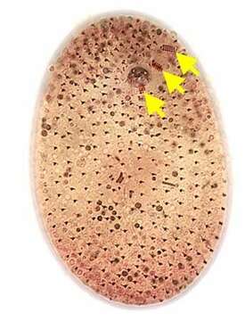
Ventral infraciliature;nassulid organelles (yellow arrows)
Опис:
Ventral infraciliature of the nassulid ciliate, Furgasonia trichocystis (Stokes, 1894) Jankowski, 1964. Synonym: Cyclogramma. The cell shape is a slightly dorsoventrally flattened ellipsoid. The left side is flattened and the right side slightly convex. The cytostome is in the anterior 1/5 of the cell in a shallow depression. It is supported by a prominent basket of obliquely oriented cytopharyngeal trichites. The somatic ciliature consists of about 32 to 26 longitudinal kineties. On the ventral surface the right kineties arch to the left anterior to the cytostome to terminate on a short but wide preoral suture. The straight left kineties terminate on this suture to the left of the cytostome. There is a short curved right paraoral membrane. There are three approximately rectangular paroral polykineties (nassulid organelles). The most anterior (M1,long arrow) is obliquely oriented in the preoral suture. The middle membrane (M2, medium arrow) is to the left of the cytostome and almost perpendicular to the long axis of the cell. The most posterior membranelle (M3, short arrow) is posterior to the cytostome, almost parallel to the long axis of the cell. These three distinctive small polykineties distinguish Furgasonia from other nassulid genera. The spherical macronucleus and adjacent micronucleus are slightly posterior to the equator. The single contractile vacuole (visible here posterior to the cytopharyngeal basket) is located in the cell center with an excretory pore on its ventral aspect. There is a prominent layer of fusiform subpellicular extrusomes (mucocysts). The cytoplasm is colorless in these bactivorous individuals. It is unclear whether this species is synonymous with F. rubens which is orange to blue colored due to ingested cyanobacteria. Morphologically the two species are quite similar aside from this coloration (see Fauré-Fremiet, E. Le Genre Cyclogramma, Perty, 1852. J. Protozool. 14: 456-464, 1967). Collected from a temporary rainwater pool with abundant decaying grass near Boise, Idaho. March, 2005. Silver carbonate (see Foissner, W. Europ. J. Protistol., 27:313-330;1991). Brightfield. This image was taken by William Bourland. He now uses a Zeiss Axioskop 2 with a Flex camera (Diagnostic Instruments).
Се јавува на следниве страници:
- Life
- Cellular
- Eukaryota (еукариот)
- SAR (Stramenopiles, Alveolates, Rhizaria)
- Alveolata
- Ciliophora (трепкари)
- Intramacronucleata
- Nassophorea
- Nassulida
- Furgasoniidae
- Furgasonia
- Furgasonia theresae
Сликата ја нема во ниедна збирка.
Информации за изворот
- лиценца
- cc-by-nc
- автор
- William Bourland
- добавувач
- micro*scope
- изворно
- изворна податотека
- посети извор
- соработничко мреж. место
- micro*scope
- ID