Image of human body louse
Description:
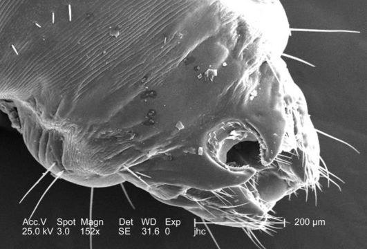
At double the magnification of PHIL #9239, this 2006 scanning electron micrograph (SEM), magnified 152x, revealed the distal tip of the abdominal region of a female body louse, Pediculus humanus var. corporis from a dorsal perspective. Some of the morphologic characteristics seen in this image include the two gonopodia, which are located dorsal to the larger two setae-bearing claspers. It is into this notch that the male would insert the aedeagus, or penis during the process of copulation. This notch, identifying the louse as a female is observable to the naked eye, whereas, in the male louse, the distal abdomen is rounded, and not concave.
Created: 2006
Included On The Following Pages:
- Life (creatures)
- Cellular (cellular organisms)
- Eukaryota (eukaryotes)
- Opisthokonta (opisthokonts)
- Metazoa (Animal)
- Bilateria
- Protostomia (protostomes)
- Ecdysozoa (ecdysozoans)
- Arthropoda (arthropods)
- Pancrustacea
- Hexapoda (hexapods)
- Insecta (insects)
- Pterygota (winged insects)
- Neoptera (neopteran)
- Paraneoptera
- Psocodea (bark lice, book lice and true lice)
- Troctomorpha (book louse)
- Pediculidae (primate body lice)
- Pediculus
- Pediculus humanus (human body louse)
- Panarthropoda
- Nanopsocetae
This image is not featured in any collections.
Source Information
- license
- cc-publicdomain
- photographer
- Janice Carr
- provider
- Public Health Image Library
- original
- original media file
- visit source
- partner site
- Public Health Image Library
- ID