Image of human body louse
Description:
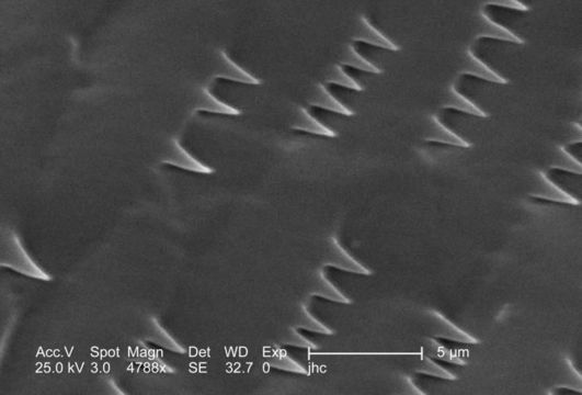
Magnified 4788x, this 2006 scanning electron micrograph (SEM) depicted an enlarged view of the chitinous, exoskeletal surface of a male louse, Pediculus humanus var. corporis. In this particular view, the exoskeletal adornments are few, consisting of small, spike-like structures. Quite often the shape and size of these skeletal adnexae are quite complex, with forms following functions, which are just as varied. The exoskeleton is composed of chitin, a molecule made up of bound units of acetylglucosamine, which is joined in such a way as to allow for increased points at which hydrogen bonding can occur. In this way chitin provides increased strength, and durability as an exoskeletal foundation.
Created: 2006
Included On The Following Pages:
- Life (creatures)
- Cellular (cellular organisms)
- Eukaryota (eukaryotes)
- Opisthokonta (opisthokonts)
- Metazoa (Animal)
- Bilateria
- Protostomia (protostomes)
- Ecdysozoa (ecdysozoans)
- Arthropoda (arthropods)
- Pancrustacea
- Hexapoda (hexapods)
- Insecta (insects)
- Pterygota (winged insects)
- Neoptera (neopteran)
- Paraneoptera
- Psocodea (bark lice, book lice and true lice)
- Troctomorpha (book louse)
- Pediculidae (primate body lice)
- Pediculus
- Pediculus humanus (human body louse)
- Panarthropoda
- Nanopsocetae
This image is not featured in any collections.
Source Information
- license
- cc-publicdomain
- photographer
- Janice Carr
- provider
- Public Health Image Library
- original
- original media file
- visit source
- partner site
- Public Health Image Library
- ID