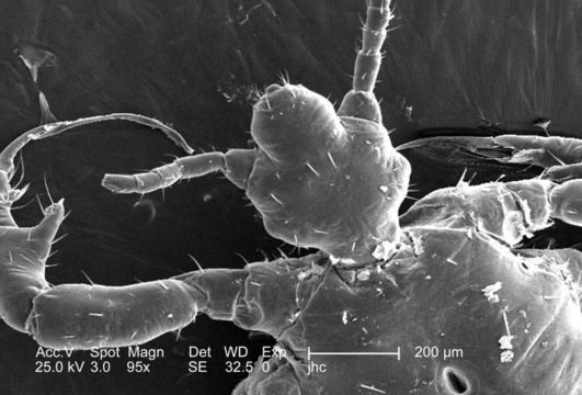
Image of human body louse
Description:
At a low magnification of 95x, this 2006 scanning electron micrograph (SEM) depicted an enlarged dorsal view of the cephalic, or head region of a male louse, Pediculus humanus var. corporis. In this particular view, the louses haustellum is withdrawn, or internalized inside the mouth region of the insects cephalic region. The haustellum is a tube-like structure, fromed from what is believed to be a modification of the labium, but is armed with teeth, which act to grab, and hold on to the skin surface of its host while obtaining its blood meal. The insect pierces the hosts skin with its sharply-pointed set of three stylets, which together for what is termed the fascicle. The pair of jointed bilateral antennae and the insects body sport sensorial hairs known as "setae, both of which provided the organism with a "picture of its environment, by taking readings in thermal, chemical, and mechanical changes encountered in its immediate surroundings.
Created: 2006
Included On The Following Pages:
- Life (creatures)
- Cellular (cellular organisms)
- Eukaryota (eukaryotes)
- Opisthokonta (opisthokonts)
- Metazoa (Animal)
- Bilateria
- Protostomia (protostomes)
- Ecdysozoa (ecdysozoans)
- Arthropoda (arthropods)
- Pancrustacea
- Hexapoda (hexapods)
- Insecta (insects)
- Pterygota (winged insects)
- Neoptera (neopteran)
- Paraneoptera
- Psocodea (bark lice, book lice and true lice)
- Troctomorpha (book louse)
- Pediculidae (primate body lice)
- Pediculus
- Pediculus humanus (human body louse)
- Panarthropoda
- Nanopsocetae
This image is not featured in any collections.
Source Information
- license
- cc-publicdomain
- photographer
- Janice Carr
- provider
- Public Health Image Library
- original
- original media file
- visit source
- partner site
- Public Health Image Library
- ID