Image of human body louse
Description:
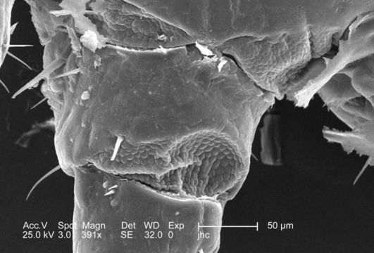
At a moderate magnification of 391x, this 2006 scanning electron micrograph (SEM) depicted an enlarged dorsal view at the proximal end of the left foreleg of a male body louse, Pediculus humanus var. corporis. The entire leg is not visible, but what is visible, includes the coxa and the trochanter, or the first and second leg segments respectively. For a complete dorsal view of the louses leg, see PHIL# 9228. The leg is composed of six segments: coxa, trochanter, femur, tibia, tarsus, and pretarsus or claw. In the case of the louse, the leg segments are very stout, and end in claws, which it used to firmly grasp clothing, or a hosts hair shafts. Note how the exoskeletal covering appears to possess an added flexibility at the coxa-trochanteric joint.
Created: 2006
Included On The Following Pages:
- Life (creatures)
- Cellular (cellular organisms)
- Eukaryota (eukaryotes)
- Opisthokonta (opisthokonts)
- Metazoa (Animal)
- Bilateria
- Protostomia (protostomes)
- Ecdysozoa (ecdysozoans)
- Arthropoda (arthropods)
- Pancrustacea
- Hexapoda (hexapods)
- Insecta (insects)
- Pterygota (winged insects)
- Neoptera (neopteran)
- Paraneoptera
- Psocodea (bark lice, book lice and true lice)
- Troctomorpha (book louse)
- Pediculidae (primate body lice)
- Pediculus
- Pediculus humanus (human body louse)
- Panarthropoda
- Nanopsocetae
This image is not featured in any collections.
Source Information
- license
- cc-publicdomain
- photographer
- Janice Carr
- provider
- Public Health Image Library
- original
- original media file
- visit source
- partner site
- Public Health Image Library
- ID