Image of human body louse
Description:
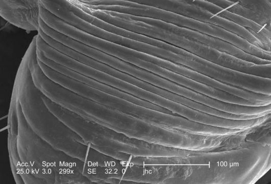
At a low magnification of 299x, this 2006 scanning electron micrograph (SEM) depicted an enlarged view of the chitinous, exoskeletal surface of a male louse, Pediculus humanus var. corporis. In this particular view, the exoskeleton seems to be configured in accordion-like convolutions, which is visible with the naked eye on the insects abdomen. Chitin is a molecule made up of bound units of acetylglucosamine, which is joined in such a way as to allow for increased points at which hydrogen bonding can occur. In this way chitin provides increased strength, and durability as an exoskeletal foundation. Note the sparse amount of setae, or sensorial hairs in this area of the abdominal surface.
Created: 2006
Included On The Following Pages:
- Life (creatures)
- Cellular (cellular organisms)
- Eukaryota (eukaryotes)
- Opisthokonta (opisthokonts)
- Metazoa (Animal)
- Bilateria
- Protostomia (protostomes)
- Ecdysozoa (ecdysozoans)
- Arthropoda (arthropods)
- Pancrustacea
- Hexapoda (hexapods)
- Insecta (insects)
- Pterygota (winged insects)
- Neoptera (neopteran)
- Paraneoptera
- Psocodea (bark lice, book lice and true lice)
- Troctomorpha (book louse)
- Pediculidae (primate body lice)
- Pediculus
- Pediculus humanus (human body louse)
- Panarthropoda
- Nanopsocetae
This image is not featured in any collections.
Source Information
- license
- cc-publicdomain
- photographer
- Janice Carr
- provider
- Public Health Image Library
- original
- original media file
- visit source
- partner site
- Public Health Image Library
- ID