Image of human body louse
Description:
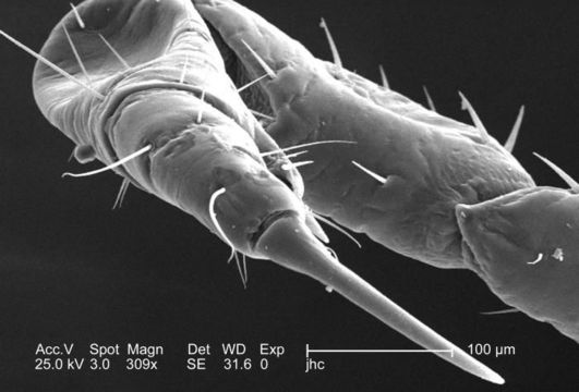
At a moderate magnification of 309x, this 2006 scanning electron micrograph (SEM) depicted an enlarged dorsal view of the right flexed foreleg of a female body louse, Pediculus humanus var. corporis. The entire leg is not quite visible, but what is visible includes the most distal segment, known as the pretarsus, followed by the more proximal tarsus, then the tibia, femur, and trochanter. The final segment, of the six segments from which each leg is composed, the coxa, is visible under reduced magnification, in PHIL# 9242. In the case of the louse, the leg segments are very stout, and end in claws, which it used to firmly grasp clothing, or a hosts hair shafts.
Created: 2006
Included On The Following Pages:
- Life (creatures)
- Cellular (cellular organisms)
- Eukaryota (eukaryotes)
- Opisthokonta (opisthokonts)
- Metazoa (Animal)
- Bilateria
- Protostomia (protostomes)
- Ecdysozoa (ecdysozoans)
- Arthropoda (arthropods)
- Pancrustacea
- Hexapoda (hexapods)
- Insecta (insects)
- Pterygota (winged insects)
- Neoptera (neopteran)
- Paraneoptera
- Psocodea (bark lice, book lice and true lice)
- Troctomorpha (book louse)
- Pediculidae (primate body lice)
- Pediculus
- Pediculus humanus (human body louse)
- Panarthropoda
- Nanopsocetae
This image is not featured in any collections.
Source Information
- license
- cc-publicdomain
- photographer
- Janice Carr
- provider
- Public Health Image Library
- original
- original media file
- visit source
- partner site
- Public Health Image Library
- ID