Image of Dasymutilla Ashmead 1899
Description:
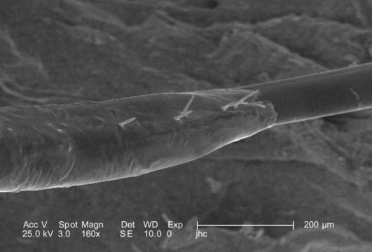
Under a moderate magnification of 160X, this scanning electron micrograph (SEM) focused on the proximal base of a female velvet ants, Dasymutilla sp., stinger. Note that the base of the stinger is encased in a sheath, with a few small sensorial hairs known as setae emanating from the surface. Also see PHIL 4638, 6363, and 6364 for photographs of the ant revealing its coloration, and velvety covering of external chitinous hairs, many of which may be seen in this image surrounding the stingers base on the insects distal abdomen.
Created: 2007
Included On The Following Pages:
- Life (creatures)
- Cellular (cellular organisms)
- Eukaryota (eukaryotes)
- Opisthokonta (opisthokonts)
- Metazoa (Animal)
- Bilateria
- Protostomia (protostomes)
- Ecdysozoa (ecdysozoans)
- Arthropoda (arthropods)
- Pancrustacea
- Hexapoda (hexapods)
- Insecta (insects)
- Pterygota (winged insects)
- Neoptera (neopteran)
- Endopterygota (endopterygotes)
- Hymenoptera (wasps, bees, and ants)
- Apocrita (wasp)
- Aculeata
- Vespoidea (Yellowjackets and Hornets, Paper Wasps; Potter, Mason and Pollen Wasps and allies)
- Mutillidae (velvet ants)
- Dasymutilla
- Panarthropoda
This image is not featured in any collections.
Source Information
- license
- cc-publicdomain
- photographer
- Janice Carr
- provider
- Public Health Image Library
- original
- original media file
- visit source
- partner site
- Public Health Image Library
- ID