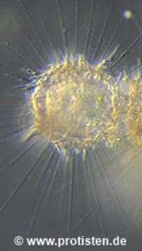
Image of Raphidocystis pallida (F. E. Schulze 1874) Zlatogursky 2018
Description:
Sampling date 10/2008.
Three specimen living close together in a feeding community in the neuston, the biofilm on the water surface. Axopodia and silicious scales are clearly visible. Diameter of one body is appr. 100 µm.
Place name: Pond Suploch, Hiddensee (Germany)
Latitude: 54.538638 Longitude: 13.097802
Microscope Zeiss Universal, camera Olympus C7070WZ.
© Wolfgang Bettighofer,
images under Creative Commons License V 3.0 (CC BY-NC-SA).
For permission to use of (high resolution) images please contact postmaster@protisten.de.
For further information about the image, please click here: Link to protisten.de page
Included On The Following Pages:
- Life (biota)
- Cellular
- Eukaryota (eukaryotes)
- Haptista
- Centroplasthelida (Centrohelid)
- Panacanthocystida
- Acanthocystida
- Raphidocystis
- Raphidocystis pallida
This image is not featured in any collections.
Source Information
- license
- cc-by-nc-sa-3.0
- copyright
- Wolfgang Bettighofer
- creator
- Wolfgang Bettighofer [email]
- original
- original media file
- visit source
- partner site
- protisten.de
- ID