in vivo, left side
Description:
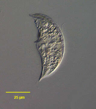
Portrait (left side) of the microthoracid ciliate Drepanomonas dentata Fresenius, 1858. The cell is crescentic in outline. The anterior and posterior ends are sharply pointed. The rigid pellicle is colorless and strongly laterally compressed. The inconspicuous cytostome is located at the midportion of the ventral surface. There is a short tooth-like projection adjacent to the cytostome. Somatic ciliature is reduced especially along the ventral surface. There are somatic kineties along the entire dorsal margin. The macronucleus is centrally located. There are two contractile vacuoles just posterior to the cytopharynx. Collected from sapropelic bottom sediments of a freshwater pond near Boise, Idaho. DIC.
Included On The Following Pages:
- Life (biota)
- Cellular
- Eukaryota (eukaryotes)
- SAR (Stramenopiles, Alveolates, Rhizaria)
- Alveolata (alveolates)
- Ciliophora (ciliates)
- Intramacronucleata
- Nassophorea
- Microthoracida
- Microthoracidae
- Drepanomonas
- Drepanomonas dentata
This image is not featured in any collections.
Source Information
- license
- cc-by-nc
- author
- William Bourland
- provider
- micro*scope
- original
- original media file
- visit source
- partner site
- micro*scope
- ID