portrait
Description:
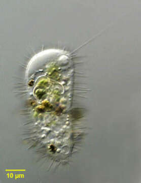
Portrait of Mastigina setosa a flagellated pelobiont. The ameboid monopodial cells have radiating "setae" which are 6-10 micron long filaments each of which has a basal granule (seen in this image). There is a single, long, lazily beating flagellum, which is closely associated with the nucleus (seen at the 12 o clock position adjacent to the cell membrane in this image). Electron microscopy shows the base of the flagellum connected to the nucleus by a cone of microtubules. The flagellum has a variable arrangement of microtubules. The nucleus contains a large endosome. Ingested algae are seen in food vacuoles. There is vigorous cytoplasmic streaming resulting in ameboid locomotion. The flagellum appears to contribute little to motility. Mastigina and the other pelobionts lack mitochondria and dictyosomes. Obvious ameboid locomotion and cytoplasmic streaming may help differentiate Mastigina from Mastigamoeba, a similar pelobiont. From slow-moving organically enriched freshwater runoff stream near Boise, Idaho. Differential interference contrast. Differential interference contrast optics.
Included On The Following Pages:
- Life (creatures)
- Cellular (cellular organisms)
- Eukaryota (eukaryotes)
- Amoebozoa (amoeboid protists)
- Evosea
- Archamoebae
- Mastigamoebida
- Mastigamoebidae
- Mastigina
- Mastigina setosa
This image is not featured in any collections.
Source Information
- license
- cc-by-nc
- author
- William Bourland
- provider
- micro*scope
- original
- original media file
- visit source
- partner site
- micro*scope
- ID