Stained specimen
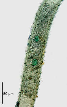
Description:
The stichotrichine ciliate, Chaetospira remex (Hudson,1875; Kahl,1932) stained with methyl green-pyronin to demonstrate the bipartite macronucleus. The animal is completely withdrawn into the tubular lorica. The two round macronuclei are stained green. The micronucleus is not seen in this image. The illustration in Kahl's compendium incorrectly depicts C. remex with a single macronucleus (Kahl,A.; Die Tierwelt Deutschlands und der angrenzenden Meeresteile. Teil 25 [Urtiere oder Protozoa I: Wimpertiere oder Ciliata (Infusoria) 3. Spirotricha. Germany:Verlag von Gustav Fischer. 1932, p. 542).Collected from a freshwater pond near Boise Idaho May 2004. Brightfield illumination with closed condenser.
Included On The Following Pages:
- Life (creatures)
- Cellular (cellular organisms)
- Eukaryota (eukaryotes)
- SAR (Stramenopiles, Alveolates, Rhizaria)
- Alveolata (alveolates)
- Ciliophora (ciliates)
- Intramacronucleata
- Spirotrichea
- Stichotrichia
- Stichotrichida
- Spirofilidae
- Chaetospira
- Chaetospira remex
This image is not featured in any collections.
Source Information
- license
- cc-by-nc
- author
- William Bourland
- provider
- micro*scope
- original
- original media file
- visit source
- partner site
- micro*scope
- ID