Fig 2

Description:
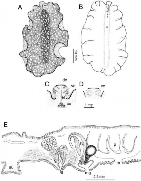
A, diagram of dorsal view. B, ventral morphology. C, dorsal view of pseudotentacles. D, ventral view of pseudotentacles. E, sagittal reconstruction of genital system portion. bl: black line; ce: cerebral eye; de: dorsal eyes of the pseudotentacle; fg: female gonopore; g: cement glands; m: mouth; mg: male gonopore; p: pharynx; su: sucker; s: stylet; sv: seminal vesicle; v: vagina; ve: ventral eyes of the pseudotentacles.
Included On The Following Pages:
- Life (creatures)
- Cellular (cellular organisms)
- Eukaryota (eukaryotes)
- Opisthokonta (opisthokonts)
- Metazoa (Animal)
- Bilateria
- Protostomia (protostomes)
- Spiralia (spiralians)
- Platyhelminthes (flatworms)
- unclassified Platyhelminthes
- Polycladida
- Pseudocerotoidea
- Pseudocerotidae
- Phrikoceros
- Phrikoceros mopsus
This image is not featured in any collections.
Source Information
- license
- cc-by-nc-sa-4.0
- copyright
- WoRMS Editorial Board
- original
- original media file
- visit source
- partner site
- World Register of Marine Species
- ID