Pseudo-nitzschia abrensis
Description:
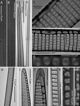
(A and B) Light micrographs. (C–M) TEM micrographs. (A and B) Whole valves. (C and D) Part of the cell in valve view. (E) Detail of the central part of a valve showing the central nodule and the striae with a row of poroids split into two sectors (seldom three or four). (F) Part of a valve showing the central nodule and the poroid arrangement. (G) Detail of the poroid structure. (H) Central part of a valve. (I) Apical part of a valve showing the proximal and distal mantles. (J) Valve end. (K) Valvocopula. (L and M) Different cingular bands. (A–D, G–L) Strain Ner-J2. (E, F, and M) Strain Ner-J3.
Included On The Following Pages:
- Life (creatures)
- Cellular (cellular organisms)
- Eukaryota (eukaryotes)
- SAR (Stramenopiles, Alveolates, Rhizaria)
- Stramenopiles (heterokont)
- Ochrophyta (Ochrophyte)
- Bacillariophyta (diatoms)
- Bacillariophyceae
- Bacillariophycidae
- Bacillariales
- Bacillariaceae
- Pseudo-nitzschia
- Pseudo-nitzschia abrensis
This image is not featured in any collections.
Source Information
- license
- cc-by-nc-sa-4.0
- copyright
- WoRMS Editorial Board
- contributor
- Orive, Emma
- original
- original media file
- visit source
- partner site
- World Register of Marine Species
- ID