Thelazia callipaeda female
Description:
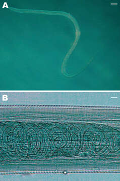
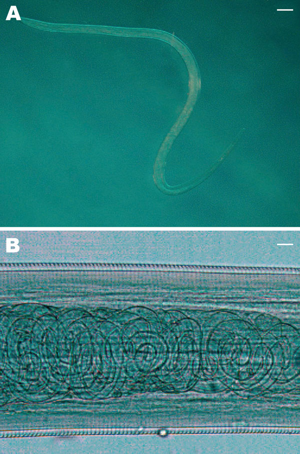
Description: English: Figure 2 A) Female Thelazia callipaeda isolated from patient 4. The posterior end is on the left and the anterior end is on the right (magnification × 200). Scale bar = 500 μm. B) T. callipaeda mature first-stage larvae in the distal uterus (magnification ×100). Scale bar = 30 μm. Date: April 2008. Source: Otranto, D. & Dutto, M. (2008). "Human thelaziasis, Europe". Emerging Infectious Diseases 14 (4): 647–649. DOI:10.3201/eid1404.071205. Author: Domenico Otranto & Moreno Dutto. Other versions: : This file supersedes the file Thelazia callipaeda female.gif. It is recommended to use this file rather than the other one. Alemannisch | العربية | беларуская (тарашкевіца) | български | čeština | dansk | Deutsch | Ελληνικά | English | Esperanto | español | euskara | suomi | français | galego | עברית | हिन्दी | hrvatski | magyar | Հայերեն | Bahasa Indonesia | italiano | 日本語 | 한국어 | lietuvių | македонски | Plattdüütsch | Nederlands | norsk nynorsk | norsk | polski | português | română | русский | slovenščina | српски / srpski | svenska | Türkçe | vèneto | 中文(简体) | 中文(繁體) | +/− :.
Included On The Following Pages:
- root
- cellular organisms
- Eukaryota (eukaryotes)
- Opisthokonta (opisthokonts)
- Metazoa (Animal)
- Eumetazoa
- Bilateria
- Protostomia (protostomes)
- Ecdysozoa (ecdysozoans)
- Nematoda (nematodes)
- Chromadorea
- Rhabditida (rhabditid)
- Spirurina
- Spiruromorpha
- Thelazioidea
- Thelaziidae
- Thelazia
- Thelazia callipaeda
This image is not featured in any collections.
Source Information
- license
- cc-publicdomain
- creator
- Domenico Otranto & Moreno Dutto
- source
- {{cite journal
- original
- original media file
- visit source
- partner site
- Wikimedia Commons
- ID