CT scan of head of Mastacembelus moorii - 177-734-1-PB-top-right
Description:
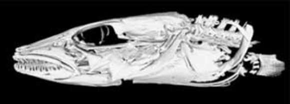
Description: English: Cropped from file:Comparison of CT scans of heads of Mastacembelus species - 177-734-1-PB.jpeg, which has the following description: These 3D scans were generated using high-resolution x-ray computed tomography. Each image is the result of over 2000 individual x-rays of the whole animal, which are then compiled to produce 3D images displaying only the bone structure underneath the skin. The skulls displayed here are different species of spiny eels that are found only in Lake Tanganyika, East Africa, where 14 species have diversified in terms of genetics, ecology and morphology. This comparison demonstrates the striking diversity in skull structure and function of four of Lake Tanganyika's spiny eels. These morphological differences are correlated to differences in feeding strategy, and, like Darwin's famous finches, are indicative of an adaptive radiation, where species rapidly evolve from a common ancestor through adaption to different environments. (Clockwise from top left: Mastacembelus albomaculatus, M. moorii, M. platysoma, M. apectoralis). Date: 19 July 2013, 02:31:32. Source: Brown, K 2012. X-ray computed tomography of spiny eels: Using state-of-the-art imaging technology to shed light on evolutionary processes. Opticon1826 7(12):21, doi:10.5334/opt.121213. Author: Brown, Katherine.
Included On The Following Pages:
- Life (creatures)
- Cellular (cellular organisms)
- Eukaryota (eukaryotes)
- Opisthokonta (opisthokonts)
- Metazoa (Animal)
- Bilateria
- Deuterostomia (deuterostomes)
- Chordata (Chordates)
- Vertebrata (vertebrates)
- Gnathostomata (jawed fish)
- Osteichthyes
- Actinopterygii (ray-finned fishes)
- Neopterygii
- Teleostei
- Euteleostei
- Neoteleostei
- Acanthopterygii
- Synbranchiformes (swamp eels)
- Mastacembeloidei
- Mastacembelidae (freshwater spiny eels)
- Mastacembelus
- Mastacembelus moorii
This image is not featured in any collections.
Source Information
- license
- cc-by-3.0
- copyright
- Brown, Katherine
- original
- original media file
- visit source
- partner site
- Wikimedia Commons
- ID