Iridovirus from Rana temporaria
Description:
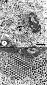
Description: English: Transmission electron micrographs of iridovirus cultured from the liver of a naturally diseased common frog (Rana temporaria) by using a fathead minnow epithelial cell line. 4a. Virus-infected cell. Large isocahedral viruses are conspicuous within the cytoplasm (arrows). Bar = 2 µm. 4b. Paracrystalline array of iridovirus. Bar = 200 µm. Date: 1999. Source: Daszak P, Berger L, Cunningham A, Hyatt A, Green D, Speare R. (1999). "Emerging Infectious Diseases and Amphibian Population Declines". Emerging Infectious Diseases 5 (6): 735–748. DOI:10.3201/eid0506.990601. Author: Peter Daszak, Lee Berger, Andrew A. Cunningham, Alex D. Hyatt, D. Earl Green, and Rick Speare.
Included On The Following Pages:
- Life (creatures)
- Viruses
- Pimascovirales
- Iridoviridae (invertebrate iridescent virus and relatives)
- Betairidovirinae
- Iridovirus
This image is not featured in any collections.
Source Information
- license
- cc-publicdomain
- creator
- Peter Daszak, Lee Berger, Andrew A. Cunningham, Alex D. Hyatt, D. Earl Green, and Rick Speare
- source
- Daszak P, Berger L, Cunningham A, Hyatt A, Green D, Speare R. (1999). "Emerging Infectious Diseases and Amphibian Population Declines". Emerging Infectious Diseases 5 (6): 735–748. DOI:10.3201/eid0506.990601.
- original
- original media file
- visit source
- partner site
- Wikimedia Commons
- ID