1937 Smithsonian miscellaneous collections Snodgrass 1936 p55 f20
Description:
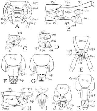
Summary.mw-parser-output table.commons-file-information-table,.mw-parser-output.fileinfotpl-type-information{border:1px solid #a2a9b1;background-color:#f8f9fa;padding:5px;font-size:95%;border-spacing:2px;box-sizing:border-box;margin:0;width:100%}.mw-parser-output table.commons-file-information-table>tbody>tr,.mw-parser-output.fileinfotpl-type-information>tbody>tr{vertical-align:top}.mw-parser-output table.commons-file-information-table>tbody>tr>td,.mw-parser-output table.commons-file-information-table>tbody>tr>th,.mw-parser-output.fileinfotpl-type-information>tbody>tr>td,.mw-parser-output.fileinfotpl-type-information>tbody>tr>th{padding:4px}.mw-parser-output.fileinfo-paramfield{background:#ccf;text-align:right;padding-right:0.4em;width:15%;font-weight:bold}.mw-parser-output.commons-file-information-table+table.commons-file-information-table,.mw-parser-output.commons-file-information-table+div.commons-file-information-table>table{border-top:0;padding-top:0;margin-top:-8px}@media only screen and (max-width:719px){.mw-parser-output table.commons-file-information-table,.mw-parser-output.commons-file-information-table.fileinfotpl-type-information{border-spacing:0;padding:0;word-break:break-word;width:100%!important}.mw-parser-output.commons-file-information-table>tbody,.mw-parser-output.fileinfotpl-type-information>tbody{display:block}.mw-parser-output.commons-file-information-table>tbody>tr>td,.mw-parser-output.commons-file-information-table>tbody>tr>th,.mw-parser-output.fileinfotpl-type-information>tbody>tr>td,.mw-parser-output.fileinfotpl-type-information>tbody>tr>th{padding:0.2em 0.4em;text-align:left;text-align:start}.mw-parser-output.commons-file-information-table>tbody>tr,.mw-parser-output.fileinfotpl-type-information>tbody>tr{display:flex;flex-direction:column}.mw-parser-output.commons-file-information-table+table.commons-file-information-table,.mw-parser-output.commons-file-information-table+div.commons-file-information-table>table{margin-top:-1px}.mw-parser-output.fileinfo-paramfield{box-sizing:border-box;flex:1 0 100%;width:100%}} Description: English: Fig. 20. — Chilopoda : external genitalia. A, Scolopendra, posterior end of body of embryo, showing two small segments in genital region between last leg-bearing segment and telson (from Heymons, 1901). B, Scolopocryptops, male, terminal part of body with penis protracted. C, same, female, genital and anal segments. D, same, male, genital and anal segments, lateral view. E, same, male, posterior end of body, ventral view. F, Lithobius, female, posterior end of body, ventral view. G, same, corresponding segments of male. H, same, female, lateral view of posterior body segments. I, same, male, ventral view of penis and ducts. J, Scutigera forceps, male, posterior end of body, ventral view. K, same, corresponding segments of female, ventral view. An, anus ; Cx, coxa ; Cxpd, coxopodite ; Dct, genital duct ; Gp, gonopod : iGp, 2Gp, first and second gonopods ; gS, genital sternum; igS, 2gS, first and second genital sterna ; igSeg, first genital, or pregenital, segment ; sgSeg, second genital, or genital, segment; gT, genital tergum; Pen, penis; Sex, sub- coxa; Stn, sternum; T, tergum; Tel, telson; Tr, trochanter. Date: 1937. Source: Snodgrass, Robert Evans. 1936. Morphology of the insect abdomen. Part III. The male genitalia (including arthropods other than insects). Smithsonian Miscellaneous Collections. 95:1–96. Author: Robert Evans Snodgrass.
Included On The Following Pages:
- Life (creatures)
- Cellular (cellular organisms)
- Eukaryota (eukaryotes)
- Opisthokonta (opisthokonts)
- Metazoa (Animal)
- Bilateria
- Protostomia (protostomes)
- Ecdysozoa (ecdysozoans)
- Arthropoda (arthropods)
- Myriapoda (myriapods)
- Chilopoda (centipedes)
- Pleurostigmophora
- Scolopendromorpha (Bark Centipedes)
- Scolopendridae
- Scolopendrinae
- Scolopendra (Centipede)
This image is not featured in any collections.